|
Preprophase
Preprophase is an additional phase during mitosis in plant cells that does not occur in other eukaryotes such as animals or fungi. It precedes prophase and is characterized by two distinct events: #The formation of the preprophase band, a dense microtubule ring underneath the plasma membrane. #The initiation of microtubule nucleation at the nuclear envelope. Function of preprophase in the cell cycle Plant cells are fixed with regards to their neighbor cells within the tissues they are growing in. In contrast to animals where certain cells can migrate within the embryo to form new tissues, the seedlings of higher plants grow entirely based on the orientation of cell division and subsequent elongation and differentiation of cells within their cell walls. Therefore, the accurate control of cell division planes and placement of the future cell wall in plant cells is crucial for the correct architecture of plant tissues and organs. The preprophase stage of somatic plant cell mit ... [...More Info...] [...Related Items...] OR: [Wikipedia] [Google] [Baidu] |
Prophase
Prophase () is the first stage of cell division in both mitosis and meiosis. Beginning after interphase, DNA has already been replicated when the cell enters prophase. The main occurrences in prophase are the condensation of the chromatin reticulum and the disappearance of the nucleolus. Staining and microscopy Microscopy can be used to visualize condensed chromosomes as they move through meiosis and mitosis. Various DNA stains are used to treat cells such that condensing chromosomes can be visualized as the move through prophase. The giemsa G-banding technique is commonly used to identify mammalian chromosomes, but utilizing the technology on plant cells was originally difficult due to the high degree of chromosome compaction in plant cells. G-banding was fully realized for plant chromosomes in 1990. During both meiotic and mitotic prophase, giemsa staining can be applied to cells to elicit G-banding in chromosomes. Silver staining, a more modern technology, in conju ... [...More Info...] [...Related Items...] OR: [Wikipedia] [Google] [Baidu] |
Preprophase Band
The preprophase band is a microtubule array found in plant cells that are about to undergo cell division and enter the preprophase stage of the plant cell cycle. Besides the phragmosome, it is the first microscopically visible sign that a plant cell is about to enter mitosis. The preprophase band was first observed and described by Jeremy Pickett-Heaps and Donald Northcote at Cambridge University in 1966. Just before mitosis starts, the preprophase band forms as a dense band of microtubules around the phragmosome and the future division plane just below the plasma membrane. It encircles the nucleus at the equatorial plane of the future mitotic spindle when dividing cells enter the G2 phase of the cell cycle after DNA replication is complete. The preprophase band consists mainly of microtubules and microfilaments (actin) and is generally 2-3 µm wide. When stained with fluorescent markers, it can be seen as two bright spots close to the cell wall on either side of the nucleu ... [...More Info...] [...Related Items...] OR: [Wikipedia] [Google] [Baidu] |
Mitosis
In cell biology, mitosis () is a part of the cell cycle in which replicated chromosomes are separated into two new nuclei. Cell division by mitosis gives rise to genetically identical cells in which the total number of chromosomes is maintained. Therefore, mitosis is also known as equational division. In general, mitosis is preceded by S phase of interphase (during which DNA replication occurs) and is often followed by telophase and cytokinesis; which divides the cytoplasm, organelles and cell membrane of one cell into two new cells containing roughly equal shares of these cellular components. The different stages of mitosis altogether define the mitotic (M) phase of an animal cell cycle—the division of the mother cell into two daughter cells genetically identical to each other. The process of mitosis is divided into stages corresponding to the completion of one set of activities and the start of the next. These stages are preprophase (specific to plant cells), p ... [...More Info...] [...Related Items...] OR: [Wikipedia] [Google] [Baidu] |
Microtubule Organizing Center
The microtubule-organizing center (MTOC) is a structure found in eukaryotic cells from which microtubules emerge. MTOCs have two main functions: the organization of eukaryotic flagella and cilia and the organization of the mitotic and meiotic spindle apparatus, which separate the chromosomes during cell division. The MTOC is a major site of microtubule nucleation and can be visualized in cells by immunohistochemical detection of γ-tubulin. The morphological characteristics of MTOCs vary between the different phyla and kingdoms. In animals, the two most important types of MTOCs are 1) the basal bodies associated with cilia and flagella and 2) the centrosome associated with spindle formation. Organization Microtubule-organizing centers function as the site where microtubule formation begins, as well as a location where free-ends of microtubules attract to. Within the cells, microtubule-organizing centers can take on many different forms. An array of microtubules can arrange t ... [...More Info...] [...Related Items...] OR: [Wikipedia] [Google] [Baidu] |
Centrosome
In cell biology, the centrosome (Latin centrum 'center' + Greek sōma 'body') (archaically cytocentre) is an organelle that serves as the main microtubule organizing center (MTOC) of the animal cell, as well as a regulator of cell-cycle progression. The centrosome provides structure for the cell. The centrosome is thought to have evolved only in the metazoan lineage of eukaryotic cells. Fungi and plants lack centrosomes and therefore use other structures to organize their microtubules. Although the centrosome has a key role in efficient mitosis in animal cells, it is not essential in certain fly and flatworm species. Centrosomes are composed of two centrioles arranged at right angles to each other, and surrounded by a dense, highly structured mass of protein termed the pericentriolar material (PCM). The PCM contains proteins responsible for microtubule nucleation and anchoring — including γ-tubulin, pericentrin and ninein. In general, each centriole of the centr ... [...More Info...] [...Related Items...] OR: [Wikipedia] [Google] [Baidu] |
Cytoplasm
In cell biology, the cytoplasm is all of the material within a eukaryotic cell, enclosed by the cell membrane, except for the cell nucleus. The material inside the nucleus and contained within the nuclear membrane is termed the nucleoplasm. The main components of the cytoplasm are cytosol (a gel-like substance), the organelles (the cell's internal sub-structures), and various cytoplasmic inclusions. The cytoplasm is about 80% water and is usually colorless. The submicroscopic ground cell substance or cytoplasmic matrix which remains after exclusion of the cell organelles and particles is groundplasm. It is the hyaloplasm of light microscopy, a highly complex, polyphasic system in which all resolvable cytoplasmic elements are suspended, including the larger organelles such as the ribosomes, mitochondria, the plant plastids, lipid droplets, and vacuoles. Most cellular activities take place within the cytoplasm, such as many metabolic pathways including glycolysis, and proc ... [...More Info...] [...Related Items...] OR: [Wikipedia] [Google] [Baidu] |
Chromosome
A chromosome is a long DNA molecule with part or all of the genetic material of an organism. In most chromosomes the very long thin DNA fibers are coated with packaging proteins; in eukaryotic cells the most important of these proteins are the histones. These proteins, aided by chaperone proteins, bind to and condense the DNA molecule to maintain its integrity. These chromosomes display a complex three-dimensional structure, which plays a significant role in transcriptional regulation. Chromosomes are normally visible under a light microscope only during the metaphase of cell division (where all chromosomes are aligned in the center of the cell in their condensed form). Before this happens, each chromosome is duplicated (S phase), and both copies are joined by a centromere, resulting either in an X-shaped structure (pictured above), if the centromere is located equatorially, or a two-arm structure, if the centromere is located distally. The joined copies are now ca ... [...More Info...] [...Related Items...] OR: [Wikipedia] [Google] [Baidu] |
Gametophyte
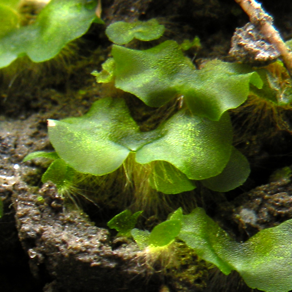
A gametophyte () is one of the two alternation of generations, alternating multicellular organism, multicellular phases in the life cycles of plants and algae. It is a haploid multicellular organism that develops from a haploid spore that has one set of chromosomes. The gametophyte is the Sexual reproduction of plants, sexual phase in the life cycle of plants and algae. It develops sex organs that produce gametes, haploid sex cells that participate in fertilization to form a diploid zygote which has a double set of chromosomes. Cell division of the zygote results in a new diploid multicellular organism, the second stage in the life cycle known as the sporophyte. The sporophyte can produce haploid spores by meiosis that on germination produce a new generation of gametophytes. Algae In some multicellular green algae (''Ulva lactuca'' is one example), red algae and brown algae, sporophytes and gametophytes may be externally indistinguishable (isomorphic). In ''Ulva (genus), Ulv ... [...More Info...] [...Related Items...] OR: [Wikipedia] [Google] [Baidu] |
Microfilament
Microfilaments, also called actin filaments, are protein filaments in the cytoplasm of eukaryotic cells that form part of the cytoskeleton. They are primarily composed of polymers of actin, but are modified by and interact with numerous other proteins in the cell. Microfilaments are usually about 7 nm in diameter and made up of two strands of actin. Microfilament functions include cytokinesis, amoeboid movement, cell motility, changes in cell shape, endocytosis and exocytosis, cell contractility, and mechanical stability. Microfilaments are flexible and relatively strong, resisting buckling by multi-piconewton compressive forces and filament fracture by nanonewton tensile forces. In inducing cell motility, one end of the actin filament elongates while the other end contracts, presumably by myosin II molecular motors. Additionally, they function as part of actomyosin-driven contractile molecular motors, wherein the thin filaments serve as tensile platforms for myosin's ATP- ... [...More Info...] [...Related Items...] OR: [Wikipedia] [Google] [Baidu] |
Cell Plate
image:Phragmoplast.png, 300px, Phragmoplast and cell plate formation in a plant cell during cytokinesis. Left side: Phragmoplast forms and cell plate starts to assemble in the center of the cell. Towards the right: Phragmoplast enlarges in a donut-shape towards the outside of the cell, leaving behind mature cell plate in the center. The cell plate will transform into the new cell wall once cytokinesis is complete. Cytokinesis in embryophyte, terrestrial plants occurs by cell plate formation. This process entails the delivery of Golgi-derived and endosomal vesicles carrying [ell wall and cell membrane components to the plane of cell division and the subsequent fusion of these vesicles within this plate. After formation of an early tubulo-vesicular network at the center of the cell, the initially labile cell plate consolidates into a tubular network and eventually a fenestrated sheet. The cell plate grows outward from the center of the cell to the parental plasma membrane with which ... [...More Info...] [...Related Items...] OR: [Wikipedia] [Google] [Baidu] |
Mitotic Spindle
In cell biology, the spindle apparatus refers to the cytoskeletal structure of eukaryotic cells that forms during cell division to separate sister chromatids between daughter cells. It is referred to as the mitotic spindle during mitosis, a process that produces genetically identical daughter cells, or the meiotic spindle during meiosis, a process that produces gametes with half the number of chromosomes of the parent cell. Besides chromosomes, the spindle apparatus is composed of hundreds of proteins. Microtubules comprise the most abundant components of the machinery. Spindle structure Attachment of microtubules to chromosomes is mediated by kinetochores, which actively monitor spindle formation and prevent premature anaphase onset. Microtubule polymerization and depolymerization dynamic drive chromosome congression. Depolymerization of microtubules generates tension at kinetochores; bipolar attachment of sister kinetochores to microtubules emanating from opposi ... [...More Info...] [...Related Items...] OR: [Wikipedia] [Google] [Baidu] |


