|
Posterior Urethral Valve
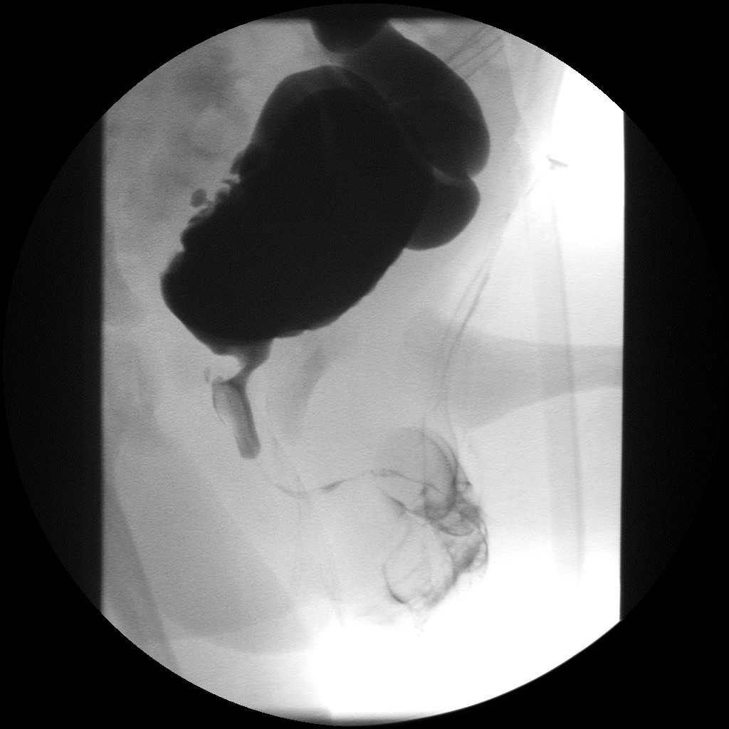
Posterior urethral valve (PUV) disorder is an obstructive developmental anomaly in the urethra and genitourinary system of male newborns. A posterior urethral valve is an obstructing membrane in the posterior male urethra as a result of abnormal '' in utero'' development. It is the most common cause of bladder outlet obstruction in male newborns. The disorder varies in degree, with mild cases presenting late due to milder symptoms. More severe cases can have renal and respiratory failure from lung underdevelopment as result of low amniotic fluid volumes, requiring intensive care and close monitoring. It occurs in about one in 8,000 babies. Presentation PUV can be diagnosed before birth, or even at birth when the ultrasound shows that the male baby has a hydronephrosis. Some babies may also have oligohydramnios due to the urinary obstruction. The later presentation can be a urinary tract infection, diurnal enuresis, or voiding pain. Complications * Incontinence * Urinary trac ... [...More Info...] [...Related Items...] OR: [Wikipedia] [Google] [Baidu] |
Urethra
The urethra (: urethras or urethrae) is the tube that connects the urinary bladder to the urinary meatus, through which Placentalia, placental mammals Urination, urinate and Ejaculation, ejaculate. The external urethral sphincter is a striated muscle that allows voluntary control over urination. The Internal urethral sphincter, internal sphincter, formed by the involuntary smooth muscles lining the bladder neck and urethra, receives its nerve supply by the Sympathetic nervous system, sympathetic division of the autonomic nervous system. The internal sphincter is present both in males and females. Structure The urethra is a fibrous and muscular tube which connects the urinary bladder to the external urethral meatus. Its length differs between the sexes, because it passes through the penis in males. Male In the human male, the urethra is on average long and opens at the end of the external urethral meatus. The urethra is divided into four parts in men, named after the lo ... [...More Info...] [...Related Items...] OR: [Wikipedia] [Google] [Baidu] |
Diverticula
In medicine or biology, a diverticulum is an outpouching of a hollow (or a fluid-filled) structure in the body. Depending upon which layers of the structure are involved, diverticula are described as being either true or false. In medicine, the term usually implies the structure is not normally present, but in embryology, the term is used for some normal structures arising from others, as for instance the thyroid diverticulum, which arises from the tongue. The word comes from Latin ''dīverticulum'', "bypath" or "byway". Classification Diverticula are described as being true or false depending upon the layers involved: *False diverticula (also known as "pseudodiverticula") do not involve muscular layers or adventitia. False diverticula, in the gastrointestinal tract for instance, involve only the submucosa and mucosa, such as Zenker's diverticulum. False diverticula are typically synonymous with Pulsion diverticulum, pulsion diverticula, which describes the mechanism of form ... [...More Info...] [...Related Items...] OR: [Wikipedia] [Google] [Baidu] |
Urinary Bladder
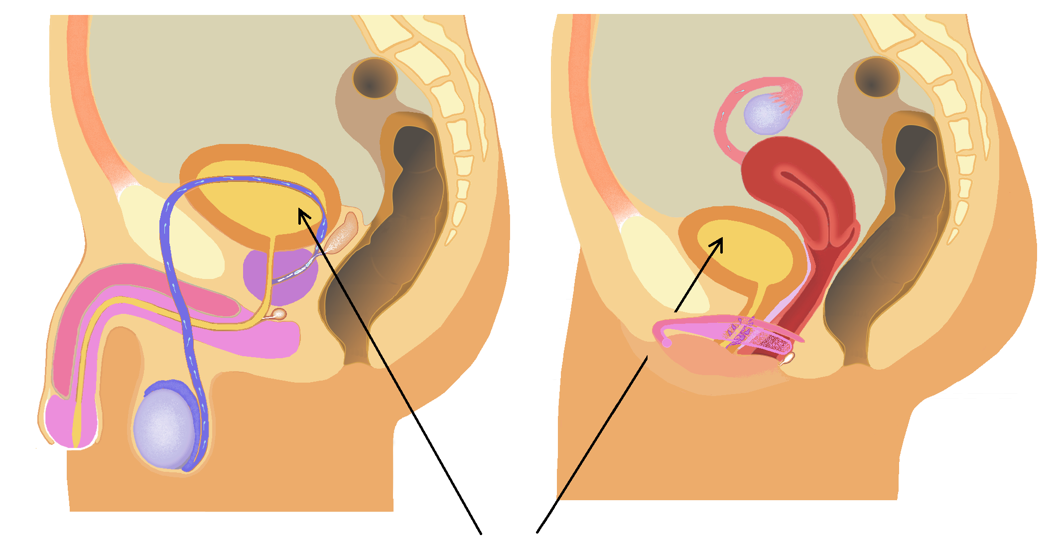
The bladder () is a hollow organ in humans and other vertebrates that stores urine from the Kidney (vertebrates), kidneys. In placental mammals, urine enters the bladder via the ureters and exits via the urethra during urination. In humans, the bladder is a distensible organ that sits on the pelvic floor. The typical adult human bladder will hold between 300 and (10 and ) before the urge to empty occurs, but can hold considerably more. The Latin phrase for "urinary bladder" is ''vesica urinaria'', and the term ''vesical'' or prefix ''vesico-'' appear in connection with associated structures such as vesical veins. The modern Latin word for "bladder" – ''cystis'' – appears in associated terms such as cystitis (inflammation of the bladder). Structure In humans, the bladder is a hollow muscular organ situated at the base of the pelvis. In gross anatomy, the bladder can be divided into a broad (base), a body, an apex, and a neck. The apex (also called the vertex) is directed ... [...More Info...] [...Related Items...] OR: [Wikipedia] [Google] [Baidu] |
Stoma
In botany, a stoma (: stomata, from Greek language, Greek ''στόμα'', "mouth"), also called a stomate (: stomates), is a pore found in the Epidermis (botany), epidermis of leaves, stems, and other organs, that controls the rate of gas exchange between the internal air spaces of the leaf and the atmosphere. The pore is bordered by a pair of specialized Ground tissue#Parenchyma, parenchyma cells known as guard cells that regulate the size of the stomatal opening. The term is usually used collectively to refer to the entire stomatal complex, consisting of the paired guard cells and the pore itself, which is referred to as the stomatal aperture. Air, containing oxygen, which is used in cellular respiration, respiration, and carbon dioxide, which is used in photosynthesis, passes through stomata by gaseous diffusion. Water vapour diffuses through the stomata into the atmosphere as part of a process called transpiration. Stomata are present in the sporophyte generation of the v ... [...More Info...] [...Related Items...] OR: [Wikipedia] [Google] [Baidu] |
Amniotic Sac
The amniotic sac, also called the bag of waters or the membranes, is the sac in which the embryo and later fetus develops in amniotes. It is a thin but tough transparent pair of biological membrane, membranes that hold a developing embryo (and later fetus) until shortly before birth. The inner of these membranes, the amnion, encloses the amniotic cavity, containing the amniotic fluid and the embryo. The outer membrane, the chorion, contains the amnion and is part of the placenta. On the outer side, the amniotic sac is connected to the yolk sac, the allantois, and via the umbilical cord, the placenta. The yolk sac, amnion, chorion, and allantois are the four extraembryonic membranes that lie outside of the embryo and are involved in providing nutrients and protection to the developing embryo. They form from the inner cell mass; the first to form is the yolk sac followed by the amnion which grows over the developing embryo. The amnion remains an important extraembryonic membrane th ... [...More Info...] [...Related Items...] OR: [Wikipedia] [Google] [Baidu] |
Amniotic Fluid
The amniotic fluid is the protective liquid contained by the amniotic sac of a gravid amniote. This fluid serves as a cushion for the growing fetus, but also serves to facilitate the exchange of nutrients, water, and biochemical products between mother and fetus. Colloquially, the amniotic fluid is commonly called water or waters (Latin liquor amnii). Development Amniotic fluid is present from the formation of the gestational sac. Amniotic fluid is in the amniotic sac. It is generated from maternal plasma, and passes through the fetal membranes by osmotic and hydrostatic forces. When fetal kidneys begin to function around week 16, fetal urine also contributes to the fluid. In earlier times, it was believed that the amniotic fluid was composed entirely of excreted fetal urine. The fluid is absorbed through the fetal tissue and skin. After 22 to 25 week of pregnancy, keratinization of an embryo's skin occurs. When this process completes around the 25th week, the fluid is pr ... [...More Info...] [...Related Items...] OR: [Wikipedia] [Google] [Baidu] |
Eponymous
An eponym is a noun after which or for which someone or something is, or is believed to be, named. Adjectives derived from the word ''eponym'' include ''eponymous'' and ''eponymic''. Eponyms are commonly used for time periods, places, innovations, biological nomenclature, astronomical objects, works of art and media, and tribal names. Various orthographic conventions are used for eponyms. Usage of the word The term ''eponym'' functions in multiple related ways, all based on an explicit relationship between two named things. ''Eponym'' may refer to a person or, less commonly, a place or thing for which someone or something is, or is believed to be, named. ''Eponym'' may also refer to someone or something named after, or believed to be named after, a person or, less commonly, a place or thing. A person, place, or thing named after a particular person share an eponymous relationship. In this way, Elizabeth I of England is the eponym of the Elizabethan era, but the Elizabethan ... [...More Info...] [...Related Items...] OR: [Wikipedia] [Google] [Baidu] |
Prostatic Urethra
The prostatic urethra, the widest and most dilatable part of the urethra canal, is about 3 cm long. It runs almost vertically through the prostate from its base to its apex, lying nearer its anterior than its posterior surface; the form of the canal is spindle-shaped, being wider in the middle than at either extremity, and narrowest below, where it joins the membranous portion. A transverse section of the canal as it lies in the prostate is horse-shoe-shaped, with the convexity directed forward. The keyhole sign, in ultrasound, is associated with a dilated bladder and prostatic urethra. Additional images File:Illu prostate lobes.jpg, Lobes of prostate File:Illu prostate zones.jpg, Zones of prostate File:Illu penis.jpg, Structure of the penis A penis (; : penises or penes) is a sex organ through which male and hermaphrodite animals expel semen during copulation (zoology), copulation, and through which male placental mammals and marsupials also Urination, urin ... [...More Info...] [...Related Items...] OR: [Wikipedia] [Google] [Baidu] |
Prostate
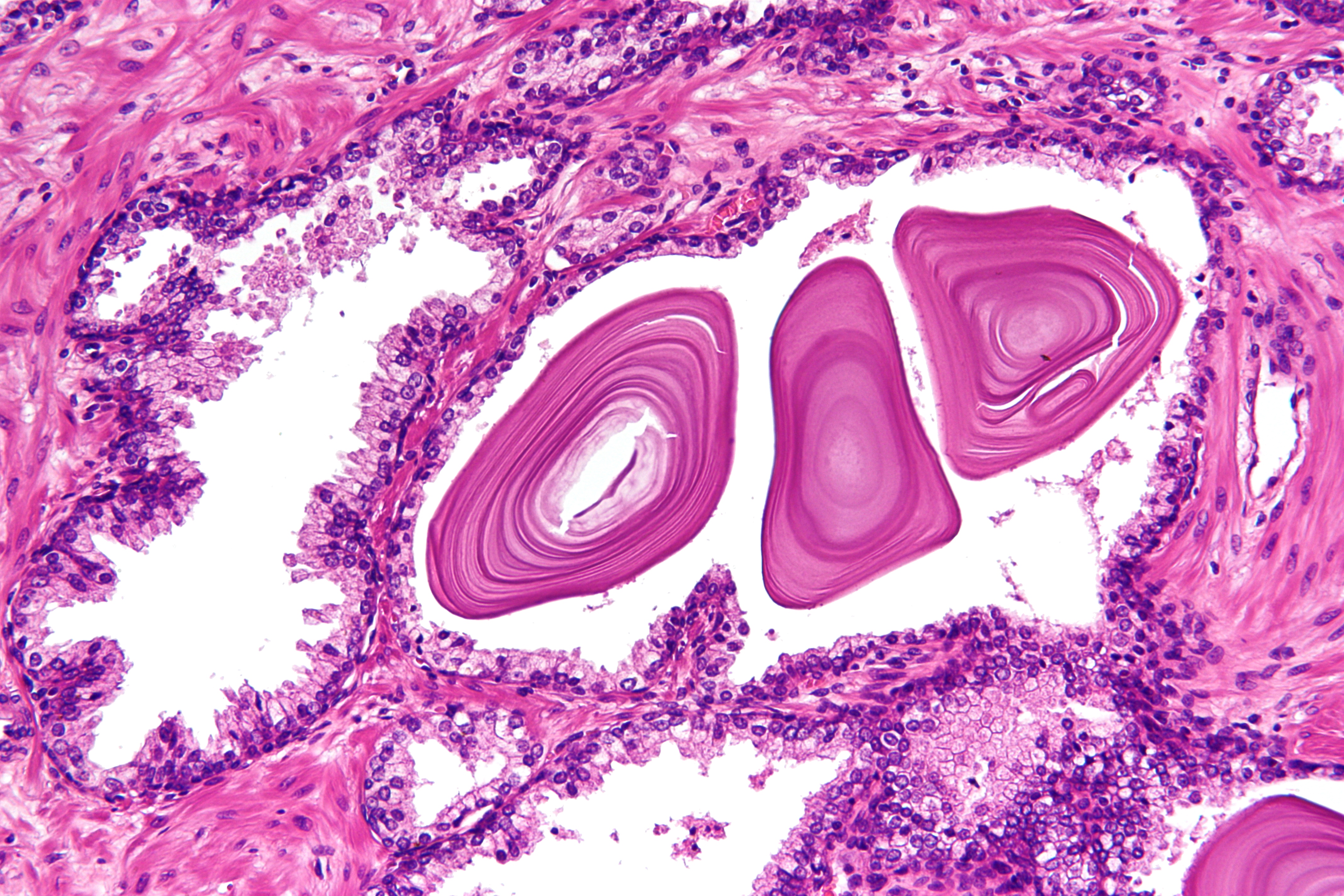
The prostate is an male accessory gland, accessory gland of the male reproductive system and a muscle-driven mechanical switch between urination and ejaculation. It is found in all male mammals. It differs between species anatomically, chemically, and physiologically. Anatomically, the prostate is found below the bladder, with the urethra passing through it. It is described in gross anatomy as consisting of lobes and in microanatomy by zone. It is surrounded by an elastic, fibromuscular capsule and contains glandular tissue, as well as connective tissue. The prostate produces and contains fluid that forms part of semen, the substance emitted during ejaculation as part of the male human sexual response cycle, sexual response. This prostatic fluid is slightly Alkalinity, alkaline, milky or white in appearance. The alkalinity of semen helps neutralize the acidity of the vagina, vaginal tract, prolonging the lifespan of sperm. The prostatic fluid is expelled in the first part of ej ... [...More Info...] [...Related Items...] OR: [Wikipedia] [Google] [Baidu] |
Verumontanum
The seminal colliculus (Latin ''colliculus seminalis''), or verumontanum, of the prostatic urethra is a landmark distal to the entrance of the ejaculatory ducts (on both sides, corresponding vas deferens and seminal vesicle feed into corresponding ejaculatory duct). ''Verumontanum'' is translated from Latin to mean 'mountain ridge', a reference to the distinctive median elevation of urothelium that characterizes the landmark on magnified views. Embryologically, it is derived from the uterovaginal primordium. The landmark is important in classification of several urethral developmental disorders. The margins of seminal colliculus are the following: * the orifices of the prostatic utricle * the slit-like openings of the ejaculatory ducts. * the openings of the prostatic ducts Posterior urethral valves The verumontanum is an important anatomic landmark for pathology in a congenital anomaly known as posterior urethral valves, in which there is a developmental obstruction of the ... [...More Info...] [...Related Items...] OR: [Wikipedia] [Google] [Baidu] |
Emedicine
eMedicine is an online clinical medical knowledge base founded in 1996 by doctors Scott Plantz and Jonathan Adler, and computer engineers Joanne Berezin and Jeffrey Berezin. The eMedicine website consists of approximately 6,800 medical topic review articles, each of which is associated with a clinical subspecialty "textbook". The knowledge base includes over 25,000 clinically multimedia files. Each article is authored by board certified specialists in the subspecialty to which the article belongs and undergoes three levels of physician peer-review, plus review by a Doctor of Pharmacy. The article's authors are identified with their current faculty appointments. Each article is updated yearly, or more frequently as changes in practice occur, and the date is published on the article. eMedicine.com was sold to WebMD in January, 2006 and is available as the Medscape Reference. History Plantz, Adler and Berezin evolved the concept for eMedicine.com in 1996 and deployed the initia ... [...More Info...] [...Related Items...] OR: [Wikipedia] [Google] [Baidu] |


