|
Pleuroperitoneal
Pleuroperitoneal is a term denoting the pleural and peritoneal serous membranes or the cavities they line. It is divided from the pericardial cavity by the transverse septum. Congenital defect or traumatic injury of pleuroperitoneal membrane can lead to diaphragmatic hernia. A congenital pleuroperitoneal hernia is called the Bochdalek hernia. This hernia is caused by an incomplete fusion of the septum transversum The septum transversum is a thick mass of cranial mesenchyme, formed in the embryo, that gives rise to parts of the thoracic diaphragm and the ventral mesentery of the foregut in the developed human being and other mammals. Origins The septum t ... with the pleuroperitoneal membranes. Membrane biology In other animals Congenital pleuroperitoneal hernias have been seen in cats and dogs, but they are rare. References [...More Info...] [...Related Items...] OR: [Wikipedia] [Google] [Baidu] |
Pleural
The pleural cavity, or pleural space (or sometimes intrapleural space), is the potential space between the pulmonary pleurae, pleurae of the pleural sac that surrounds each lung. A small amount of serous fluid, serous pleural fluid is maintained in the pleural cavity to enable lubrication between the pleurae, membranes, and also to create a pressure gradient. The serous membrane that covers the surface of the lung is the visceral pleura and is separated from the outer membrane, the parietal pleura, by just the film of pleural fluid in the pleural cavity. The visceral pleura follows the fissures of the lung and the root of the lung structures. The parietal pleura is attached to the mediastinum, the upper surface of the thoracic diaphragm, diaphragm, and to the inside of the ribcage. Structure In humans, the left and right lungs are completely separated by the mediastinum, and there is no communication between their pleural cavities. Therefore, in cases of a unilateral pneumothor ... [...More Info...] [...Related Items...] OR: [Wikipedia] [Google] [Baidu] |
Septum Transversum
The septum transversum is a thick mass of cranial mesenchyme, formed in the embryo, that gives rise to parts of the thoracic diaphragm and the ventral mesentery of the foregut in the developed human being and other mammals. Origins The septum transversum originally arises as the most cranial part of the mesenchyme on day 22. During craniocaudal folding, it assumes a position cranial to the developing heart at the level of the cervical vertebrae. During subsequent weeks the dorsal end of the embryo grows much faster than its ventral counterpart resulting in an ''apparent descent'' of the ventrally located septum transversum. At week 8, it can be found at the level of the thoracic vertebrae. Nerve supply After successful craniocaudal folding the septum transversum picks up innervation from the adjacent ventral rami of spinal nerves C3, C4 and C5, thus forming the precursor of the phrenic nerve. During the descent of the septum, the phrenic nerve is carried along and assumes its d ... [...More Info...] [...Related Items...] OR: [Wikipedia] [Google] [Baidu] |
Peritoneal
The peritoneum is the serous membrane forming the lining of the abdominal cavity or coelom in amniotes and some invertebrates, such as annelids. It covers most of the intra-abdominal (or coelomic) organs, and is composed of a layer of mesothelium supported by a thin layer of connective tissue. This peritoneal lining of the cavity supports many of the abdominal organs and serves as a conduit for their blood vessels, lymphatic vessels, and nerves. The abdominal cavity (the space bounded by the vertebrae, abdominal muscles, diaphragm, and pelvic floor) is different from the intraperitoneal space (located within the abdominal cavity but wrapped in peritoneum). The structures within the intraperitoneal space are called "intraperitoneal" (e.g., the stomach and intestines), the structures in the abdominal cavity that are located behind the intraperitoneal space are called "retroperitoneal" (e.g., the kidneys), and those structures below the intraperitoneal space are called "subperit ... [...More Info...] [...Related Items...] OR: [Wikipedia] [Google] [Baidu] |
Serous Membrane
The serous membrane (or serosa) is a smooth epithelial membrane of mesothelium lining the contents and inner walls of body cavity, body cavities, which secrete serous fluid to allow lubricated sliding (motion), sliding movements between opposing surfaces. The serous membrane that covers Viscera, internal organs (viscera) is called ''visceral'', while the one that covers the cavity wall is called ''parietal''. For instance the peritoneum, parietal peritoneum is attached to the abdominal wall and the pelvic walls. The peritoneum, visceral peritoneum is wrapped around the visceral organs. For the heart, the layers of the serous membrane are called parietal and visceral pericardium. For the lungs they are called parietal and visceral pleura. The visceral serosa of the uterus is called the perimetrium. The potential space between two opposing serosal surfaces is mostly empty except for the small amount of serous fluid. The Latin anatomical name is Tunica (biology)#Anatomy usages, tu ... [...More Info...] [...Related Items...] OR: [Wikipedia] [Google] [Baidu] |
Pericardial Cavity
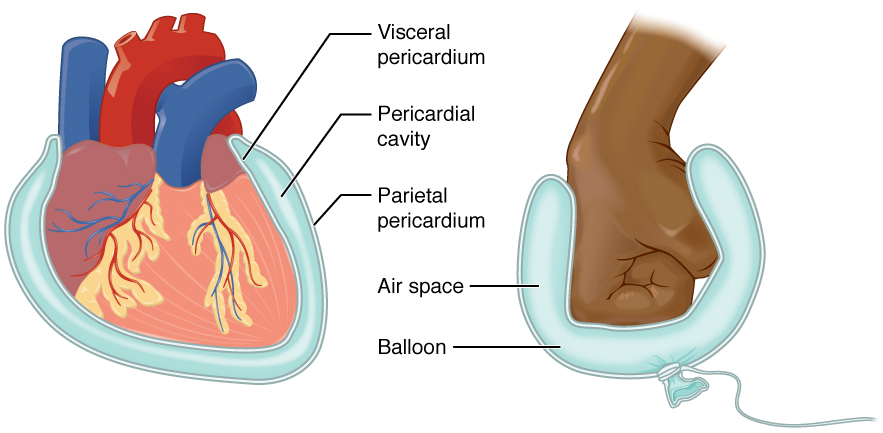
The pericardium (: pericardia), also called pericardial sac, is a double-walled sac containing the heart and the roots of the great vessels. It has two layers, an outer layer made of strong inelastic connective tissue (fibrous pericardium), and an inner layer made of serous membrane (serous pericardium). It encloses the pericardial cavity, which contains pericardial fluid, and defines the middle mediastinum. It separates the heart from interference of other structures, protects it against infection and blunt trauma, and lubricates the heart's movements. The English name originates from the Ancient Greek prefix ''peri-'' (περί) 'around' and the suffix ''-cardion'' (κάρδιον) 'heart'. Anatomy The pericardium is a tough fibroelastic sac which covers the heart from all sides except at the cardiac root (where the great vessels join the heart) and the bottom (where only the serous pericardium exists to cover the upper surface of the central tendon of diaphragm). The ... [...More Info...] [...Related Items...] OR: [Wikipedia] [Google] [Baidu] |
Diaphragmatic Hernia
Diaphragmatic hernia is a defect or hole in the diaphragm that allows the abdominal contents to move into the chest cavity. Treatment is usually surgical. Types * Congenital diaphragmatic hernia ** Morgagni's hernia ** Bochdalek hernia * Hiatal hernia * Iatrogenic diaphragmatic hernia * Traumatic diaphragmatic hernia Signs and symptoms A scaphoid abdomen (sucked inwards) may be the presenting symptom in a newborn. Diagnosis Diagnosis can be made by either CT or X-ray. Treatment Treatment for a diaphragmatic hernia usually involves surgery, with acute injuries often repaired with monofilament permanent sutures. Other animals Peritoneopericardial diaphragmatic hernia is a type of hernia more common in other mammal A mammal () is a vertebrate animal of the Class (biology), class Mammalia (). Mammals are characterised by the presence of milk-producing mammary glands for feeding their young, a broad neocortex region of the brain, fur or hair, and three ...s. ... [...More Info...] [...Related Items...] OR: [Wikipedia] [Google] [Baidu] |
Hernia
A hernia (: hernias or herniae, from Latin, meaning 'rupture') is the abnormal exit of tissue or an organ (anatomy), organ, such as the bowel, through the wall of the cavity in which it normally resides. The term is also used for the normal Development of the digestive system, development of the intestinal tract, referring to the retraction of the intestine from the extra-embryonal navel coelom into the abdomen in the healthy embryo at about 7 weeks. Various types of hernias can occur, most commonly involving the abdomen, and specifically the groin. Groin hernias are most commonly inguinal hernia, inguinal hernias but may also be femoral hernias. Other types of hernias include Hiatal hernia, hiatus, incisional hernia, incisional, and umbilical hernias. Symptoms are present in about 66% of people with groin hernias. This may include pain or discomfort in the lower abdomen, especially with coughing, exercise, or Urination, urinating or Defecation, defecating. Often, it gets worse th ... [...More Info...] [...Related Items...] OR: [Wikipedia] [Google] [Baidu] |
Bochdalek Hernia
Bochdalek hernia is one of two forms of a congenital diaphragmatic hernia, the other form being Morgagni hernia. A Bochdalek hernia is a congenital abnormality in which an opening exists in the infant's diaphragm, allowing normally intra-abdominal organs (particularly the stomach and intestines) to enter into the thoracic cavity. In the majority of people, the affected lung will be deformed, and the resulting lung compression can be life-threatening. Bochdalek hernias occur more commonly on the posterior left side (85%, versus the right side 15%). Bochdalek hernias are rare. This type of hernia was first described in 1754 by McCauley and subsequently studied and named after the Czech pathologist Vincenz Alexander Bochdalek (1801–1883). Signs and symptoms Children In normal Bochdalek hernia cases, the symptoms are often observable simultaneously with the baby's birth. A few of the symptoms of Bochdalek Hernia include difficulty breathing, fast respiration and increased heart ... [...More Info...] [...Related Items...] OR: [Wikipedia] [Google] [Baidu] |

