|
Plantaris
The plantaris is one of the superficial muscles of the superficial posterior compartment of the leg, one of the fascial compartments of the leg. It is composed of a thin muscle belly and a long thin tendon. While not as thick as the achilles tendon, the plantaris tendon (which tends to be between in length) is the longest tendon in the human body. Not including the tendon, the plantaris muscle is approximately long and is absent in 8-12% of the population. It is one of the plantar flexors in the posterior compartment of the leg, along with the gastrocnemius and soleus muscles. The plantaris is considered to have become an unimportant muscle when human ancestors switched from climbing trees to bipedalism and in anatomically modern humans it mainly acts with the gastrocnemius. Structure The plantaris muscle arises from the inferior part of the lateral supracondylar ridge of the femur at a position slightly superior to the origin of the lateral head of gastrocnemius. It passes ... [...More Info...] [...Related Items...] OR: [Wikipedia] [Google] [Baidu] |
Calcaneus
In humans and many other primates, the calcaneus (; from the Latin ''calcaneus'' or ''calcaneum'', meaning heel; : calcanei or calcanea) or heel bone is a bone of the Tarsus (skeleton), tarsus of the foot which constitutes the heel. In some other animals, it is the point of the hock (anatomy), hock. Structure In humans, the calcaneus is the largest of the tarsal bones and the largest bone of the foot. Its long axis is pointed forwards and laterally. The talus bone, calcaneus, and navicular bone are considered the proximal row of tarsal bones. In the calcaneus, several important structures can be distinguished:Platzer (2004), p 216 There is a large calcaneal tuberosity located posteriorly on plantar surface with medial and lateral tubercles on its surface. Besides, there is another peroneal tubercle on its lateral surface. On its lower edge on either side are its lateral and medial processes (serving as the origins of the Abductor hallucis muscle, abductor hallucis and Abductor di ... [...More Info...] [...Related Items...] OR: [Wikipedia] [Google] [Baidu] |
Posterior Compartment Of Leg
The posterior compartment of the leg is one of the fascial compartments of leg, fascial compartments of the leg and is divided further into deep and superficial compartments. Structure Muscles Superficial posterior compartment Deep posterior compartment Blood supply Posterior tibial artery Innervation The posterior compartment of the leg is supplied by the tibial nerve. Function * It contains the plantar flexors: Additional images File:From below - Superficial posterior compartment of leg - animation.gif, Superficial posterior compartment. Animation. File:From below - Deep posterior compartment of leg - animation.gif, Deep posterior compartment. Animation. References External links Diagram at patientcareonline.com {{DEFAULTSORT:Posterior Compartment Of Leg Muscles of the lower limb ... [...More Info...] [...Related Items...] OR: [Wikipedia] [Google] [Baidu] |
Sacral Plexus
In human anatomy, the sacral plexus is a nerve plexus which provides motor and sensory nerves for the posterior thigh, most of the lower leg and foot, and part of the pelvis. It is part of the lumbosacral plexus and emerges from the lumbar vertebrae and sacral vertebrae (L4-S4).''Thieme Atlas of Anatomy'' (2006), pp 470-471 A sacral plexopathy is a disorder affecting the nerves of the sacral plexus, usually caused by trauma, nerve compression, vascular disease, or infection. Symptoms may include pain, loss of motor control, and sensory deficits. Structure The sacral plexus is formed by: * the lumbosacral trunk * the anterior division of the first sacral nerve * portions of the anterior divisions of the second and third sacral nerves The nerves forming the sacral plexus converge toward the lower part of the greater sciatic foramen, and unite to form a flattened band, from the anterior and posterior surfaces of which several branches arise. The band itself is continued as the ... [...More Info...] [...Related Items...] OR: [Wikipedia] [Google] [Baidu] |
Femur
The femur (; : femurs or femora ), or thigh bone is the only long bone, bone in the thigh — the region of the lower limb between the hip and the knee. In many quadrupeds, four-legged animals the femur is the upper bone of the hindleg. The Femoral head, top of the femur fits into a socket in the pelvis called the hip joint, and the bottom of the femur connects to the shinbone (tibia) and kneecap (patella) to form the knee. In humans the femur is the largest and thickest bone in the body. Structure The femur is the only bone in the upper Human leg, leg. The two femurs converge Anatomical terms of location, medially toward the knees, where they articulate with the Anatomical terms of location, proximal ends of the tibiae. The angle at which the femora converge is an important factor in determining the femoral-tibial angle. In females, thicker pelvic bones cause the femora to converge more than in males. In the condition genu valgum, ''genu valgum'' (knock knee), the femurs conve ... [...More Info...] [...Related Items...] OR: [Wikipedia] [Google] [Baidu] |
Fascial Compartments Of Leg
The fascial compartments of the leg are the four fascial compartments that separate and contain the muscles of the lower leg (from the knee to the ankle). The compartments are divided by septa formed from the fascia. The compartments usually have nerve and blood supplies separate from their neighbours. All of the muscles within a compartment will generally be supplied by the same nerve. Intermuscular septa The lower leg is divided into four compartments by the interosseous membrane of the leg, the anterior intermuscular septum, the transverse intermuscular septum and the posterior intermuscular septum. Each compartment contains connective tissue, nerves and blood vessels. The septa are formed from the fascia which is made up of a strong type of connective tissue. The fascia also separates the skeletal muscles from the subcutaneous tissue. Due to the great pressure placed on the leg, from the column of blood from the heart to the feet, the fascia is very thick in order to sup ... [...More Info...] [...Related Items...] OR: [Wikipedia] [Google] [Baidu] |
Knee Joint
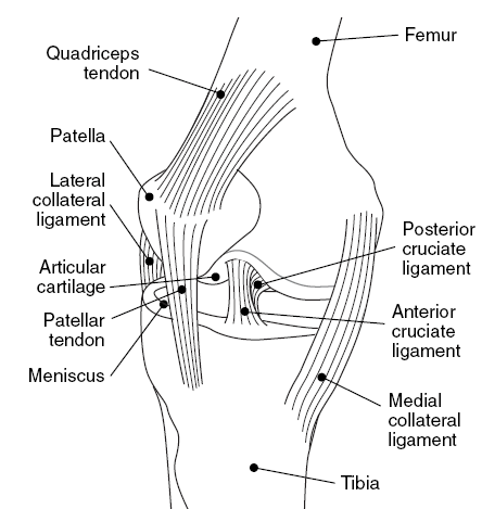
In humans and other primates, the knee joins the thigh with the leg and consists of two joints: one between the femur and tibia (tibiofemoral joint), and one between the femur and patella (patellofemoral joint). It is the largest joint in the human body. The knee is a modified hinge joint, which permits flexion and extension as well as slight internal and external rotation. The knee is vulnerable to injury and to the development of osteoarthritis. It is often termed a ''compound joint'' having tibiofemoral and patellofemoral components. (The fibular collateral ligament is often considered with tibiofemoral components.) Structure The knee is a modified hinge joint, a type of synovial joint, which is composed of three functional compartments: the patellofemoral articulation, consisting of the patella, or "kneecap", and the patellar groove on the front of the femur through which it slides; and the medial and lateral tibiofemoral articulations linking the femur, or thigh b ... [...More Info...] [...Related Items...] OR: [Wikipedia] [Google] [Baidu] |
Tibialis Anterior Muscle
The tibialis anterior muscle is a muscle of the anterior compartment of the lower leg. It originates from the upper portion of the tibia; it inserts into the medial cuneiform and first metatarsal bones of the foot. It acts to dorsiflex and invert the foot. This muscle is mostly located near the shin. It is situated on the lateral side of the tibia; it is thick and fleshy above, tendinous below. The tibialis anterior overlaps the anterior tibial vessels and deep peroneal nerve in the upper part of the leg. Structure The tibialis anterior muscle is the most medial muscle of the anterior compartment of the leg. The muscle ends in a tendon which is apparent on the anteriomedial dorsal aspect of the foot close to the ankle. Its tendon is ensheathed in a synovial sheath. The tendon passes through the medial compartment superior and inferior extensor retinacula of the foot. Origin The tibialis anterior muscle arises from the upper 2/3 of the lateral surface of the tibia and the ... [...More Info...] [...Related Items...] OR: [Wikipedia] [Google] [Baidu] |
Knee
In humans and other primates, the knee joins the thigh with the leg and consists of two joints: one between the femur and tibia (tibiofemoral joint), and one between the femur and patella (patellofemoral joint). It is the largest joint in the human body. The knee is a modified hinge joint, which permits flexion and extension (kinesiology), extension as well as slight internal and external rotation. The knee is vulnerable to injury and to the development of osteoarthritis. It is often termed a ''compound joint'' having tibiofemoral and patellofemoral components. (The fibular collateral ligament is often considered with tibiofemoral components.) Structure The knee is a modified hinge joint, a type of synovial joint, which is composed of three functional compartments: the patellofemoral articulation, consisting of the patella, or "kneecap", and the patellar groove on the front of the femur through which it slides; and the medial and lateral tibiofemoral articulations linking the ... [...More Info...] [...Related Items...] OR: [Wikipedia] [Google] [Baidu] |
Primary Motor Cortex
The primary motor cortex ( Brodmann area 4) is a brain region that in humans is located in the dorsal portion of the frontal lobe. It is the primary region of the motor system and works in association with other motor areas including premotor cortex, the supplementary motor area, posterior parietal cortex, and several subcortical brain regions, to plan and execute voluntary movements. Primary motor cortex is defined anatomically as the region of cortex that contains large neurons known as Betz cells, which, along with other cortical neurons, send long axons down the spinal cord to synapse onto the interneuron circuitry of the spinal cord and also directly onto the alpha motor neurons in the spinal cord which connect to the muscles. At the primary motor cortex, motor representation is orderly arranged (in an inverted fashion) from the toe (at the top of the cerebral hemisphere) to mouth (at the bottom) along a fold in the cortex called the central sulcus. However, some body p ... [...More Info...] [...Related Items...] OR: [Wikipedia] [Google] [Baidu] |
Pre-central Gyrus
The precentral gyrus is a prominent gyrus on the surface of the posterior frontal lobe of the brain. It is the site of the primary motor cortex that in humans is cytoarchitecturally defined as Brodmann area 4. Structure The precentral gyrus lies in front of the postcentral gyrus - mostly on the lateral (convex) side of each cerebral hemisphere - from which it is separated by the central sulcus. Its anterior border is represented by the precentral sulcus, while inferiorly it borders to the lateral sulcus (Sylvian fissure). Medially, it is contiguous with the paracentral lobule. The internal pyramidal layer (layer V) of the precentral cortex contains giant (70-100 micrometers) pyramidal neurons called Betz cells, which send long axons to the contralateral motor nuclei of the cranial nerves and to the lower motor neurons in the ventral horn of the spinal cord. These axons form the corticospinal tract. The Betz cells along with their long axons are referred to as upper motor neuro ... [...More Info...] [...Related Items...] OR: [Wikipedia] [Google] [Baidu] |
Human Brain
The human brain is the central organ (anatomy), organ of the nervous system, and with the spinal cord, comprises the central nervous system. It consists of the cerebrum, the brainstem and the cerebellum. The brain controls most of the activities of the human body, body, processing, integrating, and coordinating the information it receives from the sensory nervous system. The brain integrates sensory information and coordinates instructions sent to the rest of the body. The cerebrum, the largest part of the human brain, consists of two cerebral hemispheres. Each hemisphere has an inner core composed of white matter, and an outer surface – the cerebral cortex – composed of grey matter. The cortex has an outer layer, the neocortex, and an inner allocortex. The neocortex is made up of six Cerebral cortex#Layers of neocortex, neuronal layers, while the allocortex has three or four. Each hemisphere is divided into four lobes of the brain, lobes – the frontal lobe, frontal, pa ... [...More Info...] [...Related Items...] OR: [Wikipedia] [Google] [Baidu] |
Frontal Lobe
The frontal lobe is the largest of the four major lobes of the brain in mammals, and is located at the front of each cerebral hemisphere (in front of the parietal lobe and the temporal lobe). It is parted from the parietal lobe by a Sulcus (neuroanatomy), groove between tissues called the central sulcus and from the temporal lobe by a deeper groove called the lateral sulcus (Sylvian fissure). The most anterior rounded part of the frontal lobe (though not well-defined) is known as the frontal pole, one of the three Cerebral hemisphere#Poles, poles of the cerebrum. The frontal lobe is covered by the frontal cortex. The frontal cortex includes the premotor cortex and the primary motor cortex – parts of the motor cortex. The front part of the frontal cortex is covered by the prefrontal cortex. The nonprimary motor cortex is a functionally defined portion of the frontal lobe. There are four principal Gyrus, gyri in the frontal lobe. The precentral gyrus is directly anterior to the ... [...More Info...] [...Related Items...] OR: [Wikipedia] [Google] [Baidu] |


