|
Placental Growth Factor
Placental growth factor (PlGF) is a protein that in humans is encoded by the ''PGF'' gene. Placental growth factor is a member of the VEGF (vascular endothelial growth factor) sub-family - a key molecule in angiogenesis and vasculogenesis, in particular during embryogenesis. The main source of PlGF during pregnancy is the placental trophoblast. ''PGF'' is also expressed in many other tissues, including the villous trophoblast. The placental growth factor gene (''PGF'') is a protein-coding gene and a member of the vascular endothelial growth factor (VEGF) family. ''PGF'' is expressed only in human umbilical vein endothelial cells, and the placenta. PGF is ultimately associated with angiogenesis. Specifically, PGF plays a role in trophoblast growth and differentiation. Trophoblast cells, specifically extravillous trophoblast cells, are responsible for invading the uterine wall and the maternal spiral arteries. The extravillous trophoblast cells produce a blood vessel of lar ... [...More Info...] [...Related Items...] OR: [Wikipedia] [Google] [Baidu] |
Protein
Proteins are large biomolecules and macromolecules that comprise one or more long chains of amino acid residue (biochemistry), residues. Proteins perform a vast array of functions within organisms, including Enzyme catalysis, catalysing metabolic reactions, DNA replication, Cell signaling, responding to stimuli, providing Cytoskeleton, structure to cells and Fibrous protein, organisms, and Intracellular transport, transporting molecules from one location to another. Proteins differ from one another primarily in their sequence of amino acids, which is dictated by the Nucleic acid sequence, nucleotide sequence of their genes, and which usually results in protein folding into a specific Protein structure, 3D structure that determines its activity. A linear chain of amino acid residues is called a polypeptide. A protein contains at least one long polypeptide. Short polypeptides, containing less than 20–30 residues, are rarely considered to be proteins and are commonly called pep ... [...More Info...] [...Related Items...] OR: [Wikipedia] [Google] [Baidu] |
Perfusion
Perfusion is the passage of fluid through the circulatory system or lymphatic system to an organ (anatomy), organ or a tissue (biology), tissue, usually referring to the delivery of blood to a capillary bed in tissue. Perfusion may also refer to fixation via perfusion, used in histological studies. Perfusion is measured as the rate at which blood is delivered to tissue, or volume of blood per unit time (blood volumetric flow rate, flow) per unit tissue mass. The SI unit is m3/(s·kg), although for human organs perfusion is typically reported in ml/min/g. The word is derived from the French verb ''perfuser'', meaning to "pour over or through". All animal tissues require an adequate blood supply for health and life. Poor perfusion (malperfusion), that is, ischemia, causes health problems, as seen in cardiovascular disease, including coronary artery disease, cerebrovascular disease, peripheral artery disease, and many other conditions. Tests verifying that adequate perfusion exists ... [...More Info...] [...Related Items...] OR: [Wikipedia] [Google] [Baidu] |
Placenta
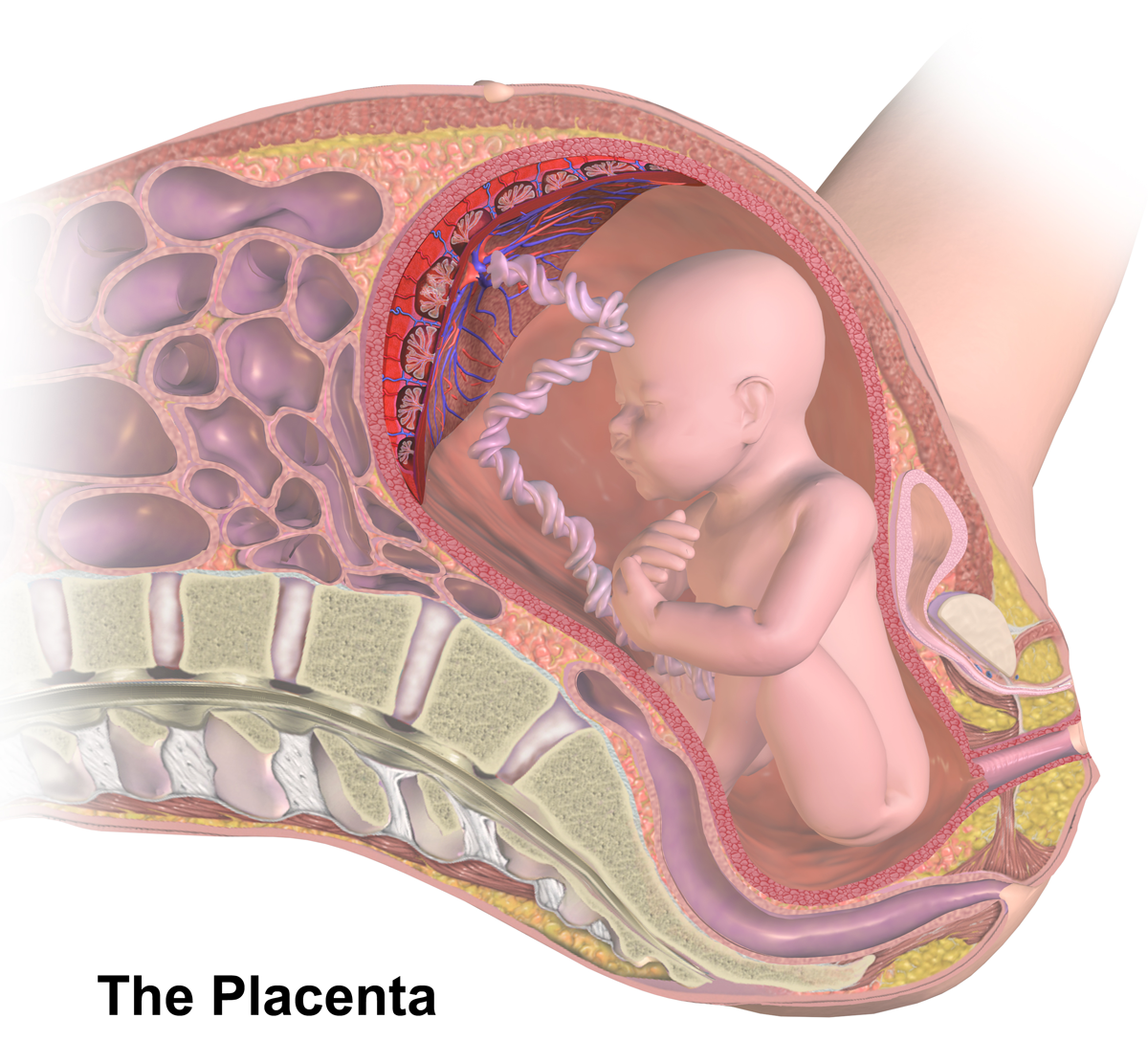
The placenta (: placentas or placentae) is a temporary embryonic and later fetal organ that begins developing from the blastocyst shortly after implantation. It plays critical roles in facilitating nutrient, gas, and waste exchange between the physically separate maternal and fetal circulations, and is an important endocrine organ, producing hormones that regulate both maternal and fetal physiology during pregnancy. The placenta connects to the fetus via the umbilical cord, and on the opposite aspect to the maternal uterus in a species-dependent manner. In humans, a thin layer of maternal decidual ( endometrial) tissue comes away with the placenta when it is expelled from the uterus following birth (sometimes incorrectly referred to as the 'maternal part' of the placenta). Placentas are a defining characteristic of placental mammals, but are also found in marsupials and some non-mammals with varying levels of development. Mammalian placentas probably first evolved abou ... [...More Info...] [...Related Items...] OR: [Wikipedia] [Google] [Baidu] |
Twin-to-twin Transfusion Syndrome
Twin-to-twin transfusion syndrome (TTTS), also known as feto-fetal transfusion syndrome (FFTS), twin oligohydramnios-polyhydramnios sequence (TOPS) and stuck twin syndrome, is a complication of monochorionic multiple pregnancies (the most common form of identical twin pregnancy) in which there is disproportionate blood supply between the fetuses. This leads to unequal levels of amniotic fluid between each fetus and usually leads to death of the undersupplied twin and, without treatment, usually death or a range of birth defects or disabilities for a surviving twin, such as underdeveloped, damaged or missing limbs, Oligodactyly, digits or organs (including the brain), especially cerebral palsy. The condition occurs when the vein–artery Anastomosis, connections within the fetuses' shared placenta allow the blood flow between each fetus to become progressively imbalanced. It usually develops between week 16 and 25 of pregnancy, during peak placental growth. The cause of the devel ... [...More Info...] [...Related Items...] OR: [Wikipedia] [Google] [Baidu] |
Pre-eclampsia
Pre-eclampsia is a multi-system disorder specific to pregnancy, characterized by the new onset of hypertension, high blood pressure and often a significant amount of proteinuria, protein in the urine or by the new onset of high blood pressure along with significant end-organ damage, with or without the proteinuria. When it arises, the condition begins after 20 Gestational age (obstetrics), weeks of pregnancy. In severe cases of the disease there may be hemolysis, red blood cell breakdown, a thrombocytopenia, low blood platelet count, impaired liver function, kidney dysfunction, edema, swelling, pulmonary edema, shortness of breath due to fluid in the lungs, or visual disturbances. Pre-eclampsia increases the risk of undesirable as well as lethal outcomes for both the mother and the fetus including preterm labor. If left untreated, it may result in seizures at which point it is known as eclampsia. Risk factors for pre-eclampsia include obesity, prior hypertension, older age, and ... [...More Info...] [...Related Items...] OR: [Wikipedia] [Google] [Baidu] |
Soluble Fms-like Tyrosine Kinase-1
Soluble fms-like tyrosine kinase-1 (sFlt-1 or sVEGFR-1) is a tyrosine kinase protein with antiangiogenic properties. A non-membrane associated splice variant of VEGF receptor, VEGF receptor 1 (Flt-1), sFlt-1 binds the angiogenic factors VEGF (vascular endothelial growth factor) and Placental growth factor, PlGF (placental growth factor), reducing blood vessel growth through reduction of free VEGF receptor, VEGF and Placental growth factor, PlGF concentrations. In humans, sFlt-1 is important in the regulation of blood vessel formation in diverse tissues, including the kidneys, cornea, and uterus. Abnormally high levels of sFlt-1 have been implicated in the pathogenesis of Pre-eclampsia, preeclampsia. Structure sFlt-1 is a truncated form of the VEGF receptor Flt-1. Though sFlt-1 contains an extracellular domain identical to that of Flt-1, it lacks both the transmembrane and intercellular domains present in Flt-1. Instead, sFlt-1 contains a novel 31 amino acid C-terminal sequence. s ... [...More Info...] [...Related Items...] OR: [Wikipedia] [Google] [Baidu] |
Neovascularization
Neovascularization is the natural formation of new blood vessels ('' neo-'' + ''vascular'' + '' -ization''), usually in the form of functional microvascular networks, capable of perfusion by red blood cells, that form to serve as collateral circulation in response to local poor perfusion or ischemia. Growth factors that inhibit neovascularization include those that affect endothelial cell division and differentiation. These growth factors often act in a paracrine or autocrine fashion; they include fibroblast growth factor, placental growth factor, insulin-like growth factor, hepatocyte growth factor, and platelet-derived endothelial growth factor. There are three different pathways that comprise neovascularization: (1) vasculogenesis, (2) angiogenesis, and (3) arteriogenesis. Three pathways of neovascularization Vasculogenesis Vasculogenesis is the de novo formation of blood vessels. This primarily occurs in the developing embryo with the development of the first primitive ... [...More Info...] [...Related Items...] OR: [Wikipedia] [Google] [Baidu] |
Atheroma
An atheroma, or atheromatous plaque, is an abnormal accumulation of material in the tunica intima, inner layer of an arterial wall. The material consists of mostly macrophage, macrophage cells, or debris, containing lipids, calcium and a variable amount of fibrous connective tissue. The accumulated material forms a swelling in the artery wall, which may intrude into the Lumen (anatomy), lumen of the artery, stenosis, narrowing it and restricting blood flow. Atheroma is the pathological basis for the disease entity atherosclerosis, a subtype of arteriosclerosis. Signs and symptoms For most people, the first symptoms result from atheroma progression within the coronary arteries, heart arteries, most commonly resulting in a myocardial infarction, heart attack and ensuing debility. The heart arteries are difficult to track because they are small (from about 5 mm down to microscopic), they are hidden deep within the chest and they never stop moving. Additionally, all mass-applie ... [...More Info...] [...Related Items...] OR: [Wikipedia] [Google] [Baidu] |
Atherosclerosis
Atherosclerosis is a pattern of the disease arteriosclerosis, characterized by development of abnormalities called lesions in walls of arteries. This is a chronic inflammatory disease involving many different cell types and is driven by elevated blood levels of cholesterol. These lesions may lead to narrowing of the arterial walls due to buildup of atheromatous plaques. At the onset, there are usually no symptoms, but if they develop, symptoms generally begin around middle age. In severe cases, it can result in coronary artery disease, stroke, peripheral artery disease, or kidney disorders, depending on which body part(s) the affected arteries are located in the body. The exact cause of atherosclerosis is unknown and is proposed to be multifactorial. Risk factors include dyslipidemia, abnormal cholesterol levels, elevated levels of inflammatory biomarkers, high blood pressure, diabetes, smoking (both active and passive smoking), obesity, genetic factors, family history, lifes ... [...More Info...] [...Related Items...] OR: [Wikipedia] [Google] [Baidu] |
Extravillous Trophoblast
Extravillous trophoblasts (EVTs), are one form of differentiated trophoblast cells of the placenta. They are invasive mesenchymal cells which function to establish critical tissue connection in the developing placental-uterine interface. EVTs derive from progenitor cytotrophoblasts (CYTs), as does the other main trophoblast subtype, syncytiotrophoblast (SYN). They are sometimes called intermediate trophoblast. EVTs that derive from CYT cells on the surface of placental chorionic villi that come into contact with the uterine wall - at the placental bed - begin to express the HLA-G antigen. Extravillous trophoblast (EVT) cells migrate from anchoring villi, and invade into the decidua basalis. Their main function is remodelling the uterine spiral arteries, to achieve an increase in the spiral artery diameter of from four to six times. This changes them from high-resistance low-flow vessels into large dilated vessels that provide good perfusion, and oxygenation to the developing plac ... [...More Info...] [...Related Items...] OR: [Wikipedia] [Google] [Baidu] |
Gene
In biology, the word gene has two meanings. The Mendelian gene is a basic unit of heredity. The molecular gene is a sequence of nucleotides in DNA that is transcribed to produce a functional RNA. There are two types of molecular genes: protein-coding genes and non-coding genes. During gene expression (the synthesis of Gene product, RNA or protein from a gene), DNA is first transcription (biology), copied into RNA. RNA can be non-coding RNA, directly functional or be the intermediate protein biosynthesis, template for the synthesis of a protein. The transmission of genes to an organism's offspring, is the basis of the inheritance of phenotypic traits from one generation to the next. These genes make up different DNA sequences, together called a genotype, that is specific to every given individual, within the gene pool of the population (biology), population of a given species. The genotype, along with environmental and developmental factors, ultimately determines the phenotype ... [...More Info...] [...Related Items...] OR: [Wikipedia] [Google] [Baidu] |
Umbilical Vein
The umbilical vein is a vein present during fetal development that carries oxygenated blood from the placenta into the growing fetus. The umbilical vein provides convenient access to the central circulation of a neonate for restoration of blood volume and for administration of glucose and drugs. The blood pressure inside the umbilical vein is approximately 20 mmHg.Wang, Y. Vascular biology of the placenta. in Colloquium Series on Integrated Systems Physiology: from Molecule to Function. 2010. Morgan & Claypool Life Sciences. Fetal circulation The unpaired umbilical vein carries oxygen and nutrient rich blood derived from fetal-maternal blood exchange at the chorionic villi. More than two-thirds of fetal hepatic circulation is via the main portal vein, while the remainder is shunted from the left portal vein via the ductus venosus to the inferior vena cava, eventually being delivered to the fetal right atrium. Closure Closure of the umbilical vein usually occurs after the umbilic ... [...More Info...] [...Related Items...] OR: [Wikipedia] [Google] [Baidu] |


