|
Pilomatricoma
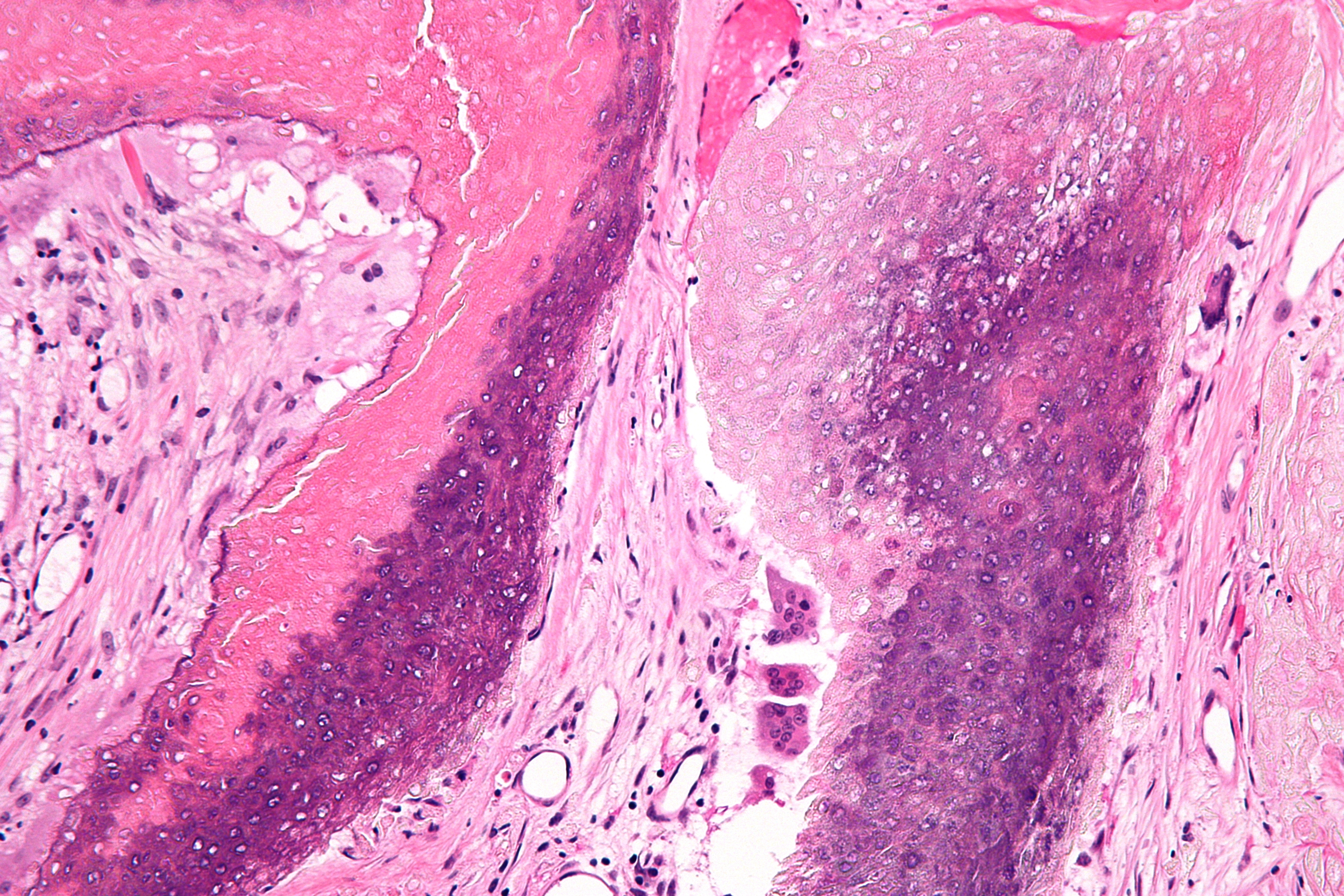
Pilomatricoma is a benign skin tumor derived from the hair matrix. These neoplasms are relatively uncommon and typically occur on the scalp, face, and upper extremities. Clinically, pilomatricomas present as a subcutaneous nodule or cyst with unremarkable overlying epidermis that can range in size from 0.5 to 3.0 cm, but the largest reported case was 24 cm. Presentation Associations Pilomatricomas have been observed in a variety of genetic disorders including Turner syndrome, myotonic dystrophy, Rubinstein–Taybi syndrome, trisomy 9, and Gardner syndrome. It has been reported that the prevalence of pilomatricomas in Turner syndrome is 2.6%. Hybrid cysts that are composed of epidermal inclusion cysts and pilomatricoma-like changes have been repeatedly observed in Gardner syndrome. This association has prognostic import, since cutaneous findings in children with Gardner syndrome generally precede colonic polyposis. Histologic features The characteristic components of ... [...More Info...] [...Related Items...] OR: [Wikipedia] [Google] [Baidu] |
List Of Cutaneous Neoplasms Associated With Systemic Syndromes
Many cutaneous neoplasms occur in the setting of systemic syndromes. See also *List of cutaneous conditions *List of contact allergens *List of cutaneous conditions associated with increased risk of nonmelanoma skin cancer *List of cutaneous conditions associated with internal malignancy *List of cutaneous conditions caused by mutations in keratins *List of cutaneous conditions caused by problems with junctional proteins *List of dental abnormalities associated with cutaneous conditions *List of genes mutated in cutaneous conditions *List of genes mutated in pigmented cutaneous lesions *List of histologic stains that aid in diagnosis of cutaneous conditions *List of immunofluorescence findings for autoimmune bullous conditions *List of inclusion bodies that aid in diagnosis of cutaneous conditions *List of keratins expressed in the human integumentary system *List of radiographic findings associated with cutaneous conditions *List of specialized glands within the human integum ... [...More Info...] [...Related Items...] OR: [Wikipedia] [Google] [Baidu] |
Malignant Pilomatricoma
Malignant pilomatricoma is a cutaneous condition characterized by a locally aggressive tumor composed of hair-matrix cells. Signs and symptoms Malignant pilomatricoma usually manifests as a single firm, painless, movable, asymptomatic dermal or subcutaneous lump. It has been shown that the underlying skin can become ulcerated and exhibit severe discoloration; the latter is thought to be one of the few significant indicators of cancer. The head, neck, and back are frequent locations of occurrence. Their sizes range from 1 to 10 cm. Diagnosis Grossly, removed tumors frequently have a grayish appearance, are cystoid, encapsulated, and have viscous fluids. Histologically, there is an abrupt shift to eosinophilic ghost or shadow cells (anucleate matrical corneocytes), which are characterized by a prominent proliferation of basaloid cells with ample transparent cytoplasm. Seldom do basaloid cells palisade or contain melanin; instead, they aggregate into nests, bands, and amorphous s ... [...More Info...] [...Related Items...] OR: [Wikipedia] [Google] [Baidu] |
Ghost Cell
A ghost cell is an enlarged eosinophilic epithelial cell with eosinophilic cytoplasm but without a nucleus. It has lost its nucleus and cytoplasmic contents, leaving behind only the cell membrane and sometimes remnants of the cell's structure. In pathology, ghost cells are often associated with certain types of tumors, such as pilomatricomas and calcifying odontogenic cysts, where they appear as pale, anucleate cells that have undergone degeneration or calcification. The ghost cells indicate coagulative necrosis where there is cell death but retainment of cellular architecture. In histologic sections ghost cells are those which appear as shadow cells. They are dead cells. For example, in peripheral blood smear preparations, the RBCs are lysed and appear as ghost cells. They are found in: * Craniopharyngioma (Rathke pouch) * Odontoma * Ameloblastic fibroma * Calcifying odontogenic cyst (Gorlin cyst) * Pilomatricoma Pilomatricoma is a benign skin tumor derived from the hair ... [...More Info...] [...Related Items...] OR: [Wikipedia] [Google] [Baidu] |
List Of Cutaneous Conditions
Many skin conditions affect the human integumentary system—the organ system covering the entire surface of the Human body, body and composed of Human skin, skin, hair, Nail (anatomy), nails, and related muscle and glands. The major function of this system is as a barrier against the external environment. The skin weighs an average of four kilograms, covers an area of two square metres, and is made of three distinct layers: the epidermis (skin), epidermis, dermis, and subcutaneous tissue. The two main types of human skin are: glabrous skin, the hairless skin on the palms and soles (also referred to as the "palmoplantar" surfaces), and hair-bearing skin.Burns, Tony; ''et al''. (2006) ''Rook's Textbook of Dermatology CD-ROM''. Wiley-Blackwell. . Within the latter type, the hairs occur in structures called pilosebaceous units, each with hair follicle, sebaceous gland, and associated arrector pili muscle. Embryology, In the embryo, the epidermis, hair, and glands form from the ectod ... [...More Info...] [...Related Items...] OR: [Wikipedia] [Google] [Baidu] |
Beta-catenin
Catenin beta-1, also known as β-catenin (''beta''-catenin), is a protein that in humans is encoded by the ''CTNNB1'' gene. β-Catenin is a dual function protein, involved in regulation and coordination of cell–cell adhesion and gene transcription. In humans, the CTNNB1 protein is encoded by the ''CTNNB1'' gene. In ''Drosophila'', the homologous protein is called ''armadillo''. β-catenin is a subunit of the cadherin protein complex and acts as an intracellular signal transducer in the Wnt signaling pathway. It is a member of the catenin protein family and homologous to Plakoglobin, γ-catenin, also known as plakoglobin. β-Catenin is widely expressed in many tissues. In cardiac muscle, β-catenin localizes to adherens junctions in intercalated disc structures, which are critical for electrical and mechanical coupling between adjacent cardiomyocytes. Mutations and overexpression of β-catenin are associated with many cancers, including hepatocellular carcinoma, colorectal cance ... [...More Info...] [...Related Items...] OR: [Wikipedia] [Google] [Baidu] |
Gardner Syndrome
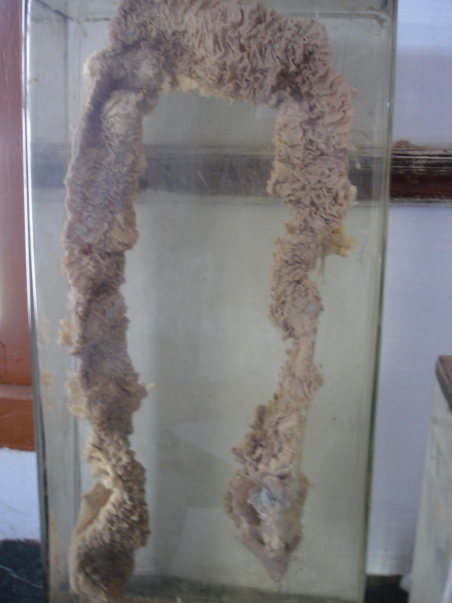
Gardner's syndrome (also known as Gardner syndrome, familial polyposis of the colon, or familial colorectal polyposis) is a subtype of familial adenomatous polyposis (FAP). Gardner syndrome is an autosomal dominant form of polyposis characterized by the presence of multiple polyps in the colon together with tumors outside the colon. The extracolonic tumors may include osteomas of the skull, thyroid cancer, epidermoid cysts, fibromas, as well as the occurrence of desmoid tumors in approximately 15% of affected individuals. Desmoid tumors are fibrous tumors that usually occur in the tissue covering the intestines and may be provoked by surgery to remove the colon. The countless polyps in the colon predispose to the development of colon cancer; if the colon is not removed, the chance of colon cancer is considered to be very significant. Polyps may also grow in the stomach, duodenum, spleen, kidneys, liver, mesentery, and small bowel. In a small number of cases, polyps have also a ... [...More Info...] [...Related Items...] OR: [Wikipedia] [Google] [Baidu] |
Adenomatous Polyposis Coli
Adenomatous polyposis coli (APC) also known as deleted in polyposis 2.5 (DP2.5) is a protein that in humans is encoded by the ''APC'' gene. The APC protein is a Down-regulation, negative regulator that controls beta-catenin concentrations and interacts with E-cadherin, which are involved in cell adhesion. Mutations in the ''APC'' gene may result in colorectal cancer and desmoid tumors. ''APC'' is classified as a tumor suppressor gene. Tumor suppressor genes prevent the uncontrolled growth of cells that may result in cancerous tumors. The protein made by the ''APC'' gene plays a critical role in several cellular processes that determine whether a cell may develop into a tumor. The APC protein helps control how often a cell divides, how it attaches to other cells within a tissue, how the cell polarizes and the morphogenesis of the 3D structures, or whether a cell moves within or away from tissue. This protein also helps ensure that the chromosome number in cells produced through c ... [...More Info...] [...Related Items...] OR: [Wikipedia] [Google] [Baidu] |
Necrosis
Necrosis () is a form of cell injury which results in the premature death of cells in living tissue by autolysis. The term "necrosis" came about in the mid-19th century and is commonly attributed to German pathologist Rudolf Virchow, who is often regarded as one of the founders of modern pathology. Necrosis is caused by factors external to the cell or tissue, such as infection, or trauma which result in the unregulated digestion of cell components. In contrast, ''apoptosis'' is a naturally occurring programmed and targeted cause of cellular death. While apoptosis often provides beneficial effects to the organism, necrosis is almost always detrimental and can be fatal. Cellular death due to necrosis does not follow the apoptotic signal transduction pathway, but rather various receptors are activated and result in the loss of cell membrane integrity and an uncontrolled release of products of cell death into the extracellular space. This initiates an inflammatory response in ... [...More Info...] [...Related Items...] OR: [Wikipedia] [Google] [Baidu] |
Eosinophilic
Eosinophilic (Greek suffix '' -phil'', meaning ''eosin-loving'') describes the staining of tissues, cells, or organelles after they have been washed with eosin, a dye commonly used in histological staining. Eosin is an acidic dye for staining cell cytoplasm, collagen, and muscle fibers. ''Eosinophilic'' describes the appearance of cells and structures seen in histological sections that take up the staining dye eosin. Such eosinophilic structures are, in general, composed of protein. Eosin is usually combined with a stain called hematoxylin to produce a hematoxylin- and eosin-stained section (also called an H&E stain, HE or H+E section). It is the most widely used histological stain for a medical diagnosis. When a pathologist examines a biopsy of a suspected cancer, they will stain the biopsy with H&E. Some structures seen inside cells are described as being eosinophilic; for example, Lewy and Mallory bodies. [...More Info...] [...Related Items...] OR: [Wikipedia] [Google] [Baidu] |
Cytoplasm
The cytoplasm describes all the material within a eukaryotic or prokaryotic cell, enclosed by the cell membrane, including the organelles and excluding the nucleus in eukaryotic cells. The material inside the nucleus of a eukaryotic cell and contained within the nuclear membrane is termed the nucleoplasm. The main components of the cytoplasm are the cytosol (a gel-like substance), the cell's internal sub-structures, and various cytoplasmic inclusions. In eukaryotes the cytoplasm also includes the nucleus, and other membrane-bound organelles.The cytoplasm is about 80% water and is usually colorless. The submicroscopic ground cell substance, or cytoplasmic matrix, that remains after the exclusion of the cell organelles and particles is groundplasm. It is the hyaloplasm of light microscopy, a highly complex, polyphasic system in which all resolvable cytoplasmic elements are suspended, including the larger organelles such as the ribosomes, mitochondria, plant plasti ... [...More Info...] [...Related Items...] OR: [Wikipedia] [Google] [Baidu] |
Stroma (tissue)
Stroma () is the part of a tissue (biology), tissue or organ (anatomy), organ with a structural or connective role. It is made up of all the parts without specific functions of the organ - for example, connective tissue, blood vessels, ducts, etc. The other part, the parenchyma, consists of the cells that perform the function of the tissue or organ. There are multiple ways of classifying tissues: one classification scheme is based on tissue functions and another analyzes their cellular components. Stromal tissue falls into the "functional" class that contributes to the body's support and movement. Stromal cell, The cells which make up stroma tissues serve as a matrix in which the other cells are embedded. Stroma is made of various types of stromal cells. Examples of stroma include: * stroma of iris * stroma of cornea * stroma of ovary * stroma of thyroid gland * stroma of thymus * stroma of bone marrow * lymph node stromal cell *Mesenchymal stem cell, multipotent stromal cell (me ... [...More Info...] [...Related Items...] OR: [Wikipedia] [Google] [Baidu] |


