|
Phosphatidic Acid
Phosphatidic acids are anionic phospholipids important to cell signaling and direct activation of lipid-gated ion channels. Hydrolysis of phosphatidic acid gives rise to one molecule each of glycerol and phosphoric acid and two molecules of fatty acids. They constitute about 0.25% of phospholipids in the bilayer. Structure Phosphatidic acid consists of a glycerol backbone, with, in general, a saturated fatty acid bonded to carbon-1, an unsaturated fatty acid bonded to carbon-2, and a phosphate group bonded to carbon-3. Formation and degradation Besides de novo synthesis, PA can be formed in three ways: * By phospholipase D (PLD), via the hydrolysis of the P-O bond of phosphatidylcholine (PC) to produce PA and choline. * By the phosphorylation of diacylglycerol (DAG) by DAG kinase (DAGK). * By the acylation of lysophosphatidic acid by lysoPA-acyltransferase (LPAAT); this is the most common pathway.Devlin, T. M. 2004. ''Bioquímica'', 4ª edición. Reverté, Barcelona. The glyc ... [...More Info...] [...Related Items...] OR: [Wikipedia] [Google] [Baidu] |
Lysophosphatidic Acid
A lysophosphatidic acid (LPA) is a phospholipid derivative that can act as a signaling molecule. Function LPA acts as a potent mitogen due to its activation of three high-affinity G-protein-coupled receptors called LPAR1, LPAR2, and LPAR3 (also known as EDG2, EDG4, and EDG7). Additional, newly identified LPA receptors include LPAR4 (P2RY9, GPR23), LPAR5 (GPR92) and LPAR6 (P2RY5, GPR87). Clinical significance Because of its ability to stimulate cell proliferation, aberrant LPA-signaling has been linked to cancer in numerous ways. Dysregulation of autotaxin or the LPA receptors can lead to hyperproliferation, which may contribute to oncogenesis and metastasis. LPA may be the cause of pruritus (itching) in individuals with cholestatic (impaired bile flow) diseases. GTPase activation Downstream of LPA receptor activation, the small GTPase Rho can be activated, subsequently activating Rho kinase. This can lead to the formation of stress fibers and cell migration through t ... [...More Info...] [...Related Items...] OR: [Wikipedia] [Google] [Baidu] |
Anionic
An ion () is an atom or molecule with a net electrical charge. The charge of an electron is considered to be negative by convention and this charge is equal and opposite to the charge of a proton, which is considered to be positive by convention. The net charge of an ion is not zero because its total number of electrons is unequal to its total number of protons. A cation is a positively charged ion with fewer electrons than protons (e.g. K+ (potassium ion)) while an anion is a negatively charged ion with more electrons than protons (e.g. Cl− (chloride ion) and OH− (hydroxide ion)). Opposite electric charges are pulled towards one another by electrostatic force, so cations and anions attract each other and readily form ionic compounds. Ions consisting of only a single atom are termed ''monatomic ions'', ''atomic ions'' or ''simple ions'', while ions consisting of two or more atoms are termed polyatomic ions or ''molecular ions''. If only a + or − is present, it indicate ... [...More Info...] [...Related Items...] OR: [Wikipedia] [Google] [Baidu] |
Sphingosine Kinase 1
Sphingosine kinase 1 is an enzyme that in humans is encoded by the ''SPHK1'' gene. Sphingosine kinase 1 phosphorylates sphingosine to sphingosine-1-phosphate (S1P). SK1 is normally a cytosolic protein but is recruited to membranes rich in phosphatidate (PA), a product of phospholipase D (PLD). Sphingosine-1-phosphate (S1P) is a novel lipid messenger with both intracellular and extracellular functions. Intracellularly, it regulates proliferation and survival, and extracellularly, it is a ligand for EDG1. Various stimuli increase cellular levels of S1P by activation of sphingosine kinase (SPHK), the enzyme that catalyzes the phosphorylation of sphingosine. Competitive inhibitors of SPHK block formation of S1P and selectively inhibit cellular proliferation induced by a variety of factors, including platelet-derived growth factor and serum. Interactions SPHK1 has been shown to interact with TRAF2 TNF receptor-associated factor 2 is a protein that in humans is encode ... [...More Info...] [...Related Items...] OR: [Wikipedia] [Google] [Baidu] |
Utrecht University
Utrecht University (UU; , formerly ''Rijksuniversiteit Utrecht'') is a public university, public research university in Utrecht, Netherlands. Established , it is one of the oldest universities in the Netherlands. In 2023, it had an enrollment of 39,769 students, and employed 8,929 faculty members and staff. More than 400 PhD degrees were awarded and 7,765 scientific articles were published. The university's 2023 budget was €2.8 billion, consisting of €1.157 billion for the university (income from work commissioned by third parties is 319 million euros) and €1.643 billion for the University Medical Center Utrecht. The university's interdisciplinary research targets life sciences, pathways to sustainability, dynamics of youth, and institutions for open societies. Utrecht University is led by the University Board, consisting of Wilco Hazeleger (Rector Magnificus), Anton Pijpers (chair), Margot van der Starre (Vice Chair) and Niels Vreeswijk (Student Assessor). Close ties are h ... [...More Info...] [...Related Items...] OR: [Wikipedia] [Google] [Baidu] |
Golgi Apparatus
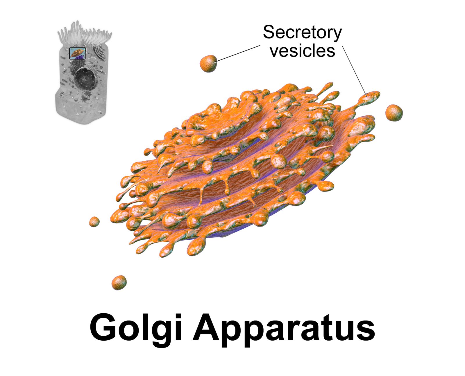
The Golgi apparatus (), also known as the Golgi complex, Golgi body, or simply the Golgi, is an organelle found in most eukaryotic Cell (biology), cells. Part of the endomembrane system in the cytoplasm, it protein targeting, packages proteins into membrane-bound Vesicle (biology and chemistry), vesicles inside the cell before the vesicles are sent to their destination. It resides at the intersection of the secretory, lysosomal, and Endocytosis, endocytic pathways. It is of particular importance in processing proteins for secretion, containing a set of glycosylation enzymes that attach various sugar monomers to proteins as the proteins move through the apparatus. The Golgi apparatus was identified in 1898 by the Italian biologist and pathologist Camillo Golgi. The organelle was later named after him in the 1910s. Discovery Because of its large size and distinctive structure, the Golgi apparatus was one of the first organelles to be discovered and observed in detail. It was d ... [...More Info...] [...Related Items...] OR: [Wikipedia] [Google] [Baidu] |
Vesicle (biology)
In cell biology, a vesicle is a structure within or outside a cell, consisting of liquid or cytoplasm enclosed by a lipid bilayer. Vesicles form naturally during the processes of secretion ( exocytosis), uptake ( endocytosis), and the transport of materials within the plasma membrane. Alternatively, they may be prepared artificially, in which case they are called liposomes (not to be confused with lysosomes). If there is only one phospholipid bilayer, the vesicles are called '' unilamellar liposomes''; otherwise they are called ''multilamellar liposomes''. The membrane enclosing the vesicle is also a lamellar phase, similar to that of the plasma membrane, and intracellular vesicles can fuse with the plasma membrane to release their contents outside the cell. Vesicles can also fuse with other organelles within the cell. A vesicle released from the cell is known as an extracellular vesicle. Vesicles perform a variety of functions. Because it is separated from the cytoso ... [...More Info...] [...Related Items...] OR: [Wikipedia] [Google] [Baidu] |
Phosphoinositide
Phosphatidylinositol or inositol phospholipid is a biomolecule. It was initially called "inosite" when it was discovered by Léon Maquenne and Johann Joseph von Scherer in the late 19th century. It was discovered in bacteria but later also found in eukaryotes, and was found to be a signaling molecule. The biomolecule can exist in 9 different isomers. It is a lipid which contains a phosphate group, two fatty acid chains, and one inositol sugar molecule. Typically, the phosphate group has a negative charge (at physiological pH values). As a result, the molecule is amphiphilic. The production of the molecule is limited to the endoplasmic reticulum. History of phospatidylinositol Phosphatidylinositol (PI) and its derivatives have a rich history dating back to their discovery by Johann Joseph von Scherer and Léon Maquenne in the late 19th century. Initially known as "inosite" based on its sweet taste, the isolation and characterization of inositol laid the groundwork for unders ... [...More Info...] [...Related Items...] OR: [Wikipedia] [Google] [Baidu] |
Phosphatidylglycerol
Phosphatidylglycerol is a glycerophospholipid found in pulmonary surfactant and in the plasma membrane where it directly activates lipid-gated ion channels. The general structure of phosphatidylglycerol consists of a L-glycerol 3-phosphate backbone ester-bonded to either saturated or unsaturated fatty acids on carbons 1 and 2. The head group substituent glycerol is bonded through a phosphomonoester. It is the precursor of surfactant and its presence (>0.3) in the amniotic fluid of the newborn indicates fetal lung maturity. Approximately 98% of alveolar wall surface area is due to the presence of type I cells, with type II cells producing pulmonary surfactant covering around 2% of the alveolar walls. Once surfactant is secreted by the type II cells, it must be spread over the remaining type I cellular surface area. Phosphatidylglycerol is thought to be important in spreading of surfactant over the Type I cellular surface area. The major surfactant deficiency in premature infants r ... [...More Info...] [...Related Items...] OR: [Wikipedia] [Google] [Baidu] |
Phosphatidylserine
Phosphatidylserine (abbreviated Ptd-L-Ser or PS) is a phospholipid and is a component of the cell membrane. It plays a key role in cell cycle signaling, specifically in relation to apoptosis. It is a key pathway for viruses to enter cells via apoptotic mimicry. Its exposure on the outer surface of a membrane marks the cell for destruction via apoptosis. Structure Phosphatidylserine is a phospholipid—more specifically a glycerophospholipid—which consists of two fatty acids attached in ester linkage to the first and second carbon of glycerol and serine attached through a phosphodiester linkage to the third carbon of the glycerol. Phosphatidylserine sourced from plants differs in fatty acid composition from that sourced from animals. It is commonly found in the inner (cytoplasmic) leaflet of biological membranes. It is almost entirely found in the inner monolayer of the membrane with only less than 10% of it in the outer monolayer. Biosynthesis Phosphatidylserine (PS) is ... [...More Info...] [...Related Items...] OR: [Wikipedia] [Google] [Baidu] |
Phosphatidylethanolamine
Phosphatidylethanolamine (PE) is a class of phospholipids found in biological membranes. They are synthesized by the addition of cytidine diphosphate-ethanolamine to diglycerides, releasing cytidine monophosphate. S-Adenosyl methionine, ''S''-Adenosyl methionine can subsequently Methylation, methylate the amine of phosphatidylethanolamines to yield phosphatidylcholines. Function In cells Phosphatidylethanolamines are found in all living cells, composing 25% of all phospholipids. In human physiology, they are found particularly in nervous tissue such as the white matter of brain, nerves, neural tissue, and in spinal cord, where they make up 45% of all phospholipids. Phosphatidylethanolamines play a role in membrane fusion and in disassembly of the contractile ring during cytokinesis in cell division. Additionally, it is thought that phosphatidylethanolamine regulates membrane curvature. Phosphatidylethanolamine is an important precursor, substrate (biochemistry), substrate, or d ... [...More Info...] [...Related Items...] OR: [Wikipedia] [Google] [Baidu] |
Mitochondria
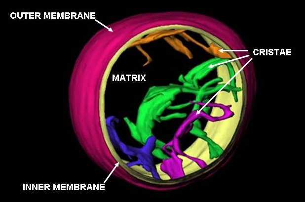
A mitochondrion () is an organelle found in the cells of most eukaryotes, such as animals, plants and fungi. Mitochondria have a double membrane structure and use aerobic respiration to generate adenosine triphosphate (ATP), which is used throughout the cell as a source of chemical energy. They were discovered by Albert von Kölliker in 1857 in the voluntary muscles of insects. The term ''mitochondrion'', meaning a thread-like granule, was coined by Carl Benda in 1898. The mitochondrion is popularly nicknamed the "powerhouse of the cell", a phrase popularized by Philip Siekevitz in a 1957 ''Scientific American'' article of the same name. Some cells in some multicellular organisms lack mitochondria (for example, mature mammalian red blood cells). The multicellular animal '' Henneguya salminicola'' is known to have retained mitochondrion-related organelles despite a complete loss of their mitochondrial genome. A large number of unicellular organisms, such as microspo ... [...More Info...] [...Related Items...] OR: [Wikipedia] [Google] [Baidu] |


