|
Pericardiocentesis
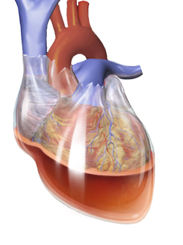
Pericardiocentesis (PCC), also called pericardial tap, is a medical procedure where fluid is aspirated from the pericardium (the sac enveloping the heart). Anatomy and physiology The pericardium is a fibrous sac surrounding the heart composed of two layers: an inner visceral pericardium and an outer parietal pericardium. The area between these two layers is known as the pericardial space and normally contains 15 to 50 mL of serous fluid. This fluid protects the heart by serving as a shock absorber and provides lubrication to the heart during contraction. The elastic nature of the pericardium allows it to accommodate a small amount of extra fluid, roughly 80 to 120 mL, in the acute setting. However, once a critical volume is reached, even small amounts of extra fluid can rapidly increase pressure within the pericardium. This pressure can significantly hinder the ability of the heart to contract, leading to cardiac tamponade. If accumulation of fluid is slow and occurs over ... [...More Info...] [...Related Items...] OR: [Wikipedia] [Google] [Baidu] |
Pericardial Effusion
A pericardial effusion is an abnormal accumulation of fluid in the pericardial cavity. The pericardium is a two-part membrane surrounding the heart: the outer fibrous Connective tissue, connective membrane and an inner two-layered serous membrane. The two layers of the serous membrane enclose the pericardial cavity (the potential space) between them. This pericardial space contains a small amount of pericardial fluid, normally 15-50 mL in volume. The pericardium, specifically the pericardial fluid provides lubrication, maintains the anatomic position of the heart in the chest (levocardia), and also serves as a barrier to protect the heart from infection and inflammation in adjacent tissues and organs. By definition, a pericardial effusion occurs when the volume of fluid in the cavity exceeds the normal limit. If large enough, it can compress the heart, causing cardiac tamponade and obstructive shock. Some of the presenting symptoms are shortness of breath, chest pain, chest pressu ... [...More Info...] [...Related Items...] OR: [Wikipedia] [Google] [Baidu] |
Pericardial Fluid
Pericardial fluid is the serous fluid secreted by the Serous membrane, serous layer of the pericardium into the pericardial cavity. The pericardium consists of two layers, an outer fibrous layer and the inner serous layer. This serous layer has two membranes which enclose the pericardial cavity into which is secreted the pericardial fluid. The fluid is similar to the cerebrospinal fluid of the brain which also serves to cushion and allow some movement of the organ. __TOC__ Function The pericardial fluid reduces friction within the pericardium by lubricating the epicardial surface allowing the membranes to glide over each other with each heart beat. Composition Ben-Horin ''et al.'' (2005) studied the composition of pericardial fluid in patients undergoing open heart surgery. They found that the fluid is made up of a high concentration of lactate dehydrogenase (LDH), protein and lymphocytes. In a healthy adult there is up to 50 ml of clear, straw-coloured fluid. However, there is l ... [...More Info...] [...Related Items...] OR: [Wikipedia] [Google] [Baidu] |
Cardiac Tamponade
Cardiac tamponade, also known as pericardial tamponade (), is a compression of the heart due to pericardial effusion (the build-up of pericardial fluid in the pericardium, sac around the heart). Onset may be rapid or gradual. Symptoms typically include those of obstructive shock including shortness of breath, weakness, lightheadedness, and cough. Other symptoms may relate to the underlying cause. Common causes of cardiac tamponade include cancer, kidney failure, chest trauma, myocardial infarction, and pericarditis. Other causes include connective tissue disease, connective tissues diseases, hypothyroidism, aortic rupture, autoimmune disease, and complications of cardiac surgery. In Africa, tuberculosis is a relatively common cause. Diagnosis may be suspected based on hypotension, low blood pressure, jugular venous distension, or quiet heart sounds (together known as Beck's triad (cardiology), Beck's triad). A pericardial rub may be present in cases due to inflammation. The dia ... [...More Info...] [...Related Items...] OR: [Wikipedia] [Google] [Baidu] |
Aortic Dissection
Aortic dissection (AD) occurs when an injury to the innermost layer of the aorta allows blood to flow between the layers of the aortic wall, forcing the layers apart. In most cases, this is associated with a sudden onset of agonizing chest or back pain, often described as "tearing" in character. Vomiting, sweating, and lightheadedness may also occur. Damage to other organs may result from the decreased blood supply, such as stroke, lower extremity ischemia, or mesenteric ischemia. Aortic dissection can quickly lead to death from insufficient blood flow to the heart or complete rupture of the aorta. AD is more common in those with a history of high blood pressure; a number of connective tissue diseases that affect blood vessel wall strength including Marfan syndrome and Ehlers–Danlos syndrome; a bicuspid aortic valve; and previous heart surgery. Major trauma, smoking, cocaine use, pregnancy, a thoracic aortic aneurysm, inflammation of arteries, and abnormal lipid ... [...More Info...] [...Related Items...] OR: [Wikipedia] [Google] [Baidu] |
Thoracic Wall
The thoracic wall or chest wall is the boundary of the thoracic cavity. Structure The bony skeletal part of the thoracic wall is the rib cage, and the rest is made up of muscle, skin, and fasciae. The chest wall has 10 layers, namely (from superficial to deep) skin (epidermis and dermis), superficial fascia, deep fascia and the invested extrinsic muscles (from the upper limbs), intrinsic muscles associated with the ribs (three layers of intercostal muscles), endothoracic fascia and parietal pleura. However, the extrinsic muscular layers vary according to the region of the chest wall. For example, the front and back sides may include attachments of large upper limb muscles like pectoralis major or latissimus dorsi, while the sides only have serratus anterior.The thoracic wall consists of a bony framework that is held together by twelve thoracic vertebrae posteriorly which give rise to ribs that encircle the lateral and anterior thoracic cavity. The first nine ribs curve a ... [...More Info...] [...Related Items...] OR: [Wikipedia] [Google] [Baidu] |
Supine Position
The supine position () means lying horizontally, with the face and torso facing up, as opposed to the prone position, which is face down. When used in surgical procedures, it grants access to the peritoneal, thoracic, and pericardium, pericardial regions; as well as the head, neck, and extremities. Using anatomical terms of location, the dorsal side is down, and the ventral side is up, when supine. Semi-supine In scientific literature "semi-supine" commonly refers to positions where the upper body is tilted (at 45° or variations) and not completely horizontal. Relation to sudden infant death syndrome The decline in death due to sudden infant death syndrome (SIDS) is said to be attributable to having babies sleep in the supine position. The realization that infants sleeping face down, or in a prone position, had an increased mortality rate re-emerged into medical awareness at the end of the 1980s when two researchers, Susan Beal in Australia and Gus De Jonge in the Nether ... [...More Info...] [...Related Items...] OR: [Wikipedia] [Google] [Baidu] |
Pacemakers
A pacemaker, also known as an artificial cardiac pacemaker, is an implanted medical device that generates electrical pulses delivered by electrodes to one or more of the chambers of the heart. Each pulse causes the targeted chamber(s) to contract and pump blood, thus regulating the function of the electrical conduction system of the heart. The primary purpose of a pacemaker is to maintain an even heart rate, either because the heart's natural cardiac pacemaker provides an inadequate or irregular heartbeat, or because there is a block in the heart's electrical conduction system. Modern pacemakers are externally programmable and allow a cardiologist to select the optimal pacing modes for individual patients. Most pacemakers are on demand, in which the stimulation of the heart is based on the dynamic demand of the circulatory system. Others send out a fixed rate of impulses. A specific type of pacemaker, called an implantable cardioverter-defibrillator, combines pacemaker and ... [...More Info...] [...Related Items...] OR: [Wikipedia] [Google] [Baidu] |
Pericardial Window
A pericardial window is a cardiac surgical procedure to create a fistula – or "window" – from the pericardial space to the pleural cavity. The purpose of the window is to allow a pericardial effusion or cardiac tamponade to drain from the space surrounding the heart into the chest cavity. Uses Pericardial window may be used to treat pericardial effusion and cardiac tamponade. It is the most common procedure to treat pericardial effusion, particularly if caused by cancer. Untreated, these can lead to death. The pericardial window decreases the incidence of postoperative pericardial tamponade and new-onset atrial fibrillation after open-heart surgery. Risks Creation of a pericardial window is a major surgical procedure. To remove pericardial fluid, other more minor techniques should be considered first, such as pericardiocentesis. Technique Pericardial window is usually performed under general anaesthetic by a cardiac surgeon. They may make an open surgical incision o ... [...More Info...] [...Related Items...] OR: [Wikipedia] [Google] [Baidu] |
Prosthetic Heart Valves
An artificial heart valve is a one-way valve implanted into a person's heart to replace a heart valve that is not functioning properly (valvular heart disease). Artificial heart valves can be separated into three broad classes: mechanical heart valves, bioprosthetic tissue valves and engineered tissue valves. The human heart contains four valves: tricuspid valve, pulmonary valve, mitral valve and aortic valve. Their main purpose is to keep blood flowing in the proper direction through the heart, and from the heart into the major blood vessels connected to it (the pulmonary artery and the aorta). Heart valves can malfunction for a variety of reasons, which can impede the flow of blood through the valve (stenosis) and/or let blood flow backwards through the valve ( regurgitation). Both processes put strain on the heart and may lead to serious problems, including heart failure. While some dysfunctional valves can be treated with drugs or repaired, others need to be replaced with a ... [...More Info...] [...Related Items...] OR: [Wikipedia] [Google] [Baidu] |
Wiley-Blackwell
Wiley-Blackwell is an international scientific, technical, medical, and scholarly publishing business of John Wiley & Sons. It was formed by the merger of John Wiley & Sons Global Scientific, Technical, and Medical business with Blackwell Publishing in 2007. Wiley-Blackwell is now an imprint that publishes a diverse range of academic and professional fields, including biology, medicine, physical sciences, technology, social science, and the humanities. Blackwell Publishing history Blackwell Publishing was formed by the 2001 merger of two Oxford-based academic publishing companies, Blackwell Science, founded in 1939 as Blackwell Scientific Publishing, and Blackwell Publishers, founded in 1922 as Basil Blackwell & Mott. Blackwell Publishers, founded in 1926, had its origins in the 19th century Blackwell's family bookshop and publishing business. The merger between the two publishing companies created the world's leading learned society publisher. The group then acquired BMJ Boo ... [...More Info...] [...Related Items...] OR: [Wikipedia] [Google] [Baidu] |


