|
Pedunculopontine
The pedunculopontine nucleus (PPN) or pedunculopontine tegmental nucleus (PPT or PPTg) is a collection of neurons located in the upper pons in the brainstem. It is involved in voluntary movements, arousal, and provides sensory feedback to the cerebral cortex and one of the main components of the ascending reticular activating system. It is a potential target for deep brain stimulation treatment for Parkinson's disease. It was first described in 1909 by Louis Jacobsohn-Lask, a German neuroanatomy, neuroanatomist. Structure and projections The pedunculopontine nucleus lies below the red nucleus, Anatomical terms of location#Caudal, caudal to the substantia nigra and adjacent to the superior cerebellar peduncle. It has two divisions of subnuclei; the pars compacta, containing mainly cholinergic neurons, and the pars dissipata, containing mainly glutamatergic neurons and some non-cholinergic neurons. Its neurons project axons to a wide range of areas in the brain, particularly parts o ... [...More Info...] [...Related Items...] OR: [Wikipedia] [Google] [Baidu] |
Ascending Reticular Activating System
The reticular formation is a set of interconnected nuclei in the brainstem that spans from the lower end of the medulla oblongata to the upper end of the midbrain. The neurons of the reticular formation make up a complex set of neural networks in the core of the brainstem. The reticular formation is made up of a diffuse net-like formation of reticular nuclei which is not well-defined. It may be seen as being made up of all the interspersed cells in the brainstem between the more compact and named structures. The reticular formation is functionally divided into the ascending reticular activating system (ARAS), ascending pathways to the cerebral cortex, and the descending reticular system, descending pathways ( reticulospinal tracts) to the spinal cord. Due to its extent along the brainstem it may be divided into different areas such as the midbrain reticular formation, the central mesencephalic reticular formation, the pontine reticular formation, the paramedian pontine reticu ... [...More Info...] [...Related Items...] OR: [Wikipedia] [Google] [Baidu] |
Deep Brain Stimulation
Deep brain stimulation (DBS) is a type of neurostimulation therapy in which an implantable pulse generator is stereotactic surgery, surgically implanted subcutaneous tissue, below the skin of the chest and connected by Lead (electronics), leads to the brain to deliver controlled electrical charge, electrical impulses. These charges therapeutically disrupt and promote dysfunctional nervous system circuits bidirectionally in both ante- and retrograde signaling, retrograde directions. Though first developed for Parkinsonian tremor, the technology has since been adapted to a wide variety of chronic neurologic disorders. The usage of electrical stimulation to treat neurologic disorders dates back thousands of years to ancient Greece and Early Dynastic Period (Egypt), dynastic Egypt. The distinguishing feature of DBS, however, is that by taking advantage of the portability of lithium-ion battery technology, it is able to be used long term without the patient having to be Electrical wir ... [...More Info...] [...Related Items...] OR: [Wikipedia] [Google] [Baidu] |
Reward System
The reward system (the mesocorticolimbic circuit) is a group of neural structures responsible for incentive salience (i.e., "wanting"; desire or craving for a reward and motivation), associative learning (primarily positive reinforcement and classical conditioning), and positively-valenced emotions, particularly ones involving pleasure as a core component (e.g., joy, euphoria and ecstasy). Reward is the attractive and motivational property of a stimulus that induces appetitive behavior, also known as approach behavior, and consummatory behavior. A rewarding stimulus has been described as "any stimulus, object, event, activity, or situation that has the potential to make us approach and consume it is by definition a reward". In operant conditioning, rewarding stimuli function as positive reinforcers; however, the converse statement also holds true: positive reinforcers are rewarding. The reward system motivates animals to approach stimuli or engage in behaviour that increase ... [...More Info...] [...Related Items...] OR: [Wikipedia] [Google] [Baidu] |
Basal Ganglia
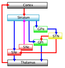
The basal ganglia (BG) or basal nuclei are a group of subcortical Nucleus (neuroanatomy), nuclei found in the brains of vertebrates. In humans and other primates, differences exist, primarily in the division of the globus pallidus into external and internal regions, and in the division of the striatum. Positioned at the base of the forebrain and the top of the midbrain, they have strong connections with the cerebral cortex, thalamus, brainstem and other brain areas. The basal ganglia are associated with a variety of functions, including regulating voluntary motor control, motor movements, procedural memory, procedural learning, habituation, habit formation, conditional learning, eye movements, cognition, and emotion. The main functional components of the basal ganglia include the striatum, consisting of both the dorsal striatum (caudate nucleus and putamen) and the ventral striatum (nucleus accumbens and olfactory tubercle), the globus pallidus, the ventral pallidum, the substa ... [...More Info...] [...Related Items...] OR: [Wikipedia] [Google] [Baidu] |
Globus Pallidus Internus
The internal globus pallidus (GPi or medial globus pallidus) is one of the two subcortical nuclei that provides inhibitory output in the basal ganglia, the other being the substantia nigra pars reticulata. Together with the external globus pallidus (GPe), it makes up one of the two segments of the globus pallidus, a structure that can decay with certain neurodegenerative disorders and is a target for medical and neurosurgical therapies. The GPi, along with the substantia nigra pars reticulata, comprise the primary output of the basal ganglia, with its outgoing GABAergic neurons having an inhibitory function in the thalamus, the centromedian complex and the pedunculopontine complex. Anatomy The efferent bundle is constituted first of the ansa and lenticular fasciculus, then crosses the internal capsule within and in parallel to the Edinger's comb system then arrives at the laterosuperior corner of the subthalamic nucleus and constitutes the field H2 of Forel, then H, and suddenl ... [...More Info...] [...Related Items...] OR: [Wikipedia] [Google] [Baidu] |
Substantia Nigra Pars Reticulata
The pars reticulata (SNpr) is a portion of the substantia nigra and is located lateral to the pars compacta. Most of the neurons that project out of the pars reticulata are inhibitory GABAergic neurons (i.e., these neurons release GABA, which is an inhibitory neurotransmitter). Anatomy Neurons in the pars reticulata are much less densely packed than those in the pars compacta (they were sometimes named pars diffusa). They are smaller and thinner than the dopaminergic neurons and conversely identical and morphologically similar to the pallidal neurons (see primate basal ganglia). Their dendrites as well as the pallidal are preferentially perpendicular to the striatal afferents. The massive striatal afferents correspond to the medial end of the nigrostriatal bundle. Nigral neurons have the same peculiar synaptology with the striatal axonal endings. They make connections with the dopamine neurons of the pars compacta whose long dendrites plunge deeply in the pars reticulata. The ... [...More Info...] [...Related Items...] OR: [Wikipedia] [Google] [Baidu] |
Louis Jacobsohn-Lask
Louis Jacobsohn-Lask (born Louis Jacobsohn; 2 March 1863, in Bromberg – 17 May 1941, in Sevastopol) was a Germans, German neurologist and neuroanatomist. He studied medicine at the University of Berlin under Heinrich Wilhelm Waldeyer, Rudolf Virchow, Emil du Bois-Reymond, Ernst Viktor von Leyden and Robert Koch. In 1899 Jacobsohn and Edward Flatau wrote ''Handbuch der Anatomie und vergleichenden Anatomie des Centralnervensystems der Säugetiere'', which included one of the first attempts to classify Sulcus (morphology), sulci and gyrus, gyri of human brain Cerebral cortex, cortex. In 1904 he wrote, together with Flatau and Lazar Salomowitch Minor, Lazar Minor, another monograph, ''Handbuch der pathologischen Anatomie der Nervensystems''. He described a finger flexion reflex called the Bekhterev-Jacobsohn reflex or Jacobsohn reflex. In 1909 he first described the pedunculopontine nucleus. In 1936 he emigrated to the Soviet Union with his wife, Berta Lask, Berta Jacobsohn-Lask, ... [...More Info...] [...Related Items...] OR: [Wikipedia] [Google] [Baidu] |
Arousal
Arousal is the physiology, physiological and psychology, psychological state of being awoken or of Five senses, sense organs stimulated to a point of perception. It involves activation of the ascending reticular activating system (ARAS) in the human brain, brain, which mediates wakefulness, the autonomic nervous system, and the endocrine system, leading to increased heart rate and blood pressure and a condition of sensory alertness, desire, mobility, and reactivity. Arousal is mediated by several neural systems. Wakefulness is regulated by the ARAS, which is composed of projections from five major neurotransmitter systems that originate in the brainstem and form connections extending throughout the Cerebral cortex, cortex; activity within the ARAS is regulated by neurons that release the neurotransmitters norepinephrine, acetylcholine, dopamine, serotonin and histamine. Activation of these neurons produces an increase in cortical activity and subsequently alertness. Arousal is ... [...More Info...] [...Related Items...] OR: [Wikipedia] [Google] [Baidu] |
Motor Cortex
The motor cortex is the region of the cerebral cortex involved in the planning, motor control, control, and execution of voluntary movements. The motor cortex is an area of the frontal lobe located in the posterior precentral gyrus immediately anterior to the central sulcus. Components The motor cortex can be divided into three areas: 1. The primary motor cortex is the main contributor to generating neural impulses that pass down to the spinal cord and control the execution of movement. However, some of the other motor areas in the brain also play a role in this function. It is located on the anterior paracentral lobule on the medial surface. 2. The premotor cortex is responsible for some aspects of motor control, possibly including the preparation for movement, the sensory guidance of movement, the spatial guidance of reaching, or the direct control of some movements with an emphasis on control of proximal and trunk muscles of the body. Located anterior to the primary mo ... [...More Info...] [...Related Items...] OR: [Wikipedia] [Google] [Baidu] |
Cerebral Cortex
The cerebral cortex, also known as the cerebral mantle, is the outer layer of neural tissue of the cerebrum of the brain in humans and other mammals. It is the largest site of Neuron, neural integration in the central nervous system, and plays a key role in attention, perception, awareness, thought, memory, language, and consciousness. The six-layered neocortex makes up approximately 90% of the Cortex (anatomy), cortex, with the allocortex making up the remainder. The cortex is divided into left and right parts by the longitudinal fissure, which separates the two cerebral hemispheres that are joined beneath the cortex by the corpus callosum and other commissural fibers. In most mammals, apart from small mammals that have small brains, the cerebral cortex is folded, providing a greater surface area in the confined volume of the neurocranium, cranium. Apart from minimising brain and cranial volume, gyrification, cortical folding is crucial for the Neural circuit, brain circuitry ... [...More Info...] [...Related Items...] OR: [Wikipedia] [Google] [Baidu] |
Cerebellum
The cerebellum (: cerebella or cerebellums; Latin for 'little brain') is a major feature of the hindbrain of all vertebrates. Although usually smaller than the cerebrum, in some animals such as the mormyrid fishes it may be as large as it or even larger. In humans, the cerebellum plays an important role in motor control and cognition, cognitive functions such as attention and language as well as emotion, emotional control such as regulating fear and pleasure responses, but its movement-related functions are the most solidly established. The human cerebellum does not initiate movement, but contributes to motor coordination, coordination, precision, and accurate timing: it receives input from sensory systems of the spinal cord and from other parts of the brain, and integrates these inputs to fine-tune motor activity. Cerebellar damage produces disorders in fine motor skill, fine movement, sense of balance, equilibrium, list of human positions, posture, and motor learning in humans. ... [...More Info...] [...Related Items...] OR: [Wikipedia] [Google] [Baidu] |
Basal Forebrain
Part of the human brain, the basal forebrain structures are located in the forebrain to the front of and below the striatum. They include the ventral basal ganglia (including nucleus accumbens and ventral pallidum), nucleus basalis, diagonal band of Broca, substantia innominata, and the medial septal nucleus. These structures are important in the production of acetylcholine, which is then distributed widely throughout the brain. The basal forebrain is considered to be the major cholinergic output of the central nervous system (CNS) centred on the output of the nucleus basalis. The presence of non-cholinergic neurons projecting to the cortex have been found to act with the cholinergic neurons to dynamically modulate activity in the cortex. Function Acetylcholine is known to promote wakefulness in the basal forebrain. Stimulating the basal forebrain gives rise to acetylcholine release, which induces wakefulness and REM sleep, whereas inhibition of acetylcholine release in the ba ... [...More Info...] [...Related Items...] OR: [Wikipedia] [Google] [Baidu] |


