|
Pedicle
Pedicle or pedicel may refer to: Human anatomy *Pedicle of vertebral arch, the segment between the transverse process and the vertebral body, and is often used as a radiographic marker and entry point in vertebroplasty and kyphoplasty procedures *Pedicle of a skin flap (medicine) * Hilum of kidney, also called the renal pedicle *Pedicel, a foot process of a some cells Non-human anatomy * Pedicle in brachiopods, a fleshy line used to attach and anchor brachiopods and some bivalve mollusks to a substrate *Pedicle (cervidae), the attachment point for antlers in cervids * Pedicel (antenna), the second segment of the antenna in the class Insecta, where the Johnston's organ is found * Pedicel or petiole (insect), the stem formed by a restricted abdominal segment which connects the thorax with the gaster (the remaining abdominal segments) in the suborder Apocrita * Pedicel (spider), the narrow segment connecting the cephalothorax with the abdomen Other *Pedicel (botany), the stalk of ... [...More Info...] [...Related Items...] OR: [Wikipedia] [Google] [Baidu] |
Zaire Pedicle
The Congo Pedicle (at one time referred to as the Zaire Pedicle; in French, ', meaning 'Katanga boot') is the southeast salient of the Haut-Katanga Province of the Democratic Republic of the Congo, which divides neighbouring Zambia into two lobes. In area, the pedicle is similar in size to Wales or New Jersey. ' Pedicle' is used in the sense of 'a little foot'. 'Congo Pedicle' or 'the Pedicle' is also used to refer to the Congo Pedicle road, which crosses it. The Congo Pedicle is an example of the boundaries imposed by European powers on Africa in the wake of the Scramble for Africa, which were set by European interests and usually did not consider pre-existing political or tribal boundaries. As it is located at the country's southeastern extremity, its eastern end is closer to the capital cities of 17 other African countries than its own, Kinshasa. British and Belgian territorial claims Cecil Rhodes's British South Africa Company (BSAC) approached Katanga from the south,and th ... [...More Info...] [...Related Items...] OR: [Wikipedia] [Google] [Baidu] |
Brachiopod
Brachiopods (), phylum (biology), phylum Brachiopoda, are a phylum of animals that have hard "valves" (shells) on the upper and lower surfaces, unlike the left and right arrangement in bivalve molluscs. Brachiopod valves are hinged at the rear end, while the front can be opened for feeding or closed for protection. Two major categories are traditionally recognized, articulate and inarticulate brachiopods. The word "articulate" is used to describe the tooth-and-groove structures of the valve-hinge which is present in the articulate group, and absent from the inarticulate group. This is the leading diagnostic skeletal feature, by which the two main groups can be readily distinguished as fossils. Articulate brachiopods have toothed hinges and simple, vertically oriented opening and closing muscles. Conversely, inarticulate brachiopods have weak, untoothed hinges and a more complex system of vertical and oblique (diagonal) muscles used to keep the two valves aligned. In many brachio ... [...More Info...] [...Related Items...] OR: [Wikipedia] [Google] [Baidu] |
Pedicle Of Vertebral Arch
Each vertebra (: vertebrae) is an irregular bone with a complex structure composed of bone and some hyaline cartilage, that make up the vertebral column or spine, of vertebrates. The proportions of the vertebrae differ according to their spinal segment and the particular species. The basic configuration of a vertebra varies; the vertebral body (also ''centrum'') is of bone and bears the load of the vertebral column. The upper and lower surfaces of the vertebra body give attachment to the intervertebral discs. The posterior part of a vertebra forms a vertebral arch, in eleven parts, consisting of two pedicles (pedicle of vertebral arch), two laminae, and seven processes. The laminae give attachment to the ligamenta flava (ligaments of the spine). There are vertebral notches formed from the shape of the pedicles, which form the intervertebral foramina when the vertebrae articulate. These foramina are the entry and exit conduits for the spinal nerves. The body of the vertebra a ... [...More Info...] [...Related Items...] OR: [Wikipedia] [Google] [Baidu] |
Skin Flap
The terms free flap, free autologous tissue transfer and microvascular free tissue transfer are synonymous terms used to describe the "transplantation" of tissue from one site of the body to another, in order to reconstruct an existing defect. "Free" implies that the tissue is completely detached from its blood supply at the original location ("donor site") and then transferred to another location ("recipient site") and the circulation in the tissue re-established by anastomosis of artery(s) and vein(s). This is in contrast to a "pedicled" flap in which the tissue is left partly attached to the donor site ("pedicle") and simply transposed to a new location; keeping the "pedicle" intact as a conduit to supply the tissue with blood. Various types of tissue may be transferred as a "free flap" including skin and fat, muscle, nerve, bone, cartilage (or any combination of these), lymph nodes and intestinal segments. An example of "free flap" could be a "free toe transfer" in which ... [...More Info...] [...Related Items...] OR: [Wikipedia] [Google] [Baidu] |
Hilum Of Kidney
The renal hilum or renal pedicle is the recessed central fissure of the kidney where its vessels, nerves and ureter pass. The medial border of the kidney is concave in the center and convex toward either extremity; it is directed forward and a little downward. Its central part presents a deep longitudinal fissure, bounded by prominent overhanging anterior and posterior lips. This fissure is a hilum that transmits the vessels, nerves, and ureter. From anterior to posterior, the renal vein exits, the renal artery enters, and the renal pelvis exits the kidney. On the left hand side the hilum is located at the L1 vertebral level and the right kidney at level L1-2. The lower border of the kidneys is usually alongside L3. Structure The superior, middle, and inferior vessels enter or leave the hilum of kidney: from anterior to posterior is renal vein, renal artery and renal pelvis, respectively. See also * Renal artery * Renal vein * Renal pyramids * Renal medulla The renal med ... [...More Info...] [...Related Items...] OR: [Wikipedia] [Google] [Baidu] |
Foot Process
Cellular extensions also known as cytoplasmic protrusions and cytoplasmic processes are those structures that project from different Cell (biology), cells, in the body, or in other organisms. Many of the extensions are cytoplasmic protrusions such as the axon and dendrite of a neuron, known also as cytoplasmic processes. Different glial cells project cytoplasmic processes. In the brain, the processes of astrocytes form terminal endfeet, foot processes that help to form protective barriers in the brain. In the kidneys specialised cells called podocytes extend processes that terminate in podocyte foot processes that cover glomerulus, capillaries in the nephron. End-processes may also be known as ''vascular footplates'', and in general may exhibit a pyramidal or finger-like morphology. Mural cells such as pericytes extend processes to wrap around capillaries. Foot-like processes are also present in Müller glia (modified astrocytes of the retina), pancreatic stellate cells, Dendri ... [...More Info...] [...Related Items...] OR: [Wikipedia] [Google] [Baidu] |
Antler
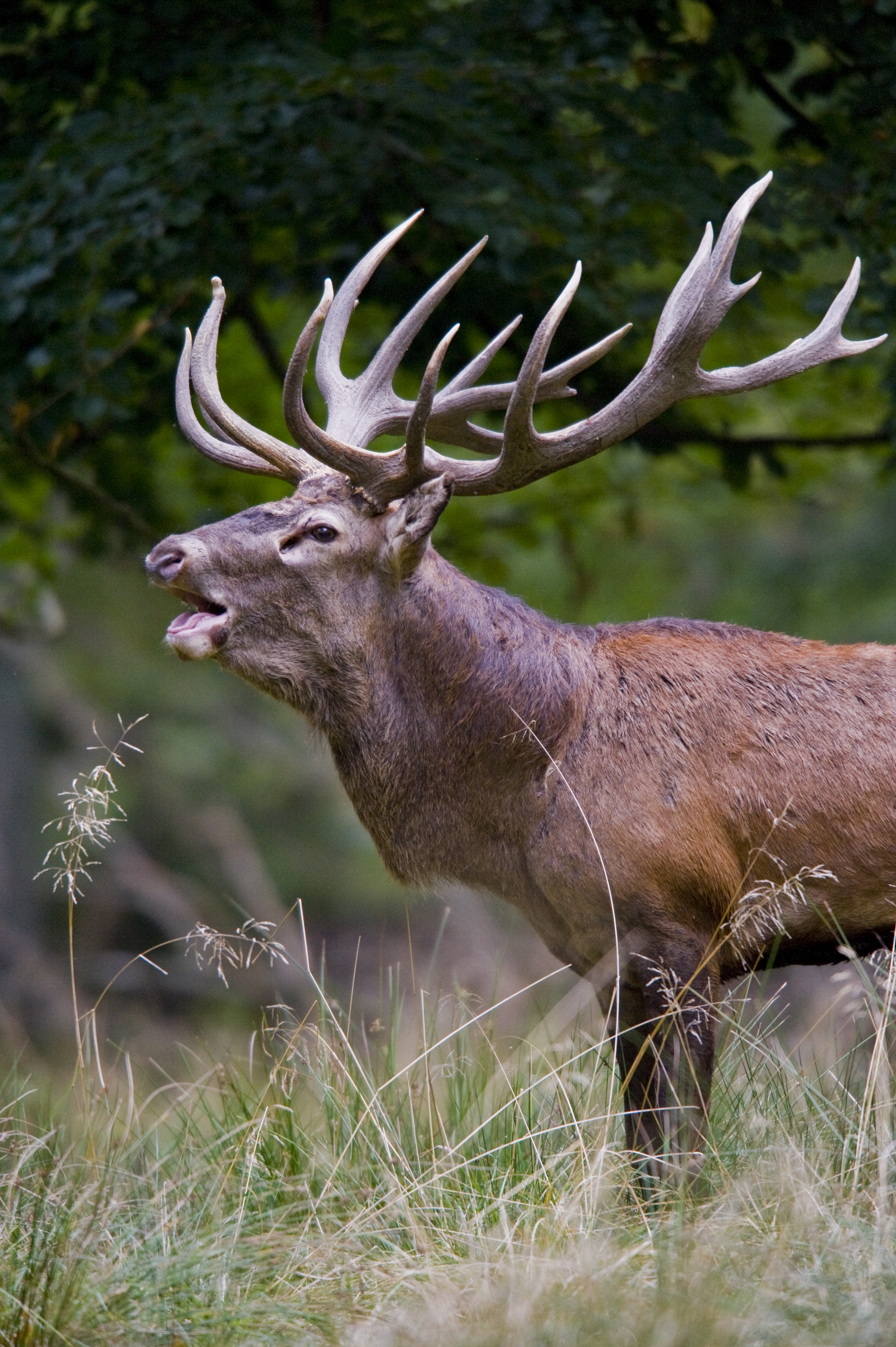
Antlers are extensions of an animal's skull found in members of the Cervidae (deer) Family (biology), family. Antlers are a single structure composed of bone, cartilage, fibrous tissue, skin, nerves, and blood vessels. They are generally found only on males, with the exception of Reindeer, reindeer/caribou. Antlers are Moulting, shed and regrown each year and function primarily as objects of sexual attraction and as Weapon (biology), weapons. Etymology Antler comes from the Old French ''antoillier ''(see present French : "Andouiller", from'' ant-, ''meaning before,'' oeil, ''meaning eye and'' -ier'', a suffix indicating an action or state of being) possibly from some form of an unattested Latin word ''*anteocularis'', "before the eye" (and applied to the word for "branch" or "horn (anatomy), horn"). Structure and development Antlers are unique to cervids. The ancestors of deer had tusks (long upper canine tooth, canine teeth). In most species, antlers appear to replace t ... [...More Info...] [...Related Items...] OR: [Wikipedia] [Google] [Baidu] |
Pedicel (antenna)
An antenna (plural: antennae) is one of a pair of appendages used for sensing in arthropods. Antennae are sometimes referred to as ''feelers''. Antennae are connected to the first one or two segments of the arthropod head. They vary widely in form but are always made of one or more jointed segments. While they are typically sensory organs, the exact nature of what they sense and how they sense it is not the same in all groups. Functions may variously include sensing touch, air motion, heat, vibration (sound), and especially smell or taste. Antennae are sometimes modified for other purposes, such as mating, brooding, swimming, and even anchoring the arthropod to a substrate. Larval arthropods have antennae that differ from those of the adult. Many crustaceans, for example, have free-swimming larvae that use their antennae for swimming. Antennae can also locate other group members if the insect lives in a group, like the ant. The common ancestor of all arthropods likely had on ... [...More Info...] [...Related Items...] OR: [Wikipedia] [Google] [Baidu] |
Petiole (insect)
In entomology, petiole is the technical term for the narrow waist of some Hymenoptera, hymenopteran insects, especially ants, bees, and wasps in the suborder Apocrita. The petiole can consist of either one or two segments, a characteristic that separates major subfamilies of ants. Structure The term 'petiole' is most commonly used to refer to the constricted first (and sometimes second) metasomal (posterior) segment of members of the hymenopteran suborder Apocrita (ants, bees, and wasps). It is sometimes also used to refer to other insects with similar body shapes, where the metasomal base is constricted. The petiole is occasionally called a wikt:pedicel, pedicel, but in entomology, that term is more correctly reserved for the second segment of the antenna (biology), antenna; while in arachnology, 'pedicel (spider), pedicel' is the accepted term to define the constriction between the cephalothorax and abdomen of spiders. The plump portion of the abdomen posterior to the petiole ... [...More Info...] [...Related Items...] OR: [Wikipedia] [Google] [Baidu] |
Pedicel (spider)
The anatomy of spiders includes many characteristics shared with other arachnids. These characteristics include bodies divided into two tagmata (sections or segments), eight jointed legs, no wings or antennae, the presence of chelicerae and pedipalps, simple eyes, and an exoskeleton, which is periodically shed. Spiders also have several adaptations that distinguish them from other arachnids. All spiders are capable of producing silk of various types, which many species use to build webs to ensnare prey. Most spiders possess venom, which is injected into prey (or defensively, when the spider feels threatened) through the fangs of the chelicerae. Male spiders have specialized pedipalps that are used to transfer sperm to the female during mating. Many species of spiders exhibit a great deal of sexual dimorphism. External anatomy Spiders, unlike insects, have only two main body parts ( tagmata) instead of three: a fused head and thorax (called a cephalothorax or prosoma) and ... [...More Info...] [...Related Items...] OR: [Wikipedia] [Google] [Baidu] |
Cephalothorax
The cephalothorax, also called prosoma in some groups, is a tagma of various arthropods, comprising the head and the thorax fused together, as distinct from the abdomen behind. (The terms ''prosoma'' and ''opisthosoma'' are equivalent to ''cephalothorax'' and ''abdomen'' in some groups. The terms ''prosoma'' and ''opisthosoma'' may be preferred by some researchers in cases such as arachnids, where there is neither fossil nor embryonic evidence animals in this class have ever had separate heads and thoraxes, and where the ''opisthosoma'' contains organs atypical of a true ''abdomen'', such as a heart and respiratory organs.) The word ''cephalothorax'' is derived from the Greek words for head (, ') and thorax (, '). This fusion of the head and thorax is seen in chelicerates and crustaceans; in other groups, such as the Hexapoda (including insect Insects (from Latin ') are Hexapoda, hexapod invertebrates of the class (biology), class Insecta. They are the largest group w ... [...More Info...] [...Related Items...] OR: [Wikipedia] [Google] [Baidu] |


