|
Pectoral Fascia
The pectoral fascia is a thin lamina, covering the surface of the pectoralis major, and sending numerous prolongations between its fasciculi: it is attached, in the middle line, to the front of the sternum; above, to the clavicle; laterally and below it is continuous with the fascia of the shoulder, axilla, and thorax. It is very thin over the upper part of the pectoralis major, but thicker in the interval between it and the latissimus dorsi, where it closes in the axillary space and forms the ''axillary fascia''; it divides at the lateral margin of the latissimus dorsi into two layers, one of which passes in front of, and the other behind it; these proceed as far as the spinous processes of the thoracic vertebrae, to which they are attached. As the fascia leaves the lower edge of the pectoralis major to cross the floor of the axilla it sends a layer upward under cover of the muscle; this lamina splits to envelop the pectoralis minor, at the upper edge of which it is continuous ... [...More Info...] [...Related Items...] OR: [Wikipedia] [Google] [Baidu] |
Muscle Fascicle
A muscle fascicle is a bundle of skeletal muscle fibers surrounded by perimysium, a type of connective tissue. Structure Muscle cells are grouped into muscle fascicles by enveloping perimysium connective tissue. Fascicles are bundled together by epimysium connective tissue. Muscle fascicles typically only contain one type of muscle cell (either type I fibres or type II fibres), but can contain a mixture of both types. Function In the heart, specialized cardiac muscle cells transmit electrical impulses from the atrioventricular node (AV node) to the Purkinje fibers – fascicles, also referred to as bundle branches. These start as a single fascicle of fibers at the AV node called the bundle of His that then splits into three bundle branches: the right fascicular branch, left anterior fascicular branch, and left posterior fascicular branch. Clinical significance Myositis may cause thickening of the muscle fascicles. This may be detected with ultrasound scans. ... [...More Info...] [...Related Items...] OR: [Wikipedia] [Google] [Baidu] |
Human Sternum
The sternum (: sternums or sterna) or breastbone is a long flat bone located in the central part of the chest. It connects to the ribs via cartilage and forms the front of the rib cage, thus helping to protect the heart, lungs, and major blood vessels from injury. Shaped roughly like a necktie, it is one of the largest and longest flat bones of the body. Its three regions are the manubrium, the body, and the xiphoid process. The word ''sternum'' originates from Ancient Greek στέρνον (''stérnon'') 'chest'. Structure The sternum is a narrow, flat bone, forming the middle portion of the front of the chest. The top of the sternum supports the clavicles (collarbones) and its edges join with the costal cartilages of the first two pairs of ribs. The inner surface of the sternum is also the attachment of the sternopericardial ligaments. Its top is also connected to the sternocleidomastoid muscle. The sternum consists of three main parts, listed from the top: * Manubr ... [...More Info...] [...Related Items...] OR: [Wikipedia] [Google] [Baidu] |
Clavicle
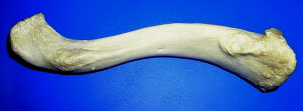
The clavicle, collarbone, or keybone is a slender, S-shaped long bone approximately long that serves as a strut between the scapula, shoulder blade and the sternum (breastbone). There are two clavicles, one on each side of the body. The clavicle is the only long bone in the body that lies horizontally. Together with the shoulder blade, it makes up the shoulder girdle. It is a palpable bone and, in people who have less fat in this region, the location of the bone is clearly visible. It receives its name from Latin ''clavicula'' 'little key' because the bone rotates along its axis like a key when the shoulder is Abduction (kinesiology), abducted. The clavicle is the most commonly fractured bone. It can easily be fractured by impacts to the shoulder from the force of falling on outstretched arms or by a direct hit. Structure The collarbone is a thin doubly curved long bone that connects the human arm, arm to the torso, trunk of the body. Located directly above the first rib, it ac ... [...More Info...] [...Related Items...] OR: [Wikipedia] [Google] [Baidu] |
Shoulder
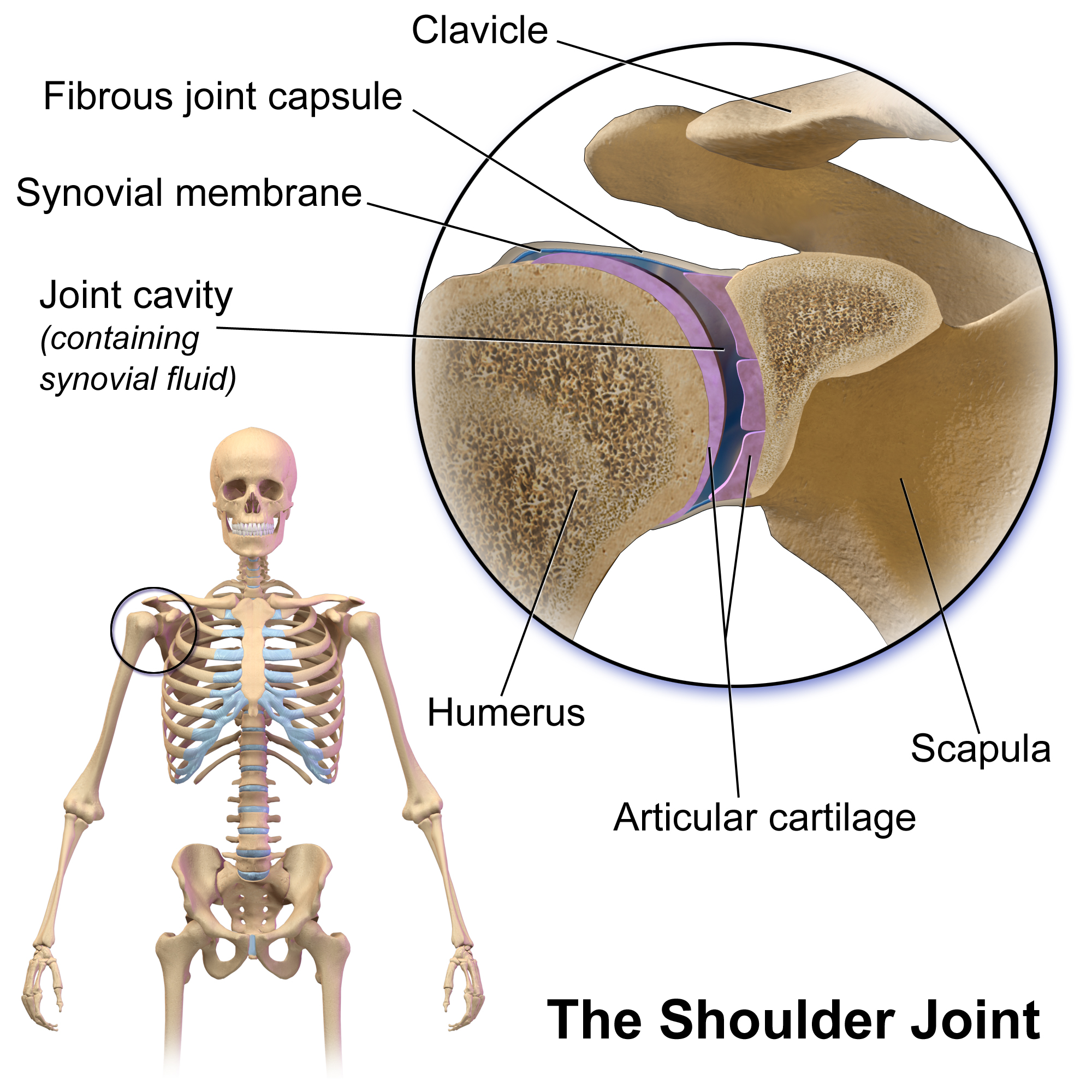
The human shoulder is made up of three bones: the clavicle (collarbone), the scapula (shoulder blade), and the humerus (upper arm bone) as well as associated muscles, ligaments and tendons. The articulations between the bones of the shoulder make up the shoulder joints. The shoulder joint, also known as the glenohumeral joint, is the major joint of the shoulder, but can more broadly include the acromioclavicular joint. In human anatomy, the shoulder joint comprises the part of the body where the humerus attaches to the scapula, and the head sits in the glenoid cavity. The shoulder is the group of structures in the region of the joint. The shoulder joint is the main joint of the shoulder. It is a ball and socket joint that allows the arm to rotate in a circular fashion or to hinge out and up away from the body. The joint capsule is a soft tissue envelope that encircles the glenohumeral joint and attaches to the scapula, humerus, and head of the biceps. It is lined by a ... [...More Info...] [...Related Items...] OR: [Wikipedia] [Google] [Baidu] |
Axilla
The axilla (: axillae or axillas; also known as the armpit, underarm or oxter) is the area on the human body directly under the shoulder joint. It includes the axillary space, an anatomical space within the shoulder girdle between the arm and the thoracic cage, bounded superiorly by the imaginary plane between the superior borders of the first rib, clavicle and scapula (above which are considered part of the neck), medially by the serratus anterior muscle and thoracolumbar fascia, anteriorly by the pectoral muscles and posteriorly by the subscapularis, teres major and latissimus dorsi muscle. The soft skin covering the lateral axilla contains many hair and sweat glands. In humans, the formation of body odor happens mostly in the axilla. These odorant substances have been suggested by some to serve as pheromones, which play a role related to mate selection, although this is a controversial topic within the scientific community. The underarms seem more importa ... [...More Info...] [...Related Items...] OR: [Wikipedia] [Google] [Baidu] |
Thorax
The thorax (: thoraces or thoraxes) or chest is a part of the anatomy of mammals and other tetrapod animals located between the neck and the abdomen. In insects, crustaceans, and the extinct trilobites, the thorax is one of the three main divisions of the body, each in turn composed of multiple segments. The human thorax includes the thoracic cavity and the thoracic wall. It contains organs including the heart, lungs, and thymus gland, as well as muscles and various other internal structures. The chest may be affected by many diseases, of which the most common symptom is chest pain. Etymology The word thorax comes from the Greek θώραξ ''thṓrax'' " breastplate, cuirass, corslet" via . Humans Structure In humans and other hominids, the thorax is the chest region of the body between the neck and the abdomen, along with its internal organs and other contents. It is mostly protected and supported by the rib cage, spine, and shoulder girdle. Contents The ... [...More Info...] [...Related Items...] OR: [Wikipedia] [Google] [Baidu] |
Latissimus Dorsi
The latissimus dorsi () is a large, flat muscle on the back that stretches to the sides, behind the arm, and is partly covered by the trapezius on the back near the midline. The word latissimus dorsi (plural: ''latissimi dorsi'') comes from Latin and means "broadest [muscle] of the back", from "latissimus" () and "dorsum" (). The pair of muscles are commonly known as "lats", especially among bodybuilders. The latissimus dorsi is responsible for Extension (kinesiology), extension, adduction, transverse extension also known as horizontal abduction (or horizontal extension), flexion from an extended position, and (medial) internal rotation of the shoulder joint. It also has a synergistic role in extension and Anatomical terms of motion, lateral flexion of the lumbar spine. Due to bypassing the scapulothoracic joints and attaching directly to the spine, the actions the latissimi dorsi have on moving the arms can also influence the movement of the scapulae, such as their downward ... [...More Info...] [...Related Items...] OR: [Wikipedia] [Google] [Baidu] |
Axillary Space
The axillary spaces are anatomic spaces. through which axillary contents leave the axilla. They consist of the quadrangular space, triangular space, and triangular interval. It is bounded by teres major, teres minor, medial border of the humerus, and long head of triceps brachii. They should not be confused with the true "axillary space" within the borders of the axilla. Structure Axilla The true axilla is a conical space with its apex at the Cervico-axillary Canal, Base at the axillary fascia and skin of the armpit. When viewed in an axillary plane (axillary cut), it is more triangle with: Medial Wall: Serratus Anterior, Anterior Wall: pectoral muscles, Posterior Wall: subscapularis muscle, where the "apex" of the triangle is the humerus Quadrangular space This space is in the posterior wall of the axilla. It is a quadrangular space bounded laterally by surgical neck of the humerus, medially by long head of triceps brachii and inferiorly by teres major. It is bounded ... [...More Info...] [...Related Items...] OR: [Wikipedia] [Google] [Baidu] |
Pectoralis Minor
Pectoralis minor muscle () is a thin, triangular muscle, situated at the upper part of the chest, beneath the pectoralis major in the human body. It arises from ribs III-V; it inserts onto the coracoid process of the scapula. It is innervated by the medial pectoral nerve. Its function is to stabilise the scapula by holding it fast in position against the chest wall. Structure Attachments From the muscle's origin, the muscle's fibers pass superiorly and laterally, converging to form a flat tendon. Origin Pectoralis minor muscle arises from the upper margins and outer surfaces of the 3rd, 4th, and 5th ribs near their costal cartilages, and from the aponeuroses covering the intercostalis. Insertion Its tendon inserts onto the medial border and upper surface of the coracoid process of the scapula. Innervation The muscle receives motor innervation from the medial pectoral nerve. Relations Pectoralis minor muscle forms part of the anterior wall of the axilla. It is ... [...More Info...] [...Related Items...] OR: [Wikipedia] [Google] [Baidu] |
Rectus Abdominis
The rectus abdominis muscle, () also known as the "abdominal muscle" or simply better known as the "abs", is a pair of segmented skeletal muscle on the ventral aspect of a person, person's abdomen. The paired muscle is separated at the midline by a band of dense connective tissue called the linea alba (abdomen), linea alba, and the connective tissue defining each lateral margin of the rectus abdominus is the linea semilunaris. The muscle extends from the pubic symphysis, pubic crest and pubic tubercle inferiorly, to the xiphoid process and costal cartilages of the 5th–7th ribs superiorly. The rectus abdominis muscle is contained in the rectus sheath, which consists of the aponeurosis, aponeuroses of the lateral abdominal muscles. Each rectus abdominus is traversed by bands of connective tissue called the tendinous intersections, which interrupt it into distinct muscle bellies. Structure The rectus abdominis is a very long flat muscle, which extends along the whole length of th ... [...More Info...] [...Related Items...] OR: [Wikipedia] [Google] [Baidu] |


