|
Pathophysiology Of Autism
The pathophysiology of autism is the study of the physiological processes that cause or are otherwise associated with autism spectrum disorders. Autism's symptoms result from maturation-related changes in various systems of the brain. How autism occurs is not yet well understood. Its mechanism can be divided into two areas: the pathophysiology of brain structures and processes associated with autism, and the neuropsychological linkages between brain structures and behaviors. The behaviors appear to have multiple pathophysiologies. There is evidence that gut–brain axis abnormalities may be involved. A 2015 review proposed that immune, gastrointestinal inflammation, malfunction of the autonomic nervous system, gut flora alterations, and food metabolites may cause brain neuroinflammation and dysfunction. A 2016 review concludes that enteric nervous system abnormalities might play a role in neurological disorders such as autism. Neural connections and the immune system are a path ... [...More Info...] [...Related Items...] OR: [Wikipedia] [Google] [Baidu] |
Biological Process
Biological processes are those processes that are vital for an organism to live, and that shape its capacities for interacting with its environment. Biological processes are made of many chemical reactions or other events that are involved in the persistence and transformation of life forms. Metabolism and homeostasis are examples. Biological processes within an organism can also work as bioindicators. Scientists are able to look at an individual's biological processes to monitor the effects of environmental changes. Regulation of biological processes occurs when any process is modulated in its frequency, rate or extent. Biological processes are regulated by many means; examples include the control of gene expression, protein modification or interaction with a protein or substrate molecule. * Homeostasis: regulation of the internal environment to maintain a constant state; for example, sweating to reduce temperature * Organization: being structurally composed of one or more cell ... [...More Info...] [...Related Items...] OR: [Wikipedia] [Google] [Baidu] |
Corpus Callosum
The corpus callosum (Latin for "tough body"), also callosal commissure, is a wide, thick nerve tract, consisting of a flat bundle of commissural fibers, beneath the cerebral cortex in the brain. The corpus callosum is only found in placental mammals. It spans part of the longitudinal fissure, connecting the left and right cerebral hemispheres, enabling communication between them. It is the largest white matter structure in the human brain, about in length and consisting of 200–300 million axonal projections. A number of separate nerve tracts, classed as subregions of the corpus callosum, connect different parts of the hemispheres. The main ones are known as the genu, the rostrum, the trunk or body, and the splenium. Structure The corpus callosum forms the floor of the longitudinal fissure that separates the two cerebral hemispheres. Part of the corpus callosum forms the roof of the lateral ventricles. The corpus callosum has four main parts – individual nerve ... [...More Info...] [...Related Items...] OR: [Wikipedia] [Google] [Baidu] |
Broca's Area
Broca's area, or the Broca area (, also , ), is a region in the frontal lobe of the dominant hemisphere, usually the left, of the brain with functions linked to speech production. Language processing has been linked to Broca's area since Pierre Paul Broca reported impairments in two patients. They had lost the ability to speak after injury to the posterior inferior frontal gyrus (pars triangularis) (BA45) of the brain. Since then, the approximate region he identified has become known as Broca's area, and the deficit in language production as Broca's aphasia, also called expressive aphasia. Broca's area is now typically defined in terms of the pars opercularis and pars triangularis of the inferior frontal gyrus, represented in Brodmann's cytoarchitectonic map as Brodmann area 44 and Brodmann area 45 of the dominant hemisphere. Functional magnetic resonance imaging (fMRI) has shown language processing to also involve the third part of the inferior frontal gyrus the pa ... [...More Info...] [...Related Items...] OR: [Wikipedia] [Google] [Baidu] |
Occipital Lobe
The occipital lobe is one of the four major lobes of the cerebral cortex in the brain of mammals. The name derives from its position at the back of the head, from the Latin ''ob'', "behind", and ''caput'', "head". The occipital lobe is the visual processing center of the mammalian brain containing most of the anatomical region of the visual cortex. The primary visual cortex is Brodmann area 17, commonly called V1 (visual one). Human V1 is located on the medial side of the occipital lobe within the calcarine sulcus; the full extent of V1 often continues onto the occipital pole. V1 is often also called striate cortex because it can be identified by a large stripe of myelin, the Stria of Gennari. Visually driven regions outside V1 are called extrastriate cortex. There are many extrastriate regions, and these are specialized for different visual tasks, such as visuospatial processing, color differentiation, and motion perception. Bilateral lesions of the occipital lobe can l ... [...More Info...] [...Related Items...] OR: [Wikipedia] [Google] [Baidu] |
Frontal Lobe
The frontal lobe is the largest of the four major lobes of the brain in mammals, and is located at the front of each cerebral hemisphere (in front of the parietal lobe and the temporal lobe). It is parted from the parietal lobe by a groove between tissues called the central sulcus and from the temporal lobe by a deeper groove called the lateral sulcus (Sylvian fissure). The most anterior rounded part of the frontal lobe (though not well-defined) is known as the frontal pole, one of the three poles of the cerebrum. The frontal lobe is covered by the frontal cortex. The frontal cortex includes the premotor cortex, and the primary motor cortex – parts of the motor cortex. The front part of the frontal cortex is covered by the prefrontal cortex. There are four principal gyri in the frontal lobe. The precentral gyrus is directly anterior to the central sulcus, running parallel to it and contains the primary motor cortex, which controls voluntary movements of specific body ... [...More Info...] [...Related Items...] OR: [Wikipedia] [Google] [Baidu] |
Spatial Visualization Ability
Spatial visualization ability or visual-spatial ability is the ability to mentally manipulate 2-dimensional and 3-dimensional figures. It is typically measured with simple cognitive tests and is predictive of user performance with some kinds of user interfaces. Measurement The cognitive tests used to measure spatial visualization ability including mental rotation tasks like the Mental Rotations Test or mental cutting tasks like the Mental Cutting Test; and cognitive tests like the VZ-1 (Form Board), VZ-2 (Paper Folding), and VZ-3 (Surface Development) tests from the Kit of Factor-Reference cognitive tests produced by Educational Testing Service. Though the descriptions of spatial visualization and mental rotation sound similar, mental rotation is a particular task that can be accomplished using spatial visualization. The Minnesota Paper Form Board Test involves giving participants a shape and a set of smaller shapes which they are then instructed to determine which combi ... [...More Info...] [...Related Items...] OR: [Wikipedia] [Google] [Baidu] |
Lateralization Of Brain Function
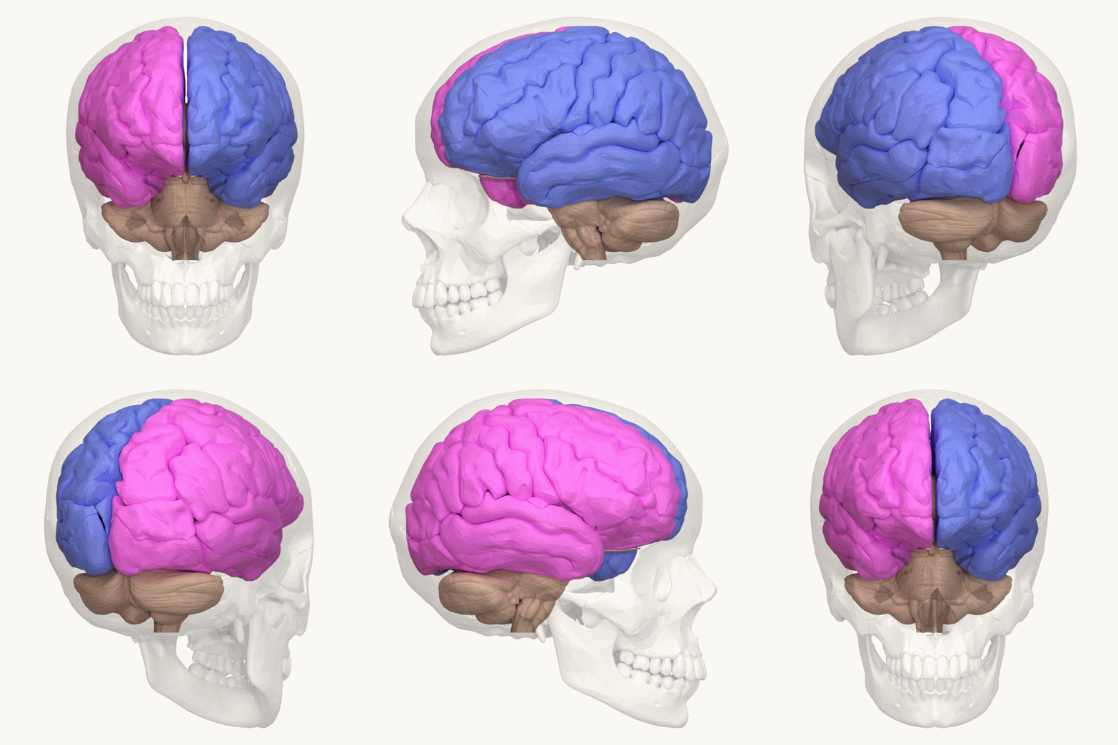
The lateralization of brain function is the tendency for some neural functions or cognitive processes to be specialized to one side of the brain or the other. The median longitudinal fissure separates the human brain into two distinct cerebral hemispheres, connected by the corpus callosum. Although the macrostructure of the two hemispheres appears to be almost identical, different composition of neuronal networks allows for specialized function that is different in each hemisphere. Lateralization of brain structures is based on general trends expressed in healthy patients; however, there are numerous counterexamples to each generalization. Each human's brain develops differently, leading to unique lateralization in individuals. This is different from specialization, as lateralization refers only to the function of one structure divided between two hemispheres. Specialization is much easier to observe as a trend, since it has a stronger anthropological history. The best exam ... [...More Info...] [...Related Items...] OR: [Wikipedia] [Google] [Baidu] |
Glutamate (neurotransmitter)
In neuroscience, glutamate refers to the anion of glutamic acid in its role as a neurotransmitter: a chemical that nerve cells use to send signals to other cells. It is by a wide margin the most abundant excitatory neurotransmitter in the vertebrate nervous system. It is used by every major excitatory function in the vertebrate brain, accounting in total for well over 90% of the synaptic connections in the human brain. It also serves as the primary neurotransmitter for some localized brain regions, such as cerebellum granule cells. Biochemical receptors for glutamate fall into three major classes, known as AMPA receptors, NMDA receptors, and metabotropic glutamate receptors. A fourth class, known as kainate receptors, are similar in many respects to AMPA receptors, but much less abundant. Many synapses use multiple types of glutamate receptors. AMPA receptors are ionotropic receptors specialized for fast excitation: in many synapses they produce excitatory electrical responses ... [...More Info...] [...Related Items...] OR: [Wikipedia] [Google] [Baidu] |
Grey Matter
Grey matter is a major component of the central nervous system, consisting of neuronal Soma (biology), cell bodies, neuropil (dendrites and unmyelinated axons), Glia, glial cells (astrocytes and oligodendrocytes), synapses, and Capillary, capillaries. Grey matter is distinguished from white matter in that it contains numerous cell bodies and relatively few myelinated axons, while white matter contains relatively few cell bodies and is composed chiefly of long-range myelinated axons. The colour difference arises mainly from the whiteness of myelin. In living tissue, grey matter actually has a very light grey colour with yellowish or pinkish hues, which come from capillary blood vessels and neuronal cell bodies. Structure Grey matter refers to unmyelinated neurons and other cells of the central nervous system The central nervous system (CNS) is the part of the nervous system consisting primarily of the brain and spinal cord. The CNS is so named because the brain integrates ... [...More Info...] [...Related Items...] OR: [Wikipedia] [Google] [Baidu] |
Control Group
In the design of experiments, hypotheses are applied to experimental units in a treatment group. In comparative experiments, members of a control group receive a standard treatment, a placebo, or no treatment at all. There may be more than one treatment group, more than one control group, or both. A placebo control group can be used to support a double-blind study, in which some subjects are given an ineffective treatment (in medical studies typically a sugar pill) to minimize differences in the experiences of subjects in the different groups; this is done in a way that ensures no participant in the experiment (subject or experimenter) knows to which group each subject belongs. In such cases, a third, non-treatment control group can be used to measure the placebo effect directly, as the difference between the responses of placebo subjects and untreated subjects, perhaps paired by age group or other factors (such as being twins). For the conclusions drawn from the results of an ... [...More Info...] [...Related Items...] OR: [Wikipedia] [Google] [Baidu] |
Cerebral Cortex
The cerebral cortex, also known as the cerebral mantle, is the outer layer of neural tissue of the cerebrum of the brain in humans and other mammals. The cerebral cortex mostly consists of the six-layered neocortex, with just 10% consisting of allocortex. It is separated into two cortices, by the longitudinal fissure that divides the cerebrum into the left and right cerebral hemispheres. The two hemispheres are joined beneath the cortex by the corpus callosum. The cerebral cortex is the largest site of neural integration in the central nervous system. It plays a key role in attention, perception, awareness, thought, memory, language, and consciousness. The cerebral cortex is part of the brain responsible for cognition. In most mammals, apart from small mammals that have small brains, the cerebral cortex is folded, providing a greater surface area in the confined volume of the cranium. Apart from minimising brain and cranial volume, cortical folding is crucial for ... [...More Info...] [...Related Items...] OR: [Wikipedia] [Google] [Baidu] |


