|
Palatopharyngeus
The palatopharyngeus (palatopharyngeal or pharyngopalatinus) muscle is a small muscle in the roof of the mouth. It is a long, fleshy fasciculus, narrower in the middle than at either end, forming, with the mucous membrane covering its surface, the palatopharyngeal arch. Structure It is separated from the palatoglossus muscle by an angular interval, in which the palatine tonsil is lodged. It arises from the soft palate, where it is divided into two fasciculi by the levator veli palatini and musculus uvulae. * The ''posterior fasciculus'' lies in contact with the mucous membrane, and joins with that of the opposite muscle in the middle line. * The ''anterior fasciculus'', the thicker, lies in the soft palate between the levator and tensor veli palatini muscles, and joins in the middle line the corresponding part of the opposite muscle. Passing laterally and downward behind the palatine tonsil, the palatopharyngeus joins the stylopharyngeus and is inserted with that muscle ... [...More Info...] [...Related Items...] OR: [Wikipedia] [Google] [Baidu] |
Palatine Aponeurosis
The palatine aponeurosis a thin, firm, fibrous lamella which gives strength and support to soft palate. It serves as the insertion for the tensor veli palatini and levator veli palatini, and the origin for the musculus uvulae, palatopharyngeus, and palatoglossus. The palatine aponeurosis is attached to the posterior margin of the hard palate. It is thicker anteriorly and thiner posteriorly. Posteriorly, it blends with the posterior muscular part of the soft palate. Posteroinferiorly, it presents a cruved free margin from which the uvula is suspended. Laterally, it is continuous with the pharyngeal aponeurosis. See also * Aponeurosis An aponeurosis (; : aponeuroses) is a flattened tendon by which muscle attaches to bone or fascia. Aponeuroses exhibit an ordered arrangement of collagen fibres, thus attaining high tensile strength in a particular direction while being vulnerable ... References Human head and neck {{musculoskeletal-stub ... [...More Info...] [...Related Items...] OR: [Wikipedia] [Google] [Baidu] |
Facial Artery
The facial artery, formerly called the external maxillary artery, is a branch of the external carotid artery that supplies blood to superficial structures of the medial regions of the face. Structure The facial artery arises in the carotid triangle from the external carotid artery, a little above the lingual artery, and sheltered by the ramus of the mandible. It passes obliquely up beneath the digastric and stylohyoid muscles, over which it arches to enter a groove on the posterior surface of the submandibular gland. It then curves upward over the body of the mandible at the antero-inferior angle of the masseter ( the antegonial notch); passes forward and upward across the cheek to the angle of the mouth, then ascends along the side of the nose, and ends at the medial commissure of the eye, under the name of the angular artery. The facial artery is remarkably tortuous. This is to accommodate itself to neck movements such as those of the pharynx in swallowing; and facia ... [...More Info...] [...Related Items...] OR: [Wikipedia] [Google] [Baidu] |
Palatopharyngeal Arch
The palatopharyngeal arch (pharyngopalatine arch, posterior pillar of fauces) is larger and projects further toward the middle line than the palatoglossal arch; it runs downward, lateralward, and backward to the side of the pharynx, and is formed by the projection of the palatopharyngeal muscle, covered by mucous membrane A mucous membrane or mucosa is a membrane that lines various cavities in the body of an organism and covers the surface of internal organs. It consists of one or more layers of epithelial cells overlying a layer of loose connective tissue. It .... References External links * * * Palate Pharynx {{anatomy-stub ... [...More Info...] [...Related Items...] OR: [Wikipedia] [Google] [Baidu] |
Stylopharyngeus
The stylopharyngeus muscle is a muscle in the head. It originates from the temporal styloid process. Some of its fibres insert onto the thyroid cartilage, while others end by intermingling with proximal structures. It is innervated by the glossopharyngeal nerve (cranial nerve IX). It acts to elevate the larynx and pharynx, and dilate the pharynx, thus facilitating swallowing. Structure The stylopharyngeus is a long, slender, tapered pharyngeal muscle. It is cylindrical superiorly, and flattened inferiorly. It passes inferior-ward along the side of the pharynx between the superior pharyngeal constrictor (situated deep to the stylopharyngeus) and the middle pharyngeal constrictor (situated superficial to the stylopharyngeus), before spreads out beneath the mucous membrane. Origin It arises from (the medial side of the base of) the temporal styloid process. It is the only muscle of the pharynx not to originate in the pharyngeal wall. Insertion Some of its fibers are lost ... [...More Info...] [...Related Items...] OR: [Wikipedia] [Google] [Baidu] |
Palate
The palate () is the roof of the mouth in humans and other mammals. It separates the oral cavity from the nasal cavity. A similar structure is found in crocodilians, but in most other tetrapods, the oral and nasal cavities are not truly separated. The palate is divided into two parts, the anterior, bony hard palate and the posterior, fleshy soft palate (or velum). Structure Innervation The maxillary nerve branch of the trigeminal nerve supplies sensory innervation to the palate. Development The hard palate forms before birth. Variation If the fusion is incomplete, a cleft palate results. Function in humans When functioning in conjunction with other parts of the mouth, the palate produces certain sounds, particularly velar, palatal, palatalized, postalveolar, alveolopalatal, and uvular consonants. History Etymology The English synonyms palate and palatum, and also the related adjective palatine (as in palatine bone), are all from the Latin ''palatum' ... [...More Info...] [...Related Items...] OR: [Wikipedia] [Google] [Baidu] |
Mucous Membrane
A mucous membrane or mucosa is a membrane that lines various cavities in the body of an organism and covers the surface of internal organs. It consists of one or more layers of epithelial cells overlying a layer of loose connective tissue. It is mostly of endodermal origin and is continuous with the skin at body openings such as the eyes, eyelids, ears, inside the nose, inside the mouth, lips, the genital areas, the urethral opening and the anus. Some mucous membranes secrete mucus, a thick protective fluid. The function of the membrane is to stop pathogens and dirt from entering the body and to prevent bodily tissues from becoming dehydrated. Structure The mucosa is composed of one or more layers of epithelial cells that secrete mucus, and an underlying lamina propria of loose connective tissue. The type of cells and type of mucus secreted vary from organ to organ and each can differ along a given tract. Mucous membranes line the digestive, respiratory and rep ... [...More Info...] [...Related Items...] OR: [Wikipedia] [Google] [Baidu] |
Muscles Of The Head And Neck
Muscle is a soft tissue, one of the four basic types of animal tissue. There are three types of muscle tissue in vertebrates: skeletal muscle, cardiac muscle, and smooth muscle. Muscle tissue gives skeletal muscles the ability to contract. Muscle tissue contains special contractile proteins called actin and myosin which interact to cause movement. Among many other muscle proteins, present are two regulatory proteins, troponin and tropomyosin. Muscle is formed during embryonic development, in a process known as myogenesis. Skeletal muscle tissue is striated consisting of elongated, multinucleate muscle cells called muscle fibers, and is responsible for movements of the body. Other tissues in skeletal muscle include tendons and perimysium. Smooth and cardiac muscle contract involuntarily, without conscious intervention. These muscle types may be activated both through the interaction of the central nervous system as well as by innervation from peripheral plexus or endocri ... [...More Info...] [...Related Items...] OR: [Wikipedia] [Google] [Baidu] |
Dissection
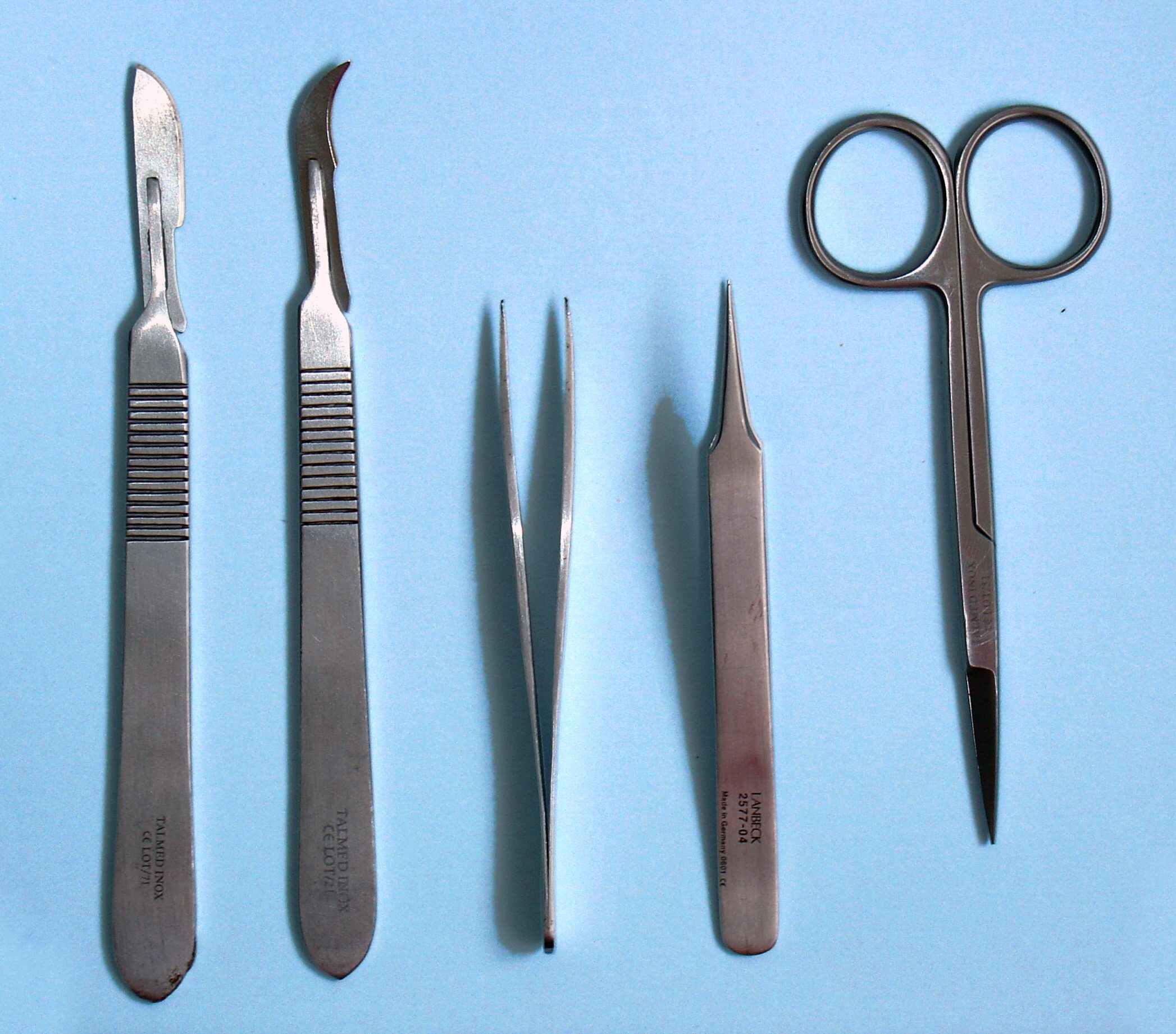
Dissection (from Latin ' "to cut to pieces"; also called anatomization) is the dismembering of the body of a deceased animal or plant to study its anatomical structure. Autopsy is used in pathology and forensic medicine to determine the cause of death in humans. Less extensive dissection of plants and smaller animals preserved in a formaldehyde solution is typically carried out or demonstrated in biology and natural science classes in middle school and high school, while extensive dissections of cadavers of adults and children, both fresh and preserved are carried out by medical students in medical schools as a part of the teaching in subjects such as anatomy, pathology and forensic medicine. Consequently, dissection is typically conducted in a morgue or in an anatomy lab. Dissection has been used for centuries to explore anatomy. Objections to the use of cadavers have led to the use of alternatives including virtual dissection of computational anatomy, computer models. In the ... [...More Info...] [...Related Items...] OR: [Wikipedia] [Google] [Baidu] |
Nasopharynx
The pharynx (: pharynges) is the part of the throat behind the mouth and nasal cavity, and above the esophagus and trachea (the tubes going down to the stomach and the lungs respectively). It is found in vertebrates and invertebrates, though its structure varies across species. The pharynx carries food to the esophagus and air to the larynx. The flap of cartilage called the epiglottis stops food from entering the larynx. In humans, the pharynx is part of the digestive system and the conducting zone of the respiratory system. (The conducting zone—which also includes the nostrils of the nose, the larynx, trachea, bronchi, and bronchioles—filters, warms, and moistens air and conducts it into the lungs). The human pharynx is conventionally divided into three sections: the nasopharynx, oropharynx, and laryngopharynx (hypopharynx). In humans, two sets of pharyngeal muscles form the pharynx and determine the shape of its lumen. They are arranged as an inner layer of longitudina ... [...More Info...] [...Related Items...] OR: [Wikipedia] [Google] [Baidu] |
Palatine Uvula
The uvula (: uvulas or uvulae), also known as the palatine uvula or staphyle, is a conic projection from the back edge of the middle of the soft palate, composed of connective tissue containing a number of racemose glands, and some muscular fibers. It also contains many serous glands, which produce thin saliva. It is only found in humans. Structure Muscle The muscular part of the uvula () shortens and broadens the uvula. This changes the contour of the posterior part of the soft palate. This change in contour allows the soft palate to adapt closely to the posterior pharyngeal wall to help close the nasopharynx during swallowing. Its muscles are controlled by the pharyngeal branch of the vagus nerve. Variation A bifid or bifurcated uvula is a split or cleft uvula. Newborns with cleft palate often also have a split uvula. The bifid uvula results from incomplete fusion of the palatine shelves but it is considered only a slight form of clefting. Bifid uvulas have less m ... [...More Info...] [...Related Items...] OR: [Wikipedia] [Google] [Baidu] |
Bolus (digestion)
In digestion, a bolus () is a ball-like mixture of food and saliva that forms in the mouth during the process of chewing (which is largely an adaptation for plant-eating mammals). It has the same color as the food being eaten, and the saliva gives it an alkaline pH. Under normal circumstances, the bolus is swallowed, and travels down the esophagus to the stomach for digestion. See also * Chyme * Chyle References Digestive system {{Digestive-stub ... [...More Info...] [...Related Items...] OR: [Wikipedia] [Google] [Baidu] |
Tensor Veli Palatini
The tensor veli palatini muscle (tensor palati or tensor muscle of the velum palatinum) is a thin, triangular muscle of the head that tenses the soft palate and opens the Eustachian tube to equalise pressure in the middle ear. Structure The tensor veli palatini muscle is thin and triangular in shape. Origin It arises from the scaphoid fossa of the pterygoid process of the sphenoid anteriorly, the (medial aspect of the) spine of sphenoid bone posteriorly, and - between the aforementioned anterior and posterior attachments - from the anterolateral aspect of the membranous wall of the pharyngotympanic tube. At the muscle's origin, some of its muscle fibres may be continuous with those of the tensor tympani muscle. Insertion Inferiorly, the muscle converges to form a tendon of attachment. This tendon winds medially around the pterygoid hamulus (with a small bursa interposed between the two) to insert into the palatine aponeurosis and into the bony surface posterior to ... [...More Info...] [...Related Items...] OR: [Wikipedia] [Google] [Baidu] |


