|
Orbital Implant
Enucleation is the removal of the eye that leaves the eye muscles and remaining orbital contents intact. This type of ocular surgery is indicated for a number of ocular tumors, in eyes that have sustained severe trauma, and in eyes that are otherwise blind and painful. Self-enucleation or auto-enucleation (oedipism) and other forms of serious self-inflicted eye injury are an extremely rare form of severe self-harm that usually results from mental illnesses involving acute psychosis. The name comes from Oedipus of Greek mythology, who gouged out his own eyes. Classification There are three types of eye removal: * Evisceration – removal of the iris, lens, and internal eye contents, but with the sclera and attached extraocular muscles left behind * Enucleation of the eye – removal of the eyeball, but with the eyelids and adjacent structures of the eye socket remaining. An intraocular tumor excision requires an enucleation, not an evisceration. * Exenteration – removal of th ... [...More Info...] [...Related Items...] OR: [Wikipedia] [Google] [Baidu] |
Eye Muscles
The extraocular muscles, or extrinsic ocular muscles, are the seven extrinsic muscles of the eye in humans and other animals. Six of the extraocular muscles, the four recti muscles, and the superior and inferior oblique muscles, control movement of the eye. The other muscle, the levator palpebrae superioris, controls eyelid elevation. The actions of the six muscles responsible for eye movement depend on the position of the eye at the time of muscle contraction. The ciliary muscle, pupillary sphincter muscle and pupillary dilator muscle sometimes are called intrinsic ocular muscles or intraocular muscles. Structure Since only a small part of the eye called the fovea provides sharp vision, the eye must move to follow a target. Eye movements must be precise and fast. This is seen in scenarios like reading, where the reader must shift gaze constantly. Although under voluntary control, most eye movement is accomplished without conscious effort. Precisely how the integration between ... [...More Info...] [...Related Items...] OR: [Wikipedia] [Google] [Baidu] |
Uveal Melanoma
Uveal melanoma is a type of eye cancer in the uvea of the eye. It is traditionally classed as originating in the iris, choroid, and ciliary body, but can also be divided into class I (low metastatic risk) and class II (high metastatic risk). Symptoms include blurred vision, loss of vision, and photopsia, but there may be no symptoms. Tumors arise from the pigment cells that reside within the uvea and give color to the eye. These melanocytes are distinct from the retinal pigment epithelium cells underlying the retina that do not form melanomas. When eye melanoma is spread to distant parts of the body, the five-year survival rate is about 15%.Eye Cancer Survival Rates American Cancer Society, Last Medical Review: December 9, 2014 Last Revised: February 5, 2016 It is the most co ... [...More Info...] [...Related Items...] OR: [Wikipedia] [Google] [Baidu] |
British Journal Of Ophthalmology
The ''British Journal of Ophthalmology'' is a peer-reviewed medical journal covering all aspects of ophthalmology. The journal was established in 1917 by the amalgamation of the ''Royal London (Moorfields) Ophthalmic Hospital Reports'' with the ''Ophthalmoscope'' and the ''Ophthalmic Record''. The journal was edited for several years by Stewart Duke-Elder. Currently, Jost Jonas, James Chodosh, and Keith Barton are editors-in-chief. Abstracting and indexing The journal is abstracted and indexed in Index Medicus, PubMed, Current Contents, Excerpta Medica, and Scopus. According to the ''Journal Citation Reports'', the journal has a 2023 impact factor The impact factor (IF) or journal impact factor (JIF) of an academic journal is a type of journal ranking. Journals with higher impact factor values are considered more prestigious or important within their field. The Impact Factor of a journa ... of 3.8. References External links *{{Official website, http://bjo.bmj.com/ Ophtha ... [...More Info...] [...Related Items...] OR: [Wikipedia] [Google] [Baidu] |
Conjunctiva
In the anatomy of the eye, the conjunctiva (: conjunctivae) is a thin mucous membrane that lines the inside of the eyelids and covers the sclera (the white of the eye). It is composed of non-keratinized, stratified squamous epithelium with goblet cells, stratified columnar epithelium and stratified cuboidal epithelium (depending on the zone). The conjunctiva is highly Angiogenesis, vascularised, with many microvessels easily accessible for imaging studies. Structure The conjunctiva is typically divided into three parts: Blood supply Blood to the bulbar conjunctiva is primarily derived from the ophthalmic artery. The blood supply to the palpebral conjunctiva (the eyelid) is derived from the external carotid artery. However, the circulations of the bulbar conjunctiva and palpebral conjunctiva are linked, so both bulbar conjunctival and palpebral conjunctival vessels are supplied by both the ophthalmic artery and the external carotid artery, to varying extents. Nerve supply Se ... [...More Info...] [...Related Items...] OR: [Wikipedia] [Google] [Baidu] |
Tenon's Capsule
Tenon's capsule (), also known as the Tenon capsule, fascial sheath of the eyeball () or the fascia bulbi, is a thin membrane which envelops the eyeball from the optic nerve to the corneal limbus, separating it from the orbital fat and forming a socket in which it moves. The inner surface of Tenon's capsule is smooth and is separated from the outer surface of the sclera by the periscleral lymph space. This lymph space is continuous with the subdural and subarachnoid cavities and is traversed by delicate bands of connective tissue which extend between the capsule and the sclera. The capsule is perforated behind by the ciliary vessels and nerves and fuses with the sheath of the optic nerve and with the sclera around the entrance of the optic nerve. In front it adheres to the conjunctiva, and both structures are attached to the ciliary region of the eyeball. The structure was named after Jacques-René Tenon (1724–1816), a French surgeon and pathologist. Structure Relations ... [...More Info...] [...Related Items...] OR: [Wikipedia] [Google] [Baidu] |
Malden, Massachusetts
Malden is a city in Middlesex County, Massachusetts, Middlesex County, Massachusetts, United States. At the time of the 2020 United States census, 2020 U.S. Census, the population was 66,263 people. History Malden is a hilly woodland area north of the Mystic River that was settled by Puritans in 1640 on land purchased in 1629 from the Naumkeag people, Mystic tribe of the Pawtucket tribe, Pawtucket Confederation, with a further grant in 1639 by the Squaw Sachem of Mistick and her husband Webcowet. The area was originally called the “Mistick Side” and was a part of Charlestown, Massachusetts, Charlestown. It was incorporated as a separate town in 1649 under the name "Mauldon". The name Malden was selected by Joseph Hills, an early settler and landholder, and was named after Maldon, Essex, Maldon, England. The city originally included the adjacent cities of Melrose, Massachusetts, Melrose (until 1850) and Everett, Massachusetts, Everett (until 1870). At the time of the Ameri ... [...More Info...] [...Related Items...] OR: [Wikipedia] [Google] [Baidu] |
Keratoplasty
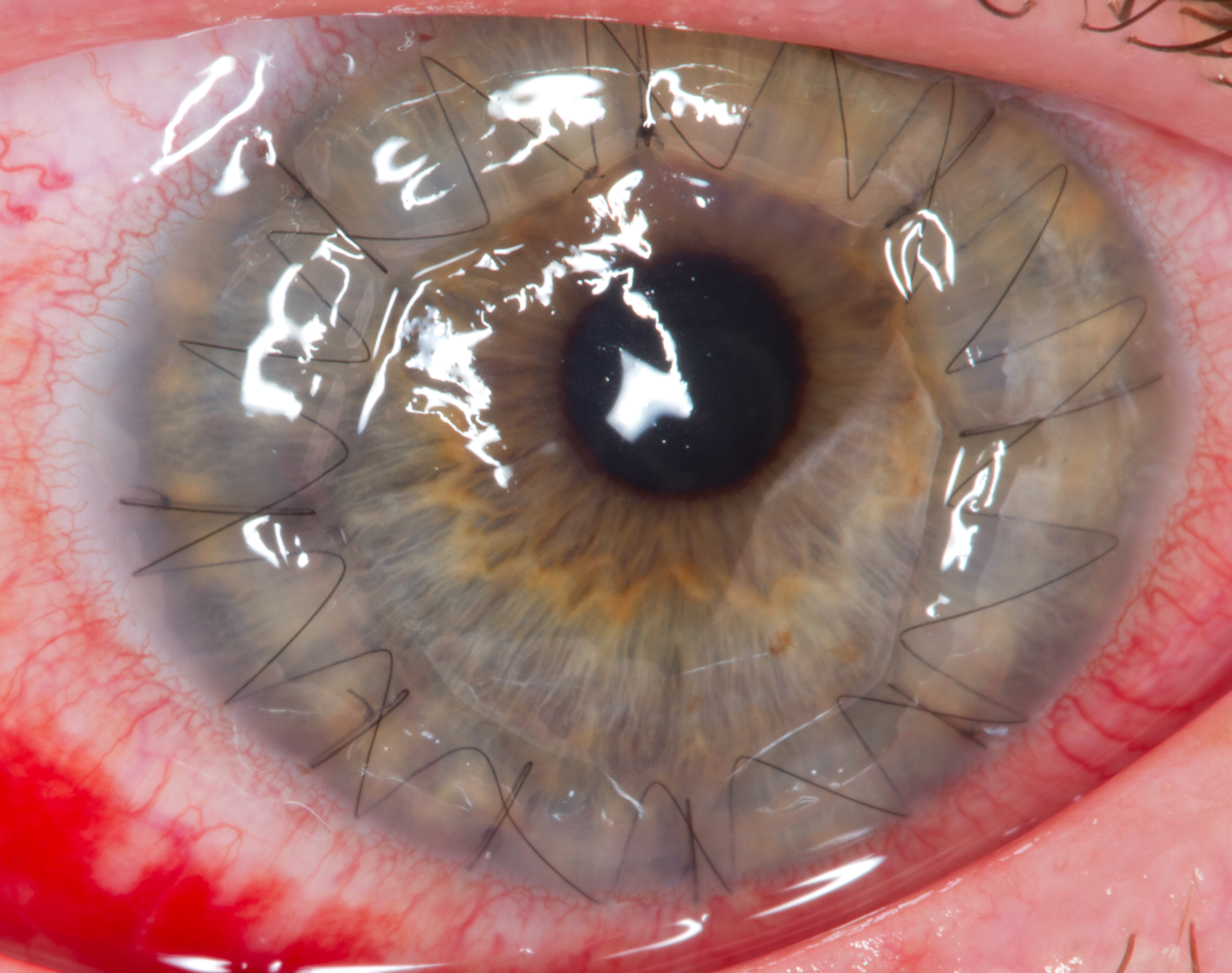
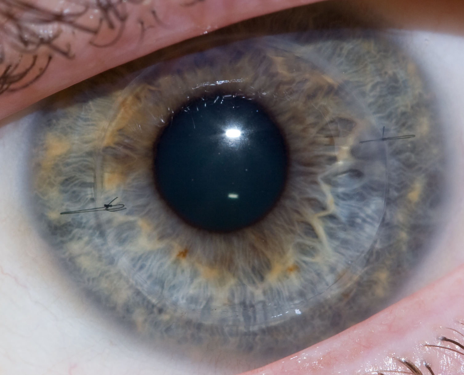
Corneal transplantation, also known as corneal grafting, is a surgical procedure where a damaged or diseased cornea is replaced by donated corneal tissue (the graft). When the entire cornea is replaced it is known as penetrating keratoplasty and when only part of the cornea is replaced it is known as lamellar keratoplasty. Keratoplasty simply means surgery to the cornea. The graft is taken from a recently deceased individual with no known diseases or other factors that may affect the chance of survival of the donated tissue or the health of the recipient. The cornea is the transparent front part of the eye that covers the iris, pupil and anterior chamber. The surgical procedure is performed by ophthalmologists, physicians who specialize in eyes, and is often done on an outpatient basis. Donors can be of any age, as is shown in the case of Janis Babson, who donated her eyes after dying at the age of 10. Corneal transplantation is performed when medicines, keratoconus conserva ... [...More Info...] [...Related Items...] OR: [Wikipedia] [Google] [Baidu] |
Corneal Transplant
Corneal transplantation, also known as corneal grafting, is a surgical procedure where a damaged or diseased cornea is replaced by Corneal button, donated corneal tissue (the graft). When the entire cornea is replaced it is known as penetrating keratoplasty and when only part of the cornea is replaced it is known as lamellar keratoplasty. Keratoplasty simply means surgery to the cornea. The graft is taken from a recently deceased individual with no known diseases or other factors that may affect the chance of survival of the donated tissue or the health of the recipient. The cornea is the transparency (optics), transparent front part of the human eye, eye that covers the Iris (anatomy), iris, pupil and anterior chamber. The surgical procedure is performed by Ophthalmology, ophthalmologists, physicians who specialize in eyes, and is often done on an outpatient basis. Donors can be of any age, as is shown in the case of Janis Babson, who donated her eyes after dying at the age of ... [...More Info...] [...Related Items...] OR: [Wikipedia] [Google] [Baidu] |
Globe Rupture
Open-globe injuries (also called globe rupture, globe laceration, globe penetration, or globe perforation) are full-thickness eye-wall wounds requiring urgent diagnosis and treatment. Classification In 1996 Kuhn et al. created the Birmingham eye trauma terminology (BETT) to standardize the language used to describe traumatic ocular injuries internationally. The BETT schema classifies open globe injuries as a laceration or a rupture. A ruptured globe occurs when rapid intraocular pressure elevation secondary to blunt trauma results in eyewall failure. The rupture site may be at the point of impact but more commonly occurs at the weakest and thinnest areas of the sclera. Regions prone to rupture are the rectus muscle insertion points, optic nerve insertion point, limbus, and prior surgical sites. Globe lacerations occur when a sharp object or projectile contacts the eye causing a full-thickness wound at the point of contact. Globe lacerations are further sub-classified into penetra ... [...More Info...] [...Related Items...] OR: [Wikipedia] [Google] [Baidu] |
Pythiosis
Pythiosis is a rare and deadly tropical disease caused by the oomycete '' Pythium insidiosum''. Long regarded as being caused by a fungus, the causative agent was not discovered until 1987. It occurs most commonly in horses, dogs, and humans, with isolated cases in other large mammals. The disease is contracted after exposure to stagnant fresh water such as swamps, ponds, lakes, and rice paddies. ''P. insidiosum'' is different from other members of the genus in that human and horse hair, skin, and decaying animal and plant tissue are chemoattractants for its zoospores. Additionally, it is the only member in the genus known to infect mammals, while other members are pathogenic to plants and are responsible for some well-known plant diseases. Epidemiology Pythiosis occurs in areas with mild winters because the organism survives in standing water that does not reach freezing temperatures. In the United States, it is most commonly found in the Southern Gulf states, especially Louisia ... [...More Info...] [...Related Items...] OR: [Wikipedia] [Google] [Baidu] |
Blindness
Visual or vision impairment (VI or VIP) is the partial or total inability of visual perception. In the absence of treatment such as corrective eyewear, assistive devices, and medical treatment, visual impairment may cause the individual difficulties with normal daily tasks, including reading and walking. The terms ''low vision'' and ''blindness'' are often used for levels of impairment which are difficult or impossible to correct and significantly impact daily life. In addition to the various permanent conditions, fleeting temporary vision impairment, amaurosis fugax, may occur, and may indicate serious medical problems. The most common causes of visual impairment globally are uncorrected refractive errors (43%), cataracts (33%), and glaucoma (2%). Refractive errors include near-sightedness, far-sightedness, presbyopia, and astigmatism (eye), astigmatism. Cataracts are the most common cause of blindness. Other disorders that may cause visual problems include age-related macular ... [...More Info...] [...Related Items...] OR: [Wikipedia] [Google] [Baidu] |
Sympathetic Ophthalmia
Sympathetic ophthalmia (SO), also called spared eye injury, is a diffuse granulomatous inflammation of the uveal layer of both eyes following trauma to one eye. It can leave the affected person completely blind. Symptoms may develop from days to several years after a penetrating eye injury. It typically results from a delayed hypersensitivity reaction. Signs and symptoms Eye floaters and loss of accommodation are among the earliest symptoms. The disease may progress to severe inflammation of the uveal layer of the eye (uveitis) with pain and sensitivity of the eyes to light. The affected eye often remains relatively painless while the inflammatory disease spreads through the uvea, where characteristic focal infiltrates in the choroid named Dalén–Fuchs nodules can be seen. The retina, however, usually remains uninvolved, although perivascular cuffing of the retinal vessels with inflammatory cells may occur. Swelling of the optic disc (papilledema), secondary glaucoma, vi ... [...More Info...] [...Related Items...] OR: [Wikipedia] [Google] [Baidu] |


