|
Oligohydramnios
Oligohydramnios is a medical condition in pregnancy characterized by a deficiency of amniotic fluid, the fluid that surrounds the fetus in the abdomen, in the amniotic sac. It is typically diagnosed by ultrasound when the amniotic fluid index (AFI) measures less than 5 cm or when the single deepest pocket (SDP) of amniotic fluid measures less than 2 cm. Amniotic fluid is necessary to allow for normal fetal movement, lung development, and cushioning from uterine compression. Low amniotic fluid can be attributed to a maternal, fetal, placental or idiopathic cause and can result in poor fetal outcomes including death. The prognosis of the fetus is dependent on the etiology, gestational age at diagnosis, and the severity of the oligohydramnios. The opposite of oligohydramnios is polyhydramnios, or an excess of amniotic fluid. Etiology The amount of amniotic fluid available is based on how much fluid is produced and how much is removed from the amniotic sac. In the first trimester, ... [...More Info...] [...Related Items...] OR: [Wikipedia] [Google] [Baidu] |
Amnioinfusion
Amnioinfusion is a method in which isotonic fluid is instilled into the uterine cavity. It was introduced in the 1960s as a means of terminating pregnancy and inducing labor in intrauterine death, but is currently used as a treatment in order to correct fetal heart rate changes caused by umbilical cord compression, indicated by Cardiotocography#Periodic or episodic decelerations, variable decelerations seen on Cardiotocography, fetal heart rate monitoring. In severe cases of oligohydramnios, amnioinfusion may be performed prophylactically to prevent umbilical cord compression. It has also been used to reduce the risk of meconium aspiration syndrome, though evidence of benefit is mixed. The UK National Institute of Health and Clinical Excellence (NICE) Guidelines recommend against the use of amnioinfusion in women with meconium stained amniotic fluid (MSAF). __TOC__ Uses Diagnostic uses Diagnostic uses for amnioinfusion are limited to pregnancies complicated by oligohydramni ... [...More Info...] [...Related Items...] OR: [Wikipedia] [Google] [Baidu] |
Prelabor Rupture Of Membranes
Prelabor rupture of membranes (PROM), previously known as premature rupture of membranes, is breakage of the amniotic sac before the onset of labor. Women usually experience a painless gush or a steady leakage of fluid from the vagina. Complications in the baby may include premature birth, cord compression, and infection. Complications in the mother may include placental abruption and postpartum endometritis. Risk factors include infection of the amniotic fluid, prior PROM, bleeding in the later parts of pregnancy, smoking, and a mother who is underweight. Diagnosis is suspected based on symptoms and speculum exam and may be supported by testing the vaginal fluid or by ultrasound. If it occurs before 37 weeks it is known as PPROM (preterm prelabor rupture of membranes) otherwise it is known as term PROM. Treatment is based on how far along a woman is in pregnancy and whether complications are present. In those at or near term without any complications, induction of labor is ... [...More Info...] [...Related Items...] OR: [Wikipedia] [Google] [Baidu] |
Posterior Urethral Valve
Posterior urethral valve (PUV) disorder is an obstructive developmental anomaly in the urethra and genitourinary system of male newborns. A posterior urethral valve is an obstructing membrane in the posterior male urethra as a result of abnormal '' in utero'' development. It is the most common cause of bladder outlet obstruction in male newborns. The disorder varies in degree, with mild cases presenting late due to milder symptoms. More severe cases can have renal and respiratory failure from lung underdevelopment as result of low amniotic fluid volumes, requiring intensive care and close monitoring. It occurs in about one in 8,000 babies. Presentation PUV can be diagnosed before birth, or even at birth when the ultrasound shows that the male baby has a hydronephrosis. Some babies may also have oligohydramnios due to the urinary obstruction. The later presentation can be a urinary tract infection, diurnal enuresis, or voiding pain. Complications * Incontinence * Urinary tract ... [...More Info...] [...Related Items...] OR: [Wikipedia] [Google] [Baidu] |
Twin-to-twin Transfusion Syndrome
Twin-to-twin transfusion syndrome (TTTS), also known as feto-fetal transfusion syndrome (FFTS), twin oligohydramnios-polyhydramnios sequence (TOPS) and stuck twin syndrome, is a complication of monochorionic multiple pregnancies (the most common form of identical twin pregnancy) in which there is disproportionate blood supply between the fetuses. This leads to unequal levels of amniotic fluid between each fetus and usually leads to death of the undersupplied twin and, without treatment, usually death or a range of birth defects or disabilities for a surviving twin, such as underdeveloped, damaged or missing limbs, digits or organs (including the brain), especially cerebral palsy. The condition occurs when the vein–artery connections within the fetuses' shared placenta allow the blood flow between each fetus to become progressively imbalanced. It usually develops between week 16 and 25 of pregnancy, during peak placental growth. The cause of the developmental effects on a sur ... [...More Info...] [...Related Items...] OR: [Wikipedia] [Google] [Baidu] |
Amniotic Fluid
The amniotic fluid is the protective liquid contained by the amniotic sac of a gravid amniote. This fluid serves as a cushion for the growing fetus, but also serves to facilitate the exchange of nutrients, water, and biochemical products between mother and fetus. For humans, the amniotic fluid is commonly called water or waters (Latin liquor amnii). Development Amniotic fluid is present from the formation of the gestational sac. Amniotic fluid is in the amniotic sac. It is generated from maternal plasma, and passes through the fetal membranes by osmotic and hydrostatic forces. When fetal kidneys begin to function around week 16, fetal urine also contributes to the fluid. In earlier times, it was believed that the amniotic fluid was composed entirely of fetal urine. The fluid is absorbed through the fetal tissue and skin. After 22 to 25 week of pregnancy, keratinization of an embryo's skin occurs. When this process completes around the 25th week, the fluid is primarily ab ... [...More Info...] [...Related Items...] OR: [Wikipedia] [Google] [Baidu] |
Amniotic Fluid Index
Amniotic fluid index (AFI) is a quantitative estimate of amniotic fluid and an indicator of fetal well-being. It is a separate measurement from the biophysical profile. AFI is the score (expressed in cm) given to the amount of amniotic fluid seen on ultrasonography of a pregnant uterus. To determine the AFI, doctors may use a four-quadrant technique, when the deepest, unobstructed, vertical length of each pocket of fluid is measured in each quadrant and then added up to the others, or the so-called "Single Deepest Pocket" technique. An AFI between 8–18 is considered normal. Median AFI level is approximately 14 from week 20 to week 35, when the amniotic fluid begins to reduce in preparation for birth. An AFI 24–25 is considered as polyhydramnios. Causes of low amniotic fluid There are many things that can cause low AFI, these include: * Leaking or rupture of membranes: Leaking or rupture of membranes may be caused by a gush of fluid or a slow constant trickle of fluid. This ... [...More Info...] [...Related Items...] OR: [Wikipedia] [Google] [Baidu] |
Renal Agenesis
Renal agenesis is a medical condition in which one (unilateral) or both (bilateral) fetal kidneys fail to develop. Unilateral and bilateral renal agenesis in humans, mice and zebra fish has been linked to mutations in the gene GREB1L. It has also been associated with mutations in the genes '' RET'' or '' UPK3A'' in humans and mice respectively. Type Bilateral Bilateral renal agenesis is a condition in which both kidneys of a fetus fail to develop during gestation. It is incompatible with life. It is one causative agent of Potter sequence. This absence of kidneys causes oligohydramnios, a deficiency of amniotic fluid in a pregnant woman, which can place extra pressure on the developing baby and cause further malformations. The condition is frequently, but not always the result of a genetic disorder, and is more common in infants born to one or more parents with a malformed or absent kidney. Unilateral This is much more common, but is not usually of any major health consequence, a ... [...More Info...] [...Related Items...] OR: [Wikipedia] [Google] [Baidu] |
Polyhydramnios
Polyhydramnios is a medical condition describing an excess of amniotic fluid in the amniotic sac. It is seen in about 1% of pregnancies. It is typically diagnosed when the amniotic fluid index (AFI) is greater than 24 cm. There are two clinical varieties of polyhydramnios: chronic polyhydramnios where excess amniotic fluid accumulates gradually, and acute polyhydramnios where excess amniotic fluid collects rapidly. The opposite to polyhydramnios is oligohydramnios, not enough amniotic fluid. Presentation Associated conditions Fetuses with polyhydramnios are at risk for a number of other problems including cord prolapse, placental abruption, premature birth and perinatal death. At delivery the baby should be checked for congenital abnormalities. Causes In most cases, the exact cause cannot be identified. A single case may have one or more causes, including intrauterine infection (TORCH), rh-isoimmunisation, or chorioangioma of the placenta. In a multiple gestation pregn ... [...More Info...] [...Related Items...] OR: [Wikipedia] [Google] [Baidu] |
Umbilical Cord Compression
Umbilical cord compression is the obstruction of blood flow through the umbilical cord secondary to pressure from an external object or misalignment of the cord itself. Cord compression happens in about one in 10 deliveries.Childbirth Complications at medicinenet.com. Last Editorial Review: 1/30/2005 Causes * Nuchal cord, when the umbilical cord is (tightly) around the neck of the fetusP02.5 Fetus and newborn affected by other compression of umbilical cordin |
External Cephalic Version
External cephalic version (ECV) is a process by which a breech baby can sometimes be turned from buttocks or foot first to head first. It is a manual procedure that is recommended by national guidelines for breech presentation of a pregnancy with a single baby, in order to enable vaginal delivery. It is usually performed late in pregnancy, that is, after 36 gestational weeks, preferably 37 weeks, and can even be performed in early labour. ECV is endorsed by the American College of Obstetricians and Gynecologists (ACOG) and Royal College of Obstetricians and Gynaecologists (RCOG) as a mode to avoid the risks associated with a vaginal breech or cesarean delivery for singleton breech presentation. ECV can be contrasted with "internal cephalic version", which involves a hand inserted through the cervix. Medical use ECV is one option of intervention should a breech position of a baby be found after 36 weeks gestation. Other options include a planned caesarian section or planned ... [...More Info...] [...Related Items...] OR: [Wikipedia] [Google] [Baidu] |
Placental Abruption
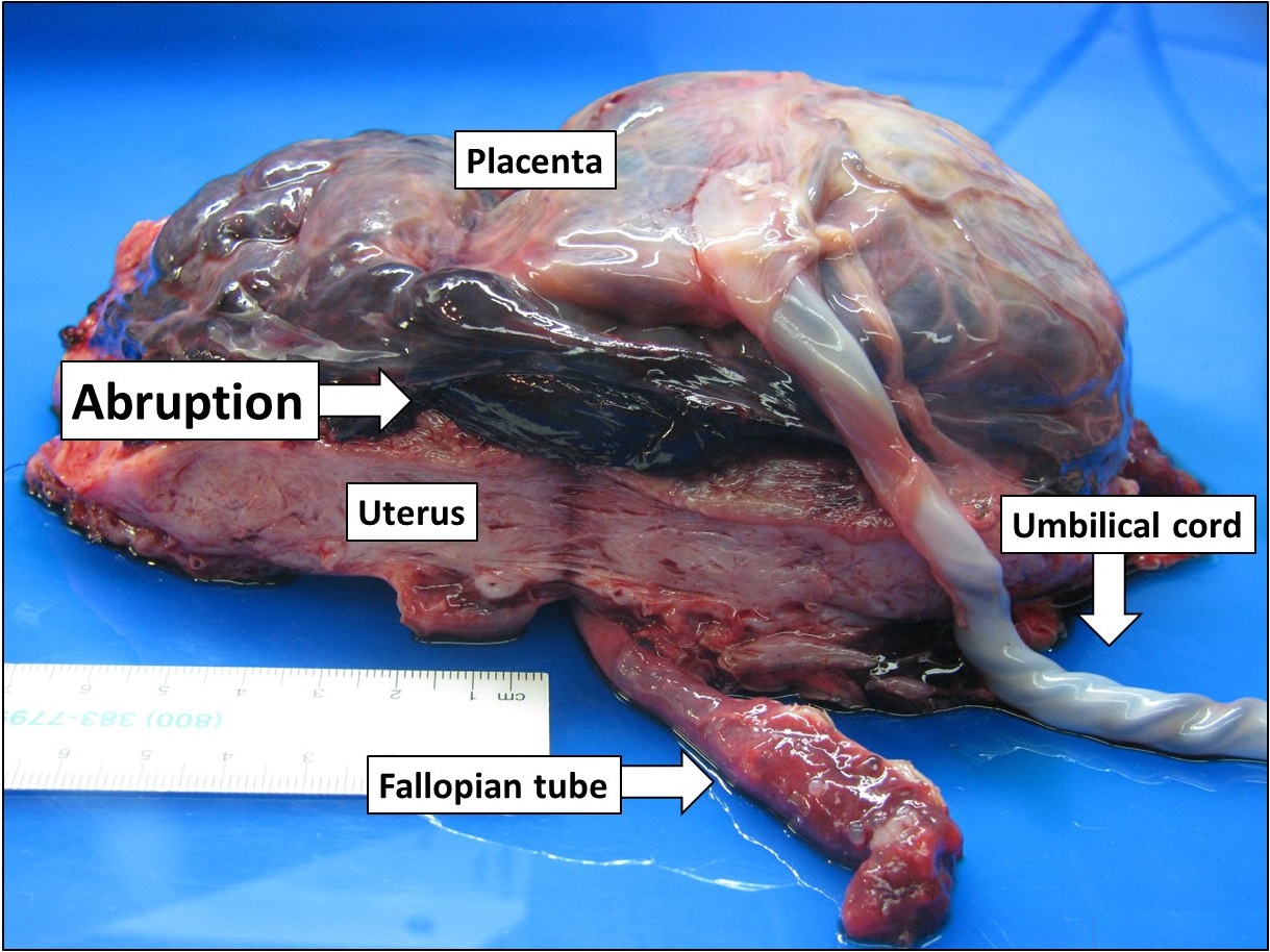
Placental abruption is when the placenta separates early from the uterus, in other words separates before childbirth. It occurs most commonly around 25 weeks of pregnancy. Symptoms may include vaginal bleeding, lower abdominal pain, and dangerously low blood pressure. Complications for the mother can include disseminated intravascular coagulopathy and kidney failure. Complications for the baby can include fetal distress, low birthweight, preterm delivery, and stillbirth. The cause of placental abruption is not entirely clear. Risk factors include smoking, pre-eclampsia, prior abruption (most important and predictive risk factor), trauma during pregnancy, cocaine use, and previous cesarean section. Diagnosis is based on symptoms and supported by ultrasound. It is classified as a complication of pregnancy. For small abruption, bed rest may be recommended, while for more significant abruptions or those that occur near term, delivery may be recommended. If everything is stab ... [...More Info...] [...Related Items...] OR: [Wikipedia] [Google] [Baidu] |
Angiotensin Converting Enzyme Inhibitors
Angiotensin-converting-enzyme inhibitors (ACE inhibitors) are a class of medication used primarily for the treatment of high blood pressure and heart failure. They work by causing relaxation of blood vessels as well as a decrease in blood volume, which leads to lower blood pressure and decreased oxygen demand from the heart. ACE inhibitors inhibit the activity of angiotensin-converting enzyme, an important component of the renin–angiotensin system which converts angiotensin I to angiotensin II, and hydrolyses bradykinin. Therefore, ACE inhibitors decrease the formation of angiotensin II, a vasoconstrictor, and increase the level of bradykinin, a peptide vasodilator. This combination is synergistic in lowering blood pressure. As a result of inhibiting the ACE enzyme in the bradykinin system, the ACE inhibitor drugs allow for increased levels of bradykinin which would normally be degraded. Bradykinin produces prostaglandin. This mechanism can explain the two most common sid ... [...More Info...] [...Related Items...] OR: [Wikipedia] [Google] [Baidu] |


