|
Occipital Vein
The occipital vein is a vein of the scalp. It originates from a plexus around the external occipital protuberance and superior nuchal line to the back part of the vertex of the skull. It usually drains into the internal jugular vein, but may also drain into the posterior auricular vein (which joins the external jugular vein). It drains part of the scalp. Structure The occipital vein is part of the scalp. It begins as a plexus at the posterior aspect of the scalp from the external occipital protuberance and superior nuchal line to the back part of the vertex of the skull. It pierces the cranial attachment of the trapezius and, dipping into the venous plexus of the suboccipital triangle, joins the deep cervical vein and the vertebral vein. Occasionally it follows the course of the occipital artery, and ends in the internal jugular vein. Alternatively, it joins the posterior auricular vein, and ends in the external jugular vein. The parietal emissary vein connects it with the ... [...More Info...] [...Related Items...] OR: [Wikipedia] [Google] [Baidu] |
Scalp
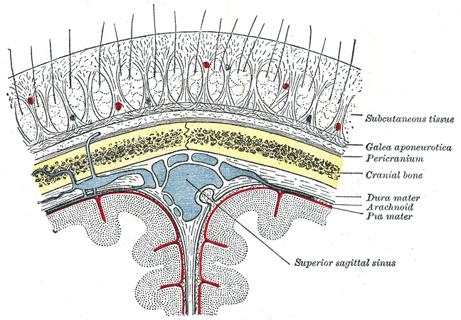
The scalp is the area of the head where head hair grows. It is made up of skin, layers of connective and fibrous tissues, and the membrane of the skull. Anatomically, the scalp is part of the epicranium, a collection of structures covering the cranium. The scalp is bordered by the face at the front, and by the neck at the sides and back. The scientific study of hair and scalp is called trichology. Structure Layers The scalp is usually described as having five layers, which can be remembered using the mnemonic 'SCALP': * S: Skin. The skin of the scalp contains numerous hair follicles and sebaceous glands. * C: Connective tissue. A dense subcutaneous layer of fat and fibrous tissue that lies beneath the skin, containing the nerves and vessels of the scalp. * A: Aponeurosis. The epicranial aponeurosis or galea aponeurotica is a tough layer of dense fibrous tissue which anchors the above layers in place. It runs from the frontalis muscle anteriorly to the occipitalis ... [...More Info...] [...Related Items...] OR: [Wikipedia] [Google] [Baidu] |
Trapezius
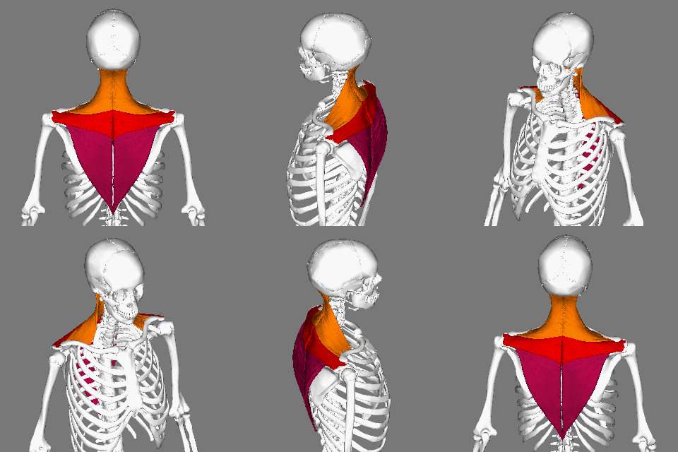
The trapezius is a large paired trapezoid-shaped surface muscle that extends longitudinally from the occipital bone to the lower thoracic vertebrae of the human spine, spine and laterally to the spine of the scapula. It moves the scapula and supports the arm. The trapezius has three functional parts: * an upper (descending) part which supports the weight of the arm; * a middle region (transverse), which retracts the scapula; and * a lower (ascending) part which medially rotates and depresses the scapula. Name and history The trapezius muscle resembles a trapezoid, trapezium, also known as a trapezoid, or diamond-shaped quadrilateral. The word "spinotrapezius" refers to the human trapezius, although it is not commonly used in modern texts. In other mammals, it refers to a portion of the analogous muscle. Structure The ''superior'' or ''upper'' (or descending) fibers of the trapezius originate from the spinous process of C7, the external occipital protuberance, the me ... [...More Info...] [...Related Items...] OR: [Wikipedia] [Google] [Baidu] |
Surgical And Radiologic Anatomy
''Surgical and Radiologic Anatomy'' is a peer-reviewed medical journal that publishes original research and review articles on the bases of medical, surgical and radiologic anatomy. According to the ''Journal Citation Reports'', it has a current impact factor The impact factor (IF) or journal impact factor (JIF) of an academic journal is a type of journal ranking. Journals with higher impact factor values are considered more prestigious or important within their field. The Impact Factor of a journa ... of 1.2, ranking it 13th out of 20 journals in the category "Anatomy & Morphology". It ranked 116th out of 135 categorized under "Surgery". References {{Reflist External links Journal website Springer Science+Business Media academic journals Academic journals established in 1978 English-language journals Anatomy journals Quarterly journals ... [...More Info...] [...Related Items...] OR: [Wikipedia] [Google] [Baidu] |
Sigmoid Sinus
The sigmoid sinuses (sigma- or s-shaped hollow curve), also known as the , are paired dural venous sinuses within the skull that receive blood from posterior transverse sinuses. Structure The sigmoid sinus is a dural venous sinus situated within the dura mater. The sigmoid sinus receives blood from the transverse sinuses, which track the posterior wall of the cranial cavity, travels inferiorly along the parietal bone, temporal bone and occipital bone, and converges with the inferior petrosal sinuses to form the internal jugular vein. Each sigmoid sinus begins beneath the temporal bone and follows a tortuous course to the jugular foramen, at which point the sinus becomes continuous with the internal jugular vein. Function The sigmoid sinus receives blood from the transverse sinuses, which receive blood from the posterior aspect of the skull. Along its course, the sigmoid sinus also receives blood from the cerebral veins, cerebellar veins, diploic vein The diploic veins are l ... [...More Info...] [...Related Items...] OR: [Wikipedia] [Google] [Baidu] |
Mastoid Emissary Vein
The emissary veins connect the extracranial venous system with the intracranial venous sinuses. They connect the veins outside the cranium to the venous sinuses inside the cranium. They drain from the scalp, through the skull, into the larger meningeal veins and dural venous sinuses. They may also connect to diploic veins within the skull. Emissary veins have an important role in selective cooling of the head. They also serve as routes where infections are carried into the cranial cavity from the extracranial veins to the intracranial veins. There are several types of emissary veins including the posterior condyloid, mastoid, occipital and parietal emissary veins. Structure There are also emissary veins passing through the foramen ovale, jugular foramen, foramen lacerum, and hypoglossal canal. Function Because the emissary veins are valveless, they are an important part in selective brain cooling through bidirectional flow of cooler blood from the evaporating surface of t ... [...More Info...] [...Related Items...] OR: [Wikipedia] [Google] [Baidu] |
Temporal Bone
The temporal bone is a paired bone situated at the sides and base of the skull, lateral to the temporal lobe of the cerebral cortex. The temporal bones are overlaid by the sides of the head known as the temples where four of the cranial bones fuse. Each temple is covered by a temporal muscle. The temporal bones house the structures of the ears. The lower seven cranial nerves and the major vessels to and from the brain traverse the temporal bone. Structure The temporal bone consists of four parts—the squamous, mastoid, petrous and tympanic parts. The squamous part is the largest and most superiorly positioned relative to the rest of the bone. The zygomatic process is a long, arched process projecting from the lower region of the squamous part and it articulates with the zygomatic bone. Posteroinferior to the squamous is the mastoid part. Fused with the squamous and mastoid parts and between the sphenoid and occipital bones lies the petrous part, which is shaped li ... [...More Info...] [...Related Items...] OR: [Wikipedia] [Google] [Baidu] |
American Journal Of Physical Anthropology
The ''American Journal of Biological Anthropology''Info pages about the renaming are: https://onlinelibrary.wiley.com/page/journal/26927691/homepage/productinformation.html and https://onlinelibrary.wiley.com/journal/26927691 (previously known as the ''American Journal of Physical Anthropology'') is a Peer review, peer-reviewed scientific journal and the official journal of the American Association of Biological Anthropologists. It was established in 1918 by Aleš Hrdlička (U.S. National Museum, now the Smithsonian Institution's National Museum of Natural History). The journal covers the field of biological anthropology, a discipline which Hrdlička defined in the first issue as "the study of racial anatomy, physiology and pathology." The Wistar Institute, Wistar Institute of Anatomy and Biology was the original publisher. Before launching publication, there were few outlets in the United States to publish scientific work in physical anthropology. Scientists hoping to learn more a ... [...More Info...] [...Related Items...] OR: [Wikipedia] [Google] [Baidu] |
Superior Sagittal Sinus
The superior sagittal sinus (also known as the superior longitudinal sinus), within the human head, is an unpaired dural venous sinus lying along the attached margin of the falx cerebri. It allows blood to drain from the lateral aspects of the anterior cerebral hemispheres to the confluence of sinuses. Cerebrospinal fluid drains through arachnoid granulations into the superior sagittal sinus and is returned to the venous circulation. Structure It is triangular in section. It is narrower anteriorly, and gradually increases in size as it passes posterior-ward. It commences at the foramen cecum, through which it receives emissary veins from the nasal cavity. It passes posterior-ward along its entire course. It is accommodated within a groove which runs across the inner surface of the frontal bone, the adjacent margins of the two parietal lobes, and the superior division of the cruciate eminence of the occipital lobe. Near the internal occipital protuberance, it deviates to e ... [...More Info...] [...Related Items...] OR: [Wikipedia] [Google] [Baidu] |
Occipital Artery
The occipital artery is a branch of the external carotid artery that provides arterial supply to the back of the scalp, sternocleidomastoid muscles, and deep muscles of the back and neck. Structure Origin The occipital artery arises from (the posterior aspect of) the external carotid artery (some 2 cm distal to the origin of the external carotid artery). Course and relations At its origin, the hypoglossal nerve (CN XII) crosses artery superficially as the nerve passes posteroanteriorly. The artery passes superoposteriorly deep to the posterior belly of the digastricus muscle. It crosses the internal carotid artery and vein, the vagus nerve (CN X), accessory nerve (CN XI), and hypoglossal nerve (CN XII). It next ascends to the interval between the transverse process of the atlas and the mastoid process of the temporal bone, and passes horizontally backward, grooving the surface of the latter bone, being covered by the sternocleidomastoideus, splenius capitis, longi ... [...More Info...] [...Related Items...] OR: [Wikipedia] [Google] [Baidu] |
Vertebral Vein
The vertebral vein is formed in the suboccipital triangle, from numerous small tributaries which spring from the internal vertebral venous plexuses and issue from the vertebral canal above the posterior arch of the atlas. They unite with small veins from the deep muscles at the upper part of the back of the neck, and form a vessel which enters the foramen in the transverse process of the atlas, and descends, forming a dense plexus around the vertebral artery, in the canal formed by the transverse foramina of the upper six cervical vertebrae In tetrapods, cervical vertebrae (: vertebra) are the vertebrae of the neck, immediately below the skull. Truncal vertebrae (divided into thoracic and lumbar vertebrae in mammals) lie caudal (toward the tail) of cervical vertebrae. In saurop .... This plexus ends in a single trunk, which emerges from the transverse foramina of the sixth cervical vertebra, and opens at the root of the neck into the back part of the innominate vein ne ... [...More Info...] [...Related Items...] OR: [Wikipedia] [Google] [Baidu] |
Deep Cervical Vein
The deep cervical vein (posterior vertebral vein or posterior deep cervical vein) is the vena comitans of the deep cervical artery. The vein is formed in the suboccipital region by the convergence of communicating branches of the occipital vein, veins draining the suboccipital muscles, and veins from the venous plexuses that surround cervical nerves. The vein and corresponding artery then pass in between the semispinalis capitis muscle and the semispinalis colli muscle. The vein passes anterior-ward in between the transverse process Each vertebra (: vertebrae) is an irregular bone with a complex structure composed of bone and some hyaline cartilage, that make up the vertebral column or spine, of vertebrates. The proportions of the vertebrae differ according to their spina ... of the 7th cervical vertebra and the neck of the first rib to terminate in the vertebral vein. References External links Veins of the head and neck {{circulatory-stub ... [...More Info...] [...Related Items...] OR: [Wikipedia] [Google] [Baidu] |
Suboccipital Triangle
The suboccipital triangle is a region of the neck bounded by the following three muscles of the suboccipital group of muscles: * Rectus capitis posterior major - above and medially * Obliquus capitis superior - above and laterally * Obliquus capitis inferior - below and laterally (Rectus capitis posterior minor is also in this region but does not form part of the triangle) It is covered by a layer of dense fibro-fatty tissue, situated beneath the semispinalis capitis. The floor is formed by the posterior atlantooccipital membrane, and the posterior arch of the atlas. In the deep groove on the upper surface of the posterior arch of the atlas are the vertebral artery and the first cervical or suboccipital nerve. In the past, the vertebral artery was accessed here in order to conduct angiography of the circle of Willis. Presently, formal angiography of the circle of Willis is performed via catheter angiography, with access usually being acquired at the common femoral artery. Alte ... [...More Info...] [...Related Items...] OR: [Wikipedia] [Google] [Baidu] |

