|
Neocardiogenesis
In cardiology neocardiogenesis is the homeostatic regeneration, repair and renewal of sections of malfunctioning adult cardiovascular tissue. This includes a combination of cardiomyogenesis (the regeneration of cardiac muscle) and angiogenesis (the regeneration of blood vessels). Definition and scope The term neocardiogenesis comes from cardiogenesis, which refers to the development of the heart in the embryo; neocardiogenesis, in turn, means the development of the heart in adults. The heart has mechanisms already in place that are responsible for small scale repair. However, these repair mechanisms are not sufficient for large scale repair, made necessary by events such as myocardial infarctions. Neocardiogenesis replaces dead cardiac muscle cells with living cells so that both the structure and function of the heart are maintained. This improves myocardial pumping of fluid around the body. Background The human heart has been thought of as a postmitotic organ. Cardiomyocytes ... [...More Info...] [...Related Items...] OR: [Wikipedia] [Google] [Baidu] |
Homeostatic
In biology, homeostasis ( British also homoeostasis) (/hɒmɪə(ʊ)ˈsteɪsɪs/) is the state of steady internal, physical, and chemical conditions maintained by living systems. This is the condition of optimal functioning for the organism and includes many variables, such as body temperature and fluid balance, being kept within certain pre-set limits (homeostatic range). Other variables include the pH of extracellular fluid, the concentrations of sodium, potassium and calcium ions, as well as that of the blood sugar level, and these need to be regulated despite changes in the environment, diet, or level of activity. Each of these variables is controlled by one or more regulators or homeostatic mechanisms, which together maintain life. Homeostasis is brought about by a natural resistance to change when already in the optimal conditions, and equilibrium is maintained by many regulatory mechanisms: it is thought to be the central motivation for all organic action. All homeostati ... [...More Info...] [...Related Items...] OR: [Wikipedia] [Google] [Baidu] |
Cardiac Muscle
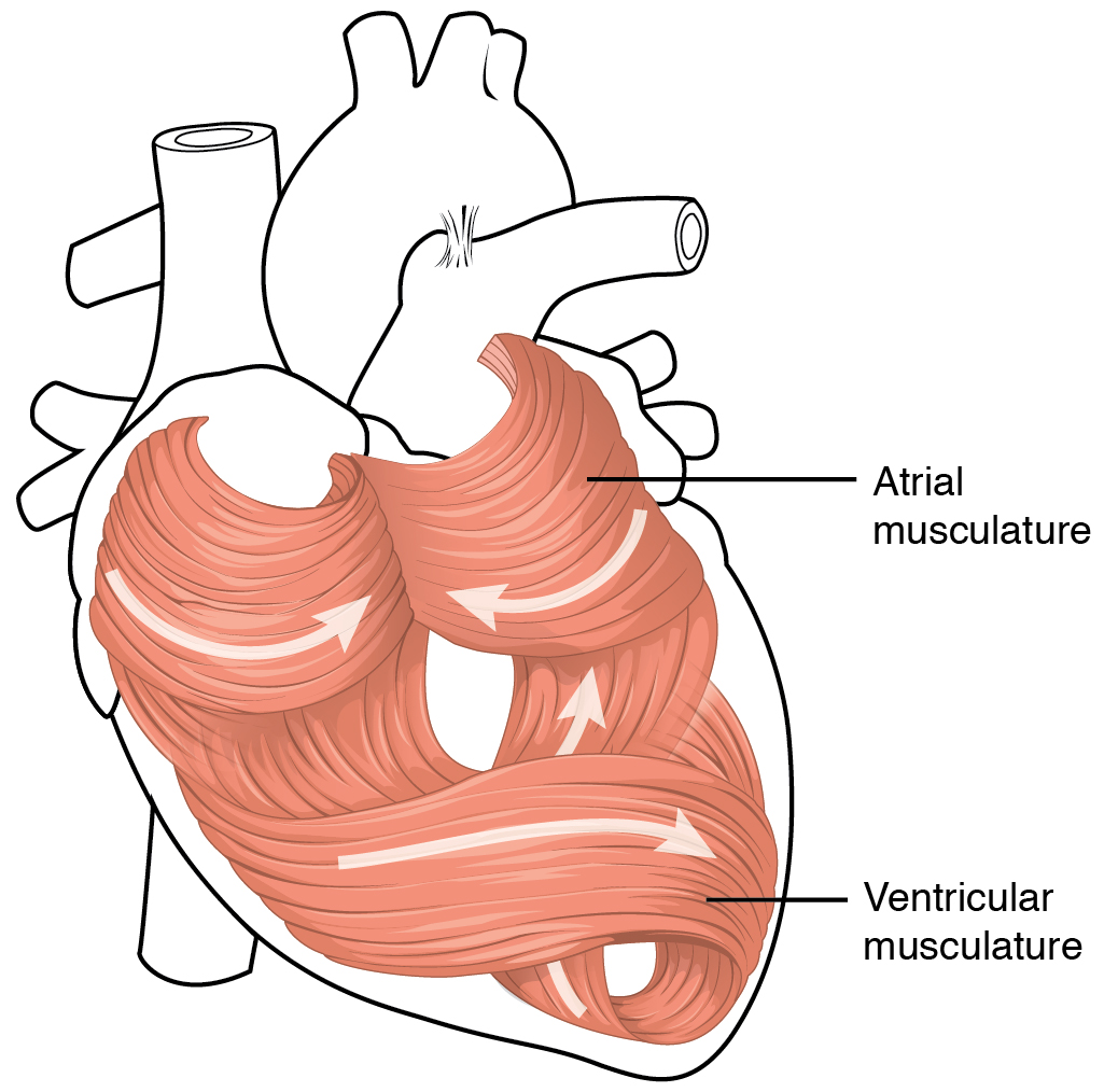
Cardiac muscle (also called heart muscle, myocardium, cardiomyocytes and cardiac myocytes) is one of three types of vertebrate muscle tissues, with the other two being skeletal muscle and smooth muscle. It is an involuntary, striated muscle that constitutes the main tissue of the wall of the heart. The cardiac muscle (myocardium) forms a thick middle layer between the outer layer of the heart wall (the pericardium) and the inner layer (the endocardium), with blood supplied via the coronary circulation. It is composed of individual cardiac muscle cells joined by intercalated discs, and encased by collagen fibers and other substances that form the extracellular matrix. Cardiac muscle contracts in a similar manner to skeletal muscle, although with some important differences. Electrical stimulation in the form of a cardiac action potential triggers the release of calcium from the cell's internal calcium store, the sarcoplasmic reticulum. The rise in calcium causes the c ... [...More Info...] [...Related Items...] OR: [Wikipedia] [Google] [Baidu] |
Angiogenesis
Angiogenesis is the physiological process through which new blood vessels form from pre-existing vessels, formed in the earlier stage of vasculogenesis. Angiogenesis continues the growth of the vasculature by processes of sprouting and splitting. Vasculogenesis is the embryonic formation of endothelial cells from mesoderm cell precursors, and from neovascularization, although discussions are not always precise (especially in older texts). The first vessels in the developing embryo form through vasculogenesis, after which angiogenesis is responsible for most, if not all, blood vessel growth during development and in disease. Angiogenesis is a normal and vital process in growth and development, as well as in wound healing and in the formation of granulation tissue. However, it is also a fundamental step in the transition of tumors from a benign state to a malignant one, leading to the use of angiogenesis inhibitors in the treatment of cancer. The essential role of angi ... [...More Info...] [...Related Items...] OR: [Wikipedia] [Google] [Baidu] |
Blood Vessels
Blood vessels are the structures of the circulatory system that transport blood throughout the human body. These vessels transport blood cells, nutrients, and oxygen to the tissues of the body. They also take waste and carbon dioxide away from the tissues. Blood vessels are needed to sustain life, because all of the body's tissues rely on their functionality. There are five types of blood vessels: the arteries, which carry the blood away from the heart; the arterioles; the capillaries, where the exchange of water and chemicals between the blood and the tissues occurs; the venules; and the veins, which carry blood from the capillaries back towards the heart. The word ''vascular'', meaning relating to the blood vessels, is derived from the Latin ''vas'', meaning vessel. Some structures – such as cartilage, the epithelium, and the lens and cornea of the eye – do not contain blood vessels and are labeled ''avascular''. Etymology * artery: late Middle English; from ... [...More Info...] [...Related Items...] OR: [Wikipedia] [Google] [Baidu] |
Cardiogenesis
Heart development, also known as cardiogenesis, refers to the prenatal development of the heart. This begins with the formation of two endocardial tubes which merge to form the tubular heart, also called the primitive heart tube. The heart is the first functional organ in vertebrate embryos. The tubular heart quickly differentiates into the truncus arteriosus, bulbus cordis, primitive ventricle, primitive atrium, and the sinus venosus. The truncus arteriosus splits into the ascending aorta and the pulmonary trunk. The bulbus cordis forms part of the ventricles. The sinus venosus connects to the fetal circulation. The heart tube elongates on the right side, looping and becoming the first visual sign of left-right asymmetry of the body. Septa form within the atria and ventricles to separate the left and right sides of the heart. Early development The heart derives from embryonic mesodermal germ layer cells that differentiate after gastrulation into mesothelium, endothelium, an ... [...More Info...] [...Related Items...] OR: [Wikipedia] [Google] [Baidu] |
Myocardial Infarctions
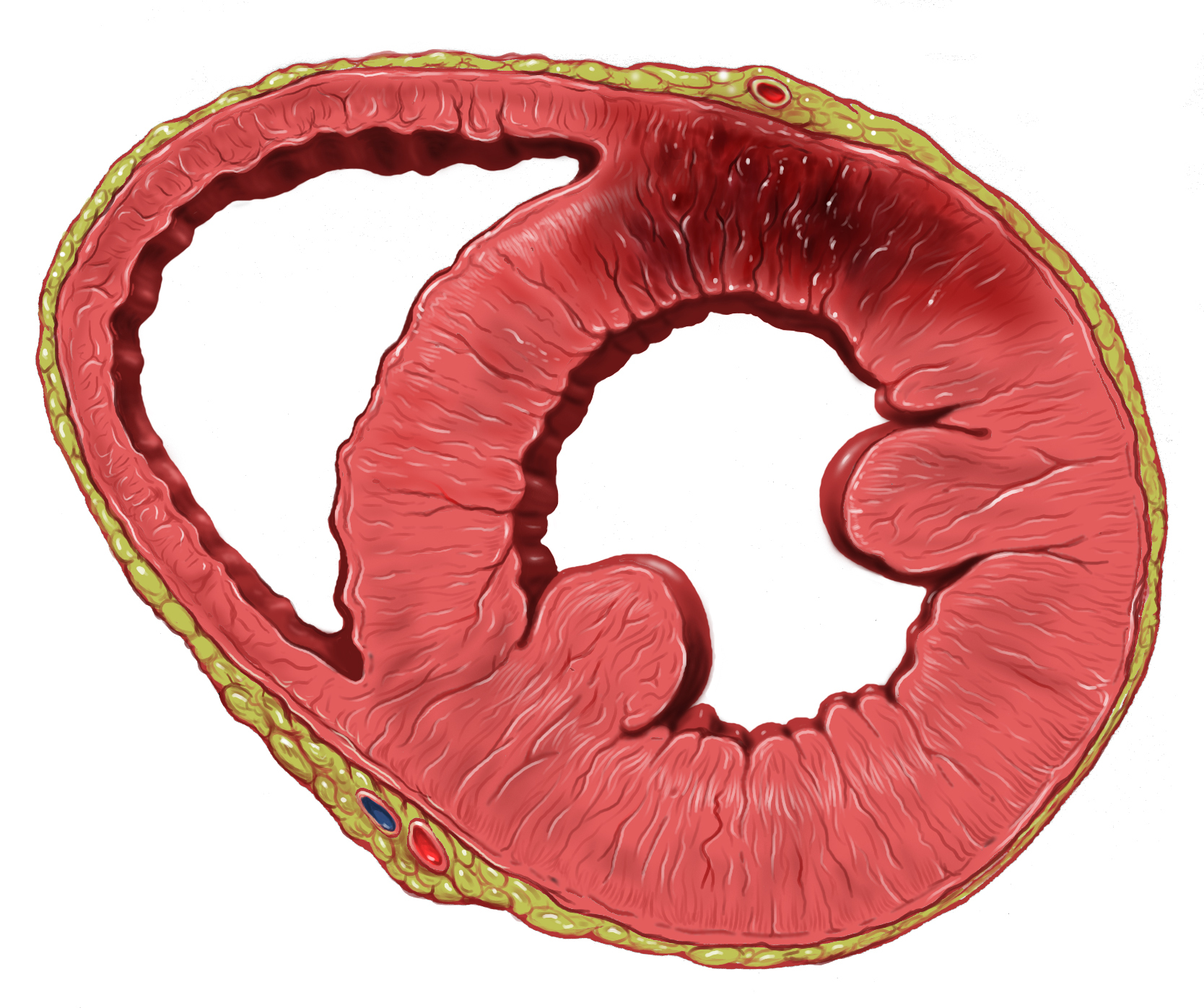
A myocardial infarction (MI), commonly known as a heart attack, occurs when blood flow decreases or stops to the coronary artery of the heart, causing damage to the heart muscle. The most common symptom is chest pain or discomfort which may travel into the shoulder, arm, back, neck or jaw. Often it occurs in the center or left side of the chest and lasts for more than a few minutes. The discomfort may occasionally feel like heartburn. Other symptoms may include shortness of breath, nausea, feeling faint, a cold sweat or feeling tired. About 30% of people have atypical symptoms. Women more often present without chest pain and instead have neck pain, arm pain or feel tired. Among those over 75 years old, about 5% have had an MI with little or no history of symptoms. An MI may cause heart failure, an irregular heartbeat, cardiogenic shock or cardiac arrest. Most MIs occur due to coronary artery disease. Risk factors include high blood pressure, smoking, diabetes, lack of ex ... [...More Info...] [...Related Items...] OR: [Wikipedia] [Google] [Baidu] |
Postmitotic
The G0 phase describes a cellular state outside of the replicative cell cycle. Classically, cells were thought to enter G0 primarily due to environmental factors, like nutrient deprivation, that limited the resources necessary for proliferation. Thus it was thought of as a ''resting phase''. G0 is now known to take different forms and occur for multiple reasons. For example, most adult neuronal cells, among the most metabolically active cells in the body, are fully differentiated and reside in a terminal G0 phase. Neurons reside in this state, not because of stochastic or limited nutrient supply, but as a part of their developmental program. G0 was first suggested as a cell state based on early cell cycle studies. When the first studies defined the four phases of the cell cycle using radioactive labeling techniques, it was discovered that not all cells in a population proliferate at similar rates. A population's “growth fraction” – or the fraction of the population that w ... [...More Info...] [...Related Items...] OR: [Wikipedia] [Google] [Baidu] |
Cardiomyocytes
Cardiac muscle (also called heart muscle, myocardium, cardiomyocytes and cardiac myocytes) is one of three types of vertebrate muscle tissues, with the other two being skeletal muscle and smooth muscle. It is an involuntary, striated muscle that constitutes the main tissue of the wall of the heart. The cardiac muscle (myocardium) forms a thick middle layer between the outer layer of the heart wall (the pericardium) and the inner layer (the endocardium), with blood supplied via the coronary circulation. It is composed of individual cardiac muscle cells joined by intercalated discs, and encased by collagen fibers and other substances that form the extracellular matrix. Cardiac muscle contracts in a similar manner to skeletal muscle, although with some important differences. Electrical stimulation in the form of a cardiac action potential triggers the release of calcium from the cell's internal calcium store, the sarcoplasmic reticulum. The rise in calcium causes the cel ... [...More Info...] [...Related Items...] OR: [Wikipedia] [Google] [Baidu] |
Ischemic
Ischemia or ischaemia is a restriction in blood supply to any tissue, muscle group, or organ of the body, causing a shortage of oxygen that is needed for cellular metabolism (to keep tissue alive). Ischemia is generally caused by problems with blood vessels, with resultant damage to or dysfunction of tissue i.e. hypoxia and microvascular dysfunction. It also implies local hypoxia in a part of a body resulting from constriction (such as vasoconstriction, thrombosis, or embolism). Ischemia causes not only insufficiency of oxygen, but also reduced availability of nutrients and inadequate removal of metabolic wastes. Ischemia can be partial (poor perfusion) or total blockage. The inadequate delivery of oxygenated blood to the organs must be resolved either by treating the cause of the inadequate delivery or reducing the oxygen demand of the system that needs it. For example, patients with myocardial ischemia have a decreased blood flow to the heart and are prescribed with ... [...More Info...] [...Related Items...] OR: [Wikipedia] [Google] [Baidu] |
Progenitor Cells
In genealogy, the progenitor (rarer: primogenitor; german: Stammvater or ''Ahnherr'') is the – sometimes legendary – founder of a family, line of descent, clan or tribe, noble house, or ethnic group.. Ebenda''Ahnherr:''"Stammvater eines Geschlechts". Genealogy (commonly known as family history) understands a progenitor to be the earliest recorded ancestor of a consanguineous family group of descendants. Progenitors are sometimes used to describe the status of a genealogical research project, or in order to compare the availability of genealogical data in different times and places. Often, progenitors are implied to be patrilineal. If a patrilineal dynasty is considered, each such dynasty has exactly one progenitor. Aristocratic and dynastic families often look back to an ancestor who is seen as the founder and progenitor of their house (i.e. family line). Even the old Roman legal concept of agnates (Latin for "descendants") was based on the idea of the unbroken ... [...More Info...] [...Related Items...] OR: [Wikipedia] [Google] [Baidu] |
Telomeres
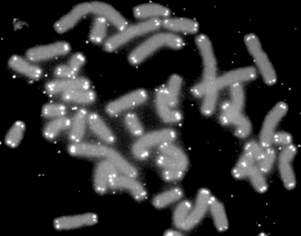
A telomere (; ) is a region of repetitive nucleotide sequences associated with specialized proteins at the ends of linear chromosomes. Although there are different architectures, telomeres, in a broad sense, are a widespread genetic feature most commonly found in eukaryotes. In most, if not all species possessing them, they protect the terminal regions of chromosomal DNA from progressive degradation and ensure the integrity of linear chromosomes by preventing DNA repair systems from mistaking the very ends of the DNA strand for a double-strand break. Discovery In the early 1970s, Soviet theorist Alexei Olovnikov first recognized that chromosomes could not completely replicate their ends; this is known as the "end replication problem". Building on this, and accommodating Leonard Hayflick's idea of limited somatic cell division, Olovnikov suggested that DNA sequences are lost every time a cell replicates until the loss reaches a critical level, at which point cell division en ... [...More Info...] [...Related Items...] OR: [Wikipedia] [Google] [Baidu] |


