|
Nasopalatine Nerve
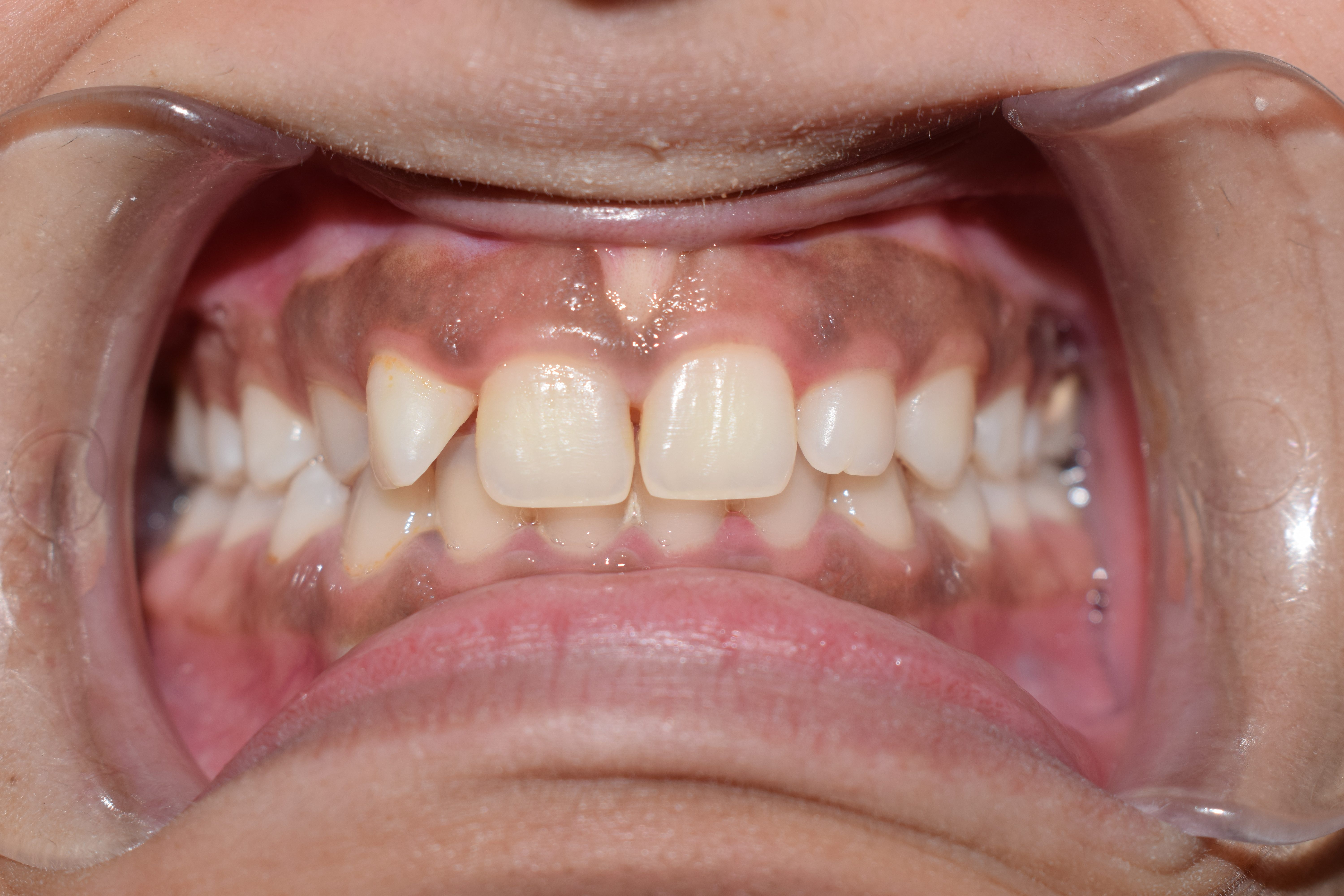
The nasopalatine nerve (also long sphenopalatine nerve) is a nerve of the head. It is a sensory branch of the maxillary nerve (CN V2) that passes through the pterygopalatine ganglion (without synapsing) and then through the sphenopalatine foramen to enter the nasal cavity, and finally out of the nasal cavity through the incisive canal and then the incisive fossa to enter the hard palate. It provides sensory innervation to the posteroinferior part of the nasal septum, and gingiva just posterior to the upper incisor teeth. The nasopalatine nerve is the largest of the medial posterior superior nasal nerves. Structure Course It exits the pterygopalatine fossa through the sphenopalatine foramen to enter the nasal cavity. It passes across the roof of the nasal cavity below the orifice of the sphenoidal sinus to reach the posterior part of the nasal septum. It passes anteroinferiorly upon the nasal septum along a groove upon the vomer, running between the periosteum and mucous membr ... [...More Info...] [...Related Items...] OR: [Wikipedia] [Google] [Baidu] |
Palate
The palate () is the roof of the mouth in humans and other mammals. It separates the oral cavity from the nasal cavity. A similar structure is found in crocodilians, but in most other tetrapods, the oral and nasal cavities are not truly separated. The palate is divided into two parts, the anterior, bony hard palate and the posterior, fleshy soft palate (or velum). Structure Innervation The maxillary nerve branch of the trigeminal nerve supplies sensory innervation to the palate. Development The hard palate forms before birth. Variation If the fusion is incomplete, a cleft palate results. Function in humans When functioning in conjunction with other parts of the mouth, the palate produces certain sounds, particularly velar, palatal, palatalized, postalveolar, alveolopalatal, and uvular consonants. History Etymology The English synonyms palate and palatum, and also the related adjective palatine (as in palatine bone), are all from the Latin ''palatum' ... [...More Info...] [...Related Items...] OR: [Wikipedia] [Google] [Baidu] |
Vomer
The vomer (; ) is one of the unpaired facial bones of the skull. It is located in the midsagittal line, and articulates with the sphenoid, the ethmoid, the left and right palatine bones, and the left and right maxillary bones. The vomer forms the inferior part of the nasal septum in humans, with the superior part formed by the perpendicular plate of the ethmoid bone. The name is derived from the Latin word for a ploughshare and the shape of the bone. In humans The vomer is situated in the median plane, but its anterior portion is frequently bent to one side. It is thin, somewhat quadrilateral in shape, and forms the hinder and lower part of the nasal septum; it has two surfaces and four borders. The surfaces are marked by small furrows for blood vessels, and on each is the nasopalatine groove, which runs obliquely downward and forward, and lodges the nasopalatine nerve and vessels. Borders The ''superior border'', the thickest, presents a deep furrow, bounded on eith ... [...More Info...] [...Related Items...] OR: [Wikipedia] [Google] [Baidu] |
Domenico Cotugno
Domenico Felice Antonio Cotugno (January 29, 1736 – October 6, 1822) was an Italian people, Italian physician. Biography Born at Ruvo di Puglia (Province of Bari, Apulia) into a family of humble means, Cotugno underwent physical and economic hardships to get an education. He was sent to nearby Molfetta for training in Latin, returning to Ruvo for work in logic, metaphysics, mathematics, physics, and the natural sciences. He soon found his natural bent in medicine and continued his studies from 1753 at the University of Naples, and in 1756 graduated from Schola Medica Salernitana, Salerno medical school. He received his doctorate in philosophy and physics in 1755, and became an assistant at the ''Ospedale degli Incurabili, Naples, Ospedale degli Incurabili'' (Neapolitan Hospital for Incurables). In 1761 he became professor of surgery at that hospital, and was subsequently for 30 years professor of anatomy at the high school of Naples. In 1808 he was appointed royal physician to ... [...More Info...] [...Related Items...] OR: [Wikipedia] [Google] [Baidu] |
Soft Palate
The soft palate (also known as the velum, palatal velum, or muscular palate) is, in mammals, the soft biological tissue, tissue constituting the back of the roof of the mouth. The soft palate is part of the palate of the mouth; the other part is the hard palate. The soft palate is distinguished from the hard palate at the front of the mouth in that it does not contain bone. Structure Muscles The five muscles of the soft palate play important roles in swallowing and breathing. The muscles are: # Tensor veli palatini, which is involved in swallowing # Palatoglossus, involved in swallowing # Palatopharyngeus, involved in breathing # Levator veli palatini, involved in swallowing # Musculus uvulae, which moves the palatine uvula, uvula These muscles are innervated by the pharyngeal plexus of vagus nerve, pharyngeal plexus via the vagus nerve, with the exception of the tensor veli palatini. The tensor veli palatini is innervated by the mandibular division of the trigeminal nerve (V ... [...More Info...] [...Related Items...] OR: [Wikipedia] [Google] [Baidu] |
Oral And Maxillofacial Surgery
Oral and maxillofacial surgery (OMFS) is a surgical specialty focusing on reconstructive surgery of the face, facial trauma surgery, the Human mouth, mouth, Human head, head and neck, and jaws, as well as facial plastic surgery including cleft lip and cleft palate surgery. Specialty An oral and maxillofacial surgeon is a specialist surgery, surgeon who treats the entire Craniofacial, craniomaxillofacial complex: Anatomy, anatomical area of the Human mouth, mouth, jaws, face, and Human skull, skull, head and neck as well as associated structures. Depending upon the national jurisdiction, oral and maxillofacial surgery may require a degree in medicine, dentistry or both. United States In the U.S., oral and maxillofacial surgeons, whether possessing a single or dual degree, may further specialise after residency, undergoing additional one or two year sub-specialty oral and maxillofacial surgery fellowship training in the following areas: *Cosmetic surgery#Cosmetic surgery, C ... [...More Info...] [...Related Items...] OR: [Wikipedia] [Google] [Baidu] |
Anesthesia
Anesthesia (American English) or anaesthesia (British English) is a state of controlled, temporary loss of sensation or awareness that is induced for medical or veterinary purposes. It may include some or all of analgesia (relief from or prevention of pain), paralysis (muscle relaxation), amnesia (loss of memory), and unconsciousness. An individual under the effects of anesthetic drugs is referred to as being anesthetized. Anesthesia enables the painless performance of procedures that would otherwise require physical restraint in a non-anesthetized individual, or would otherwise be technically unfeasible. Three broad categories of anesthesia exist: * ''General anesthesia'' suppresses central nervous system activity and results in unconsciousness and total lack of Sensation (psychology), sensation, using either injected or inhaled drugs. * ''Sedation'' suppresses the central nervous system to a lesser degree, inhibiting both anxiolysis, anxiety and creation of long-term memory, ... [...More Info...] [...Related Items...] OR: [Wikipedia] [Google] [Baidu] |
Greater Palatine Nerve
The greater palatine nerve is a branch of the pterygopalatine ganglion. This nerve is also referred to as the anterior palatine nerve, due to its location anterior to the lesser palatine nerve. It carries both general sensory fibres from the maxillary nerve, and parasympathetic fibers from the nerve of the pterygoid canal. It may be anaesthetised for procedures of the mouth and maxillary (upper) teeth. Structure The greater palatine nerve is a branch of the pterygopalatine ganglion. It descends through the greater palatine canal, moving anteriorly and inferiorly. Here, it is accompanied by the descending palatine artery. It emerges upon the hard palate through the greater palatine foramen. It then passes forward in a groove in the hard palate, nearly as far as the incisor teeth. While in the pterygopalatine canal, it gives off lateral posterior inferior nasal branches, which enter the nasal cavity through openings in the palatine bone, and ramify over the inferior nasal ... [...More Info...] [...Related Items...] OR: [Wikipedia] [Google] [Baidu] |
Incisor
Incisors (from Latin ''incidere'', "to cut") are the front teeth present in most mammals. They are located in the premaxilla above and on the mandible below. Humans have a total of eight (two on each side, top and bottom). Opossums have 18, whereas armadillos, anteaters and other animals in the order Edentata have none. Structure Adult humans normally have eight incisors, two of each type. The types of incisors are: * maxillary central incisor (upper jaw, closest to the center of the lips) * maxillary lateral incisor (upper jaw, beside the maxillary central incisor) * mandibular central incisor (lower jaw, closest to the center of the lips) * mandibular lateral incisor (lower jaw, beside the mandibular central incisor) Children with a full set of deciduous teeth (primary teeth) also have eight incisors, named the same way as in permanent teeth. Young children may have from zero to eight incisors depending on the stage of their tooth eruption and tooth development. Typic ... [...More Info...] [...Related Items...] OR: [Wikipedia] [Google] [Baidu] |
Gingiva
The gums or gingiva (: gingivae) consist of the mucosal tissue that lies over the mandible and maxilla inside the mouth. Gum health and disease can have an effect on general health. Structure The gums are part of the soft tissue lining of the mouth. They surround the teeth and provide a seal around them. Unlike the soft tissue linings of the lips and cheeks, most of the gums are tightly bound to the underlying bone which helps resist the friction of food passing over them. Thus when healthy, it presents an effective barrier to the barrage of periodontal insults to deeper tissue. Healthy gums are usually coral pink in light skinned people, and may be naturally darker with melanin pigmentation. Changes in color, particularly increased redness, together with swelling and an increased tendency to bleed, suggest an inflammation that is possibly due to the accumulation of bacterial plaque. Overall, the clinical appearance of the tissue reflects the underlying histology, both in hea ... [...More Info...] [...Related Items...] OR: [Wikipedia] [Google] [Baidu] |
Palate
The palate () is the roof of the mouth in humans and other mammals. It separates the oral cavity from the nasal cavity. A similar structure is found in crocodilians, but in most other tetrapods, the oral and nasal cavities are not truly separated. The palate is divided into two parts, the anterior, bony hard palate and the posterior, fleshy soft palate (or velum). Structure Innervation The maxillary nerve branch of the trigeminal nerve supplies sensory innervation to the palate. Development The hard palate forms before birth. Variation If the fusion is incomplete, a cleft palate results. Function in humans When functioning in conjunction with other parts of the mouth, the palate produces certain sounds, particularly velar, palatal, palatalized, postalveolar, alveolopalatal, and uvular consonants. History Etymology The English synonyms palate and palatum, and also the related adjective palatine (as in palatine bone), are all from the Latin ''palatum' ... [...More Info...] [...Related Items...] OR: [Wikipedia] [Google] [Baidu] |
Incisive Canal
The incisive canals (also: "''nasopalatine canals''") are two bony canals of the anterior hard palate connecting the nasal cavity and the oral cavity. An incisive canal courses through each maxilla. Below, the two incisive canals typically converge medially. Each incisive canal transmits a nasopalatine nerve, and an anastomosis of the greater palatine artery and a posterior septal branch of the sphenopalatine artery. Anatomy An incisive canal has an average length of 10 mm, and an average width of up to 6 mm at the incisive fossa (the dimensions of the canal change with age, trauma, and loss of teeth). Course and openings The two incisive canals usually (in 60% of individuals) have a characteristic Y-shaped or V-shaped morphology: above, each incisive canal opens into the nasal cavity on either side of the nasal septum as the nasal foramina; below, the two incisive canals converge medially to open into the oral cavity at midline at the incisive fossa as several incisive f ... [...More Info...] [...Related Items...] OR: [Wikipedia] [Google] [Baidu] |
Periosteum
The periosteum is a membrane that covers the outer surface of all bones, except at the articular surfaces (i.e. the parts within a joint space) of long bones. (At the joints of long bones the bone's outer surface is lined with "articular cartilage", a type of hyaline cartilage.) Endosteum lines the inner surface of the medullary cavity of all long bones. Structure The periosteum consists of an outer fibrous layer, and an inner ''cambium layer'' (or osteogenic layer). The fibrous layer is of dense irregular connective tissue, containing fibroblasts, while the cambium layer is highly cellular containing progenitor cells that develop into osteoblasts. These osteoblasts are responsible for increasing the width of a long bone (the length of a long bone is controlled by the epiphyseal plate) and the overall size of the other bone types. After a bone fracture A bone fracture (abbreviated FRX or Fx, Fx, or #) is a medical condition in which there is a partial or complete break ... [...More Info...] [...Related Items...] OR: [Wikipedia] [Google] [Baidu] |


