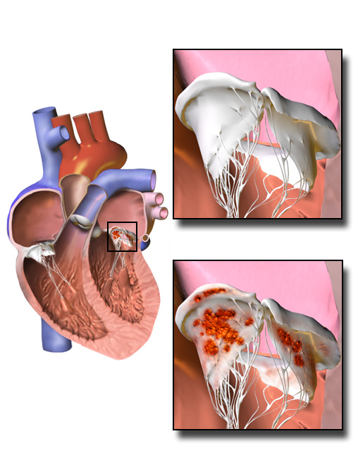
|
Myxomatous Mitral Valve
Mitral valve prolapse (MVP) is a valvular heart disease characterized by the displacement of an abnormally thickened mitral valve leaflet into the left atrium during systole. It is the primary form of myxomatous degeneration of the valve. There are various types of MVP, broadly classified as classic and nonclassic. In severe cases of classic MVP, complications include mitral regurgitation, infective endocarditis, congestive heart failure, and, in rare circumstances, cardiac arrest. The diagnosis of MVP primarily relies on echocardiography, which uses ultrasound to visualize the mitral valve. MVP is the most common valvular abnormality, and is estimated to affect 2–3% of the population and 1 in 40 people might have it. The condition was first described by John Brereton Barlow in 1966. It was subsequently termed ''mitral valve prolapse'' by J. Michael Criley. Although mid-systolic click (the sound produced by the prolapsing mitral leaflet) and systolic murmur associated with M ... [...More Info...] [...Related Items...] OR: [Wikipedia] [Google] [Baidu] |
Mitral Valve
The mitral valve ( ), also known as the bicuspid valve or left atrioventricular valve, is one of the four heart valves. It has two Cusps of heart valves, cusps or flaps and lies between the atrium (heart), left atrium and the ventricle (heart), left ventricle of the heart. The heart valves are all one-way valves allowing blood flow in just one direction. The mitral valve and the tricuspid valve are known as the Heart valve#Atrioventricular valves, atrioventricular valves because they lie between the atria and the ventricles. In normal conditions, blood flows through an open mitral valve during diastole with contraction of the left atrium, and the mitral valve closes during systole with contraction of the left ventricle. The valve opens and closes because of pressure differences, opening when there is greater pressure in the left atrium than ventricle and closing when there is greater pressure in the left ventricle than atrium. In abnormal conditions, blood may flow backward thro ... [...More Info...] [...Related Items...] OR: [Wikipedia] [Google] [Baidu] |
Auscultation
Auscultation (based on the Latin verb ''auscultare'' "to listen") is listening to the internal sounds of the body, usually using a stethoscope. Auscultation is performed for the purposes of examining the circulatory system, circulatory and respiratory systems (heart sounds, heart and breath sounds), as well as the alimentary canal. The term was introduced by René Laennec. The act of listening to body sounds for diagnostic purposes has its origin further back in history, possibly as early as Ancient Egypt. Auscultation and palpation go together in physical examination and are alike in that both have ancient roots, both require skill, and both are still important today. Laënnec's contributions were refining the procedure, linking sounds with specific pathological changes in the chest, and inventing a suitable instrument (the stethoscope) to mediate between the patient's body and the clinician's ear. Auscultation is a skill that requires substantial clinical experience, a fine ... [...More Info...] [...Related Items...] OR: [Wikipedia] [Google] [Baidu] |
Stethoscope
The stethoscope is a medicine, medical device for auscultation, or listening to internal sounds of an animal or human body. It typically has a small disc-shaped resonator that is placed against the skin, with either one or two tubes connected to two earpieces. A stethoscope can be used to listen to the sounds made by the heart sounds, heart, lungs or intestines, as well as blood flow in artery, arteries and veins. In combination with a manual sphygmomanometer, it is commonly used when measuring blood pressure. It was invented in 1816 by René Laennec and the binaural version by Arthur Leared in 1851. Less commonly, "mechanic's stethoscopes", equipped with rod shaped chestpieces, are used to listen to internal sounds made by machines (for example, sounds and vibrations emitted by worn ball bearings), such as diagnosing a malfunctioning automobile engine by listening to the sounds of its internal parts. Stethoscopes can also be used to check scientific vacuum chambers for leaks an ... [...More Info...] [...Related Items...] OR: [Wikipedia] [Google] [Baidu] |
John Brereton Barlow
John Brereton Barlow (24 October 1924 – 10 December 2008) was a world-renowned South African cardiologist. He qualified as a doctor in 1951, gained experience as a registrar in Hammersmith Hospital and the Royal Postgraduate Medical School in London. In the late 1950s he returned to South Africa to Johannesburg Hospital where he became Professor of Cardiology in the research unit and carried out significant studies on cardiac disorders as well as discovering the cause of a well known mitral valve disorder. He gives his name to Barlow's Syndrome. Professional life Barlow commenced medical studies at the University of the Witwatersrand. However, shortly afterwards, in 1940 when South Africa became involved in World War II he enlisted in the military and spent time attached to British forces in North Africa and later the Fifth US Army in Italy. He returned to medical school in 1946 and graduated with MBBCh. in November 1951. Barlow served his internship and registrar post ... [...More Info...] [...Related Items...] OR: [Wikipedia] [Google] [Baidu] |
Ultrasound
Ultrasound is sound with frequency, frequencies greater than 20 Hertz, kilohertz. This frequency is the approximate upper audible hearing range, limit of human hearing in healthy young adults. The physical principles of acoustic waves apply to any frequency range, including ultrasound. Ultrasonic devices operate with frequencies from 20 kHz up to several gigahertz. Ultrasound is used in many different fields. Ultrasonic devices are used to detect objects and measure distances. Ultrasound imaging or sonography is often used in medicine. In the nondestructive testing of products and structures, ultrasound is used to detect invisible flaws. Industrially, ultrasound is used for cleaning, mixing, and accelerating chemical processes. Animals such as bats and porpoises use ultrasound for locating prey and obstacles. History Acoustics, the science of sound, starts as far back as Pythagoras in the 6th century BC, who wrote on the mathematical properties of String instrument ... [...More Info...] [...Related Items...] OR: [Wikipedia] [Google] [Baidu] |
Echocardiography
Echocardiography, also known as cardiac ultrasound, is the use of ultrasound to examine the heart. It is a type of medical imaging, using standard ultrasound or Doppler ultrasound. The visual image formed using this technique is called an echocardiogram, a cardiac echo, or simply an echo. Echocardiography is routinely used in the diagnosis, management, and follow-up of patients with any suspected or known heart diseases. It is one of the most widely used diagnostic imaging modalities in cardiology. It can provide a wealth of helpful information, including the size and shape of the heart (internal chamber size quantification), pumping capacity, location and extent of any tissue damage, and assessment of valves. An echocardiogram can also give physicians other estimates of heart function, such as a calculation of the cardiac output, ejection fraction, and diastolic function (how well the heart relaxes). Echocardiography is an important tool in assessing wall motion abnorma ... [...More Info...] [...Related Items...] OR: [Wikipedia] [Google] [Baidu] |
Cardiac Arrest
Cardiac arrest (also known as sudden cardiac arrest [SCA]) is when the heart suddenly and unexpectedly stops beating. When the heart stops beating, blood cannot properly Circulatory system, circulate around the body and the blood flow to the brain and other organs is decreased. When the brain does not receive enough blood, this can cause a person to lose consciousness and brain cells can start to die due to lack of oxygen. Coma and persistent vegetative state may result from cardiac arrest. Cardiac arrest is also identified by a lack of Pulse, central pulses and respiratory arrest, abnormal or absent breathing. Cardiac arrest and resultant hemodynamic collapse often occur due to arrhythmias (irregular heart rhythms). Ventricular fibrillation and ventricular tachycardia are most commonly recorded. However, as many incidents of cardiac arrest occur out-of-hospital or when a person is not having their cardiac activity monitored, it is difficult to identify the specific mechanism ... [...More Info...] [...Related Items...] OR: [Wikipedia] [Google] [Baidu] |
Congestive Heart Failure
Heart failure (HF), also known as congestive heart failure (CHF), is a syndrome caused by an impairment in the heart's ability to fill with and pump blood. Although symptoms vary based on which side of the heart is affected, HF typically presents with shortness of breath, excessive fatigue, and bilateral leg swelling. The severity of the heart failure is mainly decided based on ejection fraction and also measured by the severity of symptoms. Other conditions that have symptoms similar to heart failure include obesity, kidney failure, liver disease, anemia, and thyroid disease. Common causes of heart failure include coronary artery disease, heart attack, high blood pressure, atrial fibrillation, valvular heart disease, excessive alcohol consumption, infection, and cardiomyopathy. These cause heart failure by altering the structure or the function of the heart or in some cases both. There are different types of heart failure: right-sided heart failure, which affect ... [...More Info...] [...Related Items...] OR: [Wikipedia] [Google] [Baidu] |
Infective Endocarditis
Infective endocarditis is an infection of the inner surface of the heart (endocardium), usually the heart valve, valves. Signs and symptoms may include fever, petechia, small areas of bleeding into the skin, heart murmur, feeling tired, and anemia, low red blood cell count. Complications may include valvular insufficiency, backward blood flow in the heart, heart failure – the heart struggling to pump a sufficient amount of blood to meet the body's needs, Heart block, abnormal electrical conduction in the heart, stroke, and kidney failure. The cause is typically a bacterial infection and less commonly a fungal infection. Risk factors include valvular heart disease, including rheumatic heart disease, rheumatic disease, congenital heart disease, artificial valves, hemodialysis, intravenous drug use, and electronic pacemakers. The bacteria most commonly involved are streptococci or staphylococci. Diagnosis is suspected based on symptoms and supported by blood cultures or Echocardi ... [...More Info...] [...Related Items...] OR: [Wikipedia] [Google] [Baidu] |
Mitral Regurgitation
Mitral regurgitation (MR), also known as mitral insufficiency or mitral incompetence, is a form of valvular heart disease in which the mitral valve is insufficient and does not close properly when the heart pumps out blood. Section: Valvular Heart Disease Rupture or dysfunction of the papillary muscle are also common causes in acute cases, dysfunction, which can include mitral valve prolapse.VOC=VITIUM ORGANICUM CORDIS, a compendium of the Department of Cardiology at Uppsala Academic Hospital. By Per Kvidal September 1999, with revision by Erik Björklund May 2008 Pathophysiology The pathophysiology of MR can be broken into three phases of the disease process: the acute phase, the chronic compensated phase, and the chronic decompensated phase. Acute phase Acute MR (as may occur due to the sudden rupture of the chordae tendinae or papillary muscle) causes a sudden volume overload of both the left atrium and the left ventricle. The left ventricle develops volume overload because ... [...More Info...] [...Related Items...] OR: [Wikipedia] [Google] [Baidu] |
Myxomatous Degeneration
A myxoma (New Latin from Greek 'mucus') is a myxoid tumor of primitive connective tissue. It is most commonly found in the heart (and is the most common primary tumor of the heart in adults) but can also occur in other locations. Types Table below: 1.SMA, smooth muscle actin. 2.MSA, muscle-specific actin. 3.EMA, epithelial membrane antigen. Symptoms and signs Symptoms associated with cardiac myxomas are typically due to the effect of the mass of the tumor obstructing the normal flow of blood within the chambers of the heart. Because pedunculated myxomas are somewhat mobile, symptoms may only occur when the patient is in a particular position. Some symptoms of myxoma may be associated with the release of interleukin 6 (IL-6) by the myxoma. High levels of IL-6 may be associated with a higher risk of embolism of the myxoma. Symptoms of a cardiac myxoma include: * Dyspnea on exertion * Paroxysmal nocturnal dyspnea * Fever * Weight loss (see cachexia) * Lightheadedness or sy ... [...More Info...] [...Related Items...] OR: [Wikipedia] [Google] [Baidu] |
Systole (medicine)
Systole ( ) is the part of the cardiac cycle during which some chambers of the heart contract after refilling with blood. Its contrasting phase is diastole, the relaxed phase of the cardiac cycle when the chambers of the heart are refilling with blood. Etymology The term originates, via Neo-Latin, from Ancient Greek (''sustolē''), from (''sustéllein'' 'to contract'; from ''sun'' 'together' + ''stéllein'' 'to send'), and is similar to the use of the English term ''to squeeze''. Terminology, general explanation The mammalian heart has four chambers: the left atrium above the left ventricle (lighter pink, see graphic), which two are connected through the mitral (or bicuspid) valve; and the right atrium above the right ventricle (lighter blue), connected through the tricuspid valve. The atria are the receiving blood chambers for the circulation of blood and the ventricles are the discharging chambers. In late ventricular diastole, the atrial chambers contract and send ... [...More Info...] [...Related Items...] OR: [Wikipedia] [Google] [Baidu] |


