|
Myenteric
The myenteric plexus (or Auerbach's plexus) provides motor innervation to both layers of the muscular layer of the gut, having both parasympathetic and sympathetic input (although present ganglion cell bodies belong to parasympathetic innervation, fibers from sympathetic innervation also reach the plexus), whereas the submucous plexus provides secretomotor innervation to the mucosa nearest the lumen of the gut. It arises from cells in the vagal trigone also known as the nucleus ala cinerea, the parasympathetic nucleus of origin for the tenth cranial nerve (vagus nerve), located in the medulla oblongata. The fibers are carried by both the anterior and posterior vagal nerves. The myenteric plexus is the major nerve supply to the gastrointestinal tract and controls GI tract motility. According to preclinical studies, 30% of myenteric plexus' neurons are enteric sensory neurons, thus Auerbach's plexus has also a sensory component. Structure A part of the enteric nervous system, ... [...More Info...] [...Related Items...] OR: [Wikipedia] [Google] [Baidu] |
Submucous Plexus
The submucosal plexus (Meissner's plexus, plexus of the submucosa, plexus submucosus) lies in the submucosa of the intestinal wall. The nerves of this plexus are derived from the myenteric plexus which itself is derived from the plexuses of parasympathetic nerves around the superior mesenteric artery. Branches from the myenteric plexus perforate the circular muscle fibers to form the submucosal plexus. Ganglia from the plexus extend into the muscularis mucosae and also extend into the mucous membrane A mucous membrane or mucosa is a membrane that lines various cavities in the body of an organism and covers the surface of internal organs. It consists of one or more layers of epithelial cells overlying a layer of loose connective tissue. It .... They contain Dogiel cells. The nerve bundles of the submucosal plexus are finer than those of the myenteric plexus. Its function is to innervate cells in the epithelial layer and the smooth muscle of the muscularis mucosae. 14% o ... [...More Info...] [...Related Items...] OR: [Wikipedia] [Google] [Baidu] |
Enteric Nervous System
The enteric nervous system (ENS) is one of the three divisions of the autonomic nervous system (ANS), the others being the sympathetic nervous system (SNS) and parasympathetic nervous system (PSNS). It consists of a mesh-like system of neurons that governs the function of the gastrointestinal tract. The ENS is nicknamed the "second brain". It is derived from neural crest cells. The enteric nervous system is capable of operating independently of the brain and spinal cord, but is thought to rely on innervation from the vagus nerve and prevertebral ganglia in healthy subjects. However, studies have shown that the system is operable with a severed vagus nerve. The neurons of the enteric nervous system control the motor functions of the system, in addition to the secretion of gastrointestinal enzymes. These neurons communicate through many neurotransmitters similar to the CNS, including acetylcholine, dopamine, and serotonin. The large presence of serotonin and dopamine in the intest ... [...More Info...] [...Related Items...] OR: [Wikipedia] [Google] [Baidu] |
Dogiel Cells
Dogiel cells, also known as cells of Dogiel, are a type of multipolar neuronal cells within the prevertebral sympathetic ganglia. They are named after the Russian anatomist and physiologist Alexandre Dogiel (1852–1922). Dogiel cells play a role in the enteric nervous system The enteric nervous system (ENS) is one of the three divisions of the autonomic nervous system (ANS), the others being the sympathetic nervous system (SNS) and parasympathetic nervous system (PSNS). It consists of a mesh-like system of neurons th .... Types There are seven types of cells of Dogiel. References ;Further reading * * * * {{Nervous tissue Sympathetic nervous system Enteric nervous system ... [...More Info...] [...Related Items...] OR: [Wikipedia] [Google] [Baidu] |
Leopold Auerbach
Leopold Auerbach (27 April 1828 – 30 September 1897) was a Jewish German anatomy, anatomist and neuropathology, neuropathologist born in Breslau. He is best known for discovering the myenteric plexus aka Auerbach's plexus, which helps control the GI tract. Education and career Auerbach studied medicine at the Universities of University of Breslau, Breslau, University of Berlin, Berlin and the University of Leipzig, Leipzig. He became a physician in 1849, obtained his habilitation in 1863. From 1872 he was an associate professor of neuropathology at the University of Breslau. Discoveries Auerbach was among the first physicians to diagnose the nervous system using histology, histological staining methods. He published a number of papers on neuropathological problems and muscle-related disorders. He is credited with the discovery of ''Plexus myentericus Auerbachi'', or Auerbach's plexus, a layer of ganglion cells that provide control of movements of the gastro-intestinal tract, al ... [...More Info...] [...Related Items...] OR: [Wikipedia] [Google] [Baidu] |
Hirschsprung's Disease
Hirschsprung's disease (HD or HSCR) is a birth defect in which nerves are missing from parts of the intestine. The most prominent symptom is constipation. Other symptoms may include vomiting, abdominal pain, diarrhea and slow growth. Most children develop signs and symptoms shortly after birth. However, others may be diagnosed later in infancy or early childhood. About half of all children with Hirschsprung's disease are diagnosed in the first year of life. Complications may include enterocolitis, megacolon, bowel obstruction and intestinal perforation. The disorder may occur by itself or in association with other genetic disorders such as Down syndrome or Waardenburg syndrome. About half of isolated cases are linked to a specific genetic mutation, and about 20% occur within families. Some of these occur in an autosomal dominant manner. The cause of the remaining cases is unclear. If otherwise normal parents have one child with the condition, the next child has a 4% risk o ... [...More Info...] [...Related Items...] OR: [Wikipedia] [Google] [Baidu] |
Digestive System
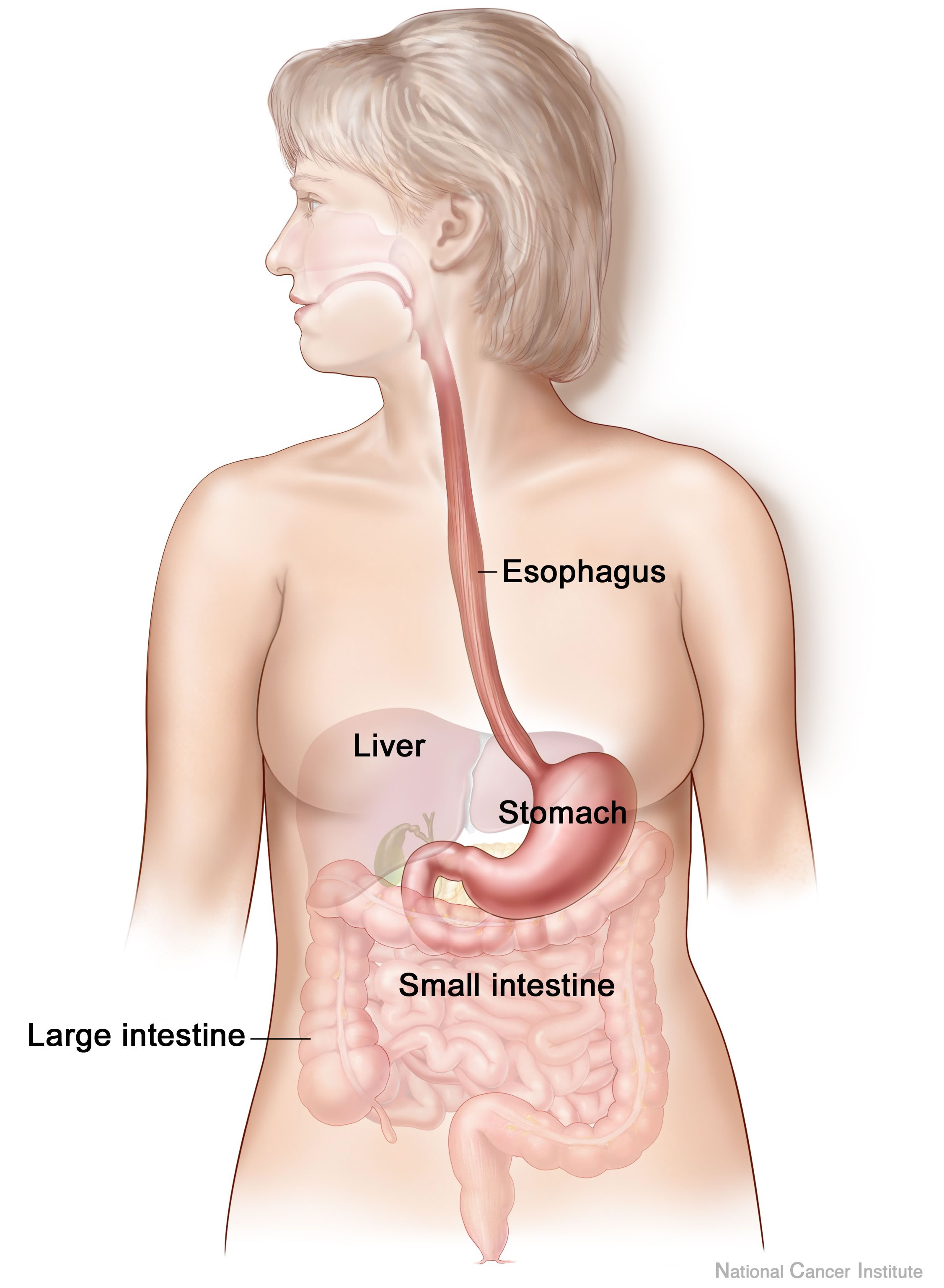
The human digestive system consists of the gastrointestinal tract plus the accessory organs of digestion (the tongue, salivary glands, pancreas, liver, and gallbladder). Digestion involves the breakdown of food into smaller and smaller components, until they can be absorbed and assimilated into the body. The process of digestion has three stages: the cephalic phase, the gastric phase, and the intestinal phase. The first stage, the cephalic phase of digestion, begins with secretions from gastric glands in response to the sight and smell of food, and continues in the human mouth, mouth with the mechanical breakdown of food by chewing, and the chemical breakdown by digestive enzymes in the saliva. Saliva contains amylase, and lingual lipase, secreted by the salivary glands, and serous glands on the tongue. Chewing mixes the food with saliva to produce a Bolus (digestion), bolus to be Swallowing, swallowed down the esophagus to enter the stomach. The second stage, the gastric phase ... [...More Info...] [...Related Items...] OR: [Wikipedia] [Google] [Baidu] |
Gastrointestinal
The gastrointestinal tract (GI tract, digestive tract, alimentary canal) is the tract or passageway of the digestive system that leads from the mouth to the anus. The tract is the largest of the body's systems, after the cardiovascular system. The GI tract contains all the major organs of the digestive system, in humans and other animals, including the esophagus, stomach, and intestines. Food taken in through the mouth is digested to extract nutrients and absorb energy, and the waste expelled at the anus as feces. ''Gastrointestinal'' is an adjective meaning of or pertaining to the stomach and intestines. Most animals have a "through-gut" or complete digestive tract. Exceptions are more primitive ones: sponges have small pores ( ostia) throughout their body for digestion and a larger dorsal pore ( osculum) for excretion, comb jellies have both a ventral mouth and dorsal anal pores, while cnidarians and acoels have a single pore for both digestion and excretion. The h ... [...More Info...] [...Related Items...] OR: [Wikipedia] [Google] [Baidu] |
Enteric Sensory Neurons
The gastrointestinal tract (GI tract, digestive tract, alimentary canal) is the tract or passageway of the digestive system that leads from the mouth to the anus. The tract is the largest of the body's systems, after the cardiovascular system. The GI tract contains all the major organs of the digestive system, in humans and other animals, including the esophagus, stomach, and intestines. Food taken in through the mouth is digested to extract nutrients and absorb energy, and the waste expelled at the anus as feces. ''Gastrointestinal'' is an adjective meaning of or pertaining to the stomach and intestines. Most animals have a "through-gut" or complete digestive tract. Exceptions are more primitive ones: sponges have small pores ( ostia) throughout their body for digestion and a larger dorsal pore (osculum) for excretion, comb jellies have both a ventral mouth and dorsal anal pores, while cnidarians and acoels have a single pore for both digestion and excretion. The human gastroi ... [...More Info...] [...Related Items...] OR: [Wikipedia] [Google] [Baidu] |
Achalasia
Esophageal achalasia, often referred to simply as achalasia, is a failure of smooth muscle fibers to relax, which can cause the lower esophageal sphincter to remain closed. Without a modifier, "achalasia" usually refers to achalasia of the esophagus. Achalasia can happen at various points along the human gastrointestinal tract, gastrointestinal tract; achalasia of the rectum, for instance, may occur in Hirschsprung's disease. The lower esophageal sphincter is a muscle between the esophagus and stomach that opens when food comes in. It closes to avoid Gastric acid, stomach acids from coming back up. A fully understood cause to the disease is unknown, as are factors that increase the risk of its appearance. Suggestions of a Genetic disorder, genetically transmittable form of achalasia exist, but this is neither fully understood, nor agreed upon. Esophageal achalasia is an esophageal motility disorder involving the smooth muscle cell, smooth muscle layer of the esophagus and the lowe ... [...More Info...] [...Related Items...] OR: [Wikipedia] [Google] [Baidu] |
Muscular Layer
The muscular layer (muscular coat, muscular fibers, muscularis propria, muscularis externa) is a region of muscle in many organs in the vertebrate body, adjacent to the submucosa. It is responsible for gut movement such as peristalsis. The Latin, tunica muscularis, may also be used. Structure It usually has two layers of smooth muscle: * inner and "circular" * outer and "longitudinal" However, there are some exceptions to this pattern. * In the stomach, there are three layers to the muscular layer. Stomach contains an additional oblique muscle layer just interior to circular muscle layer. * In the upper esophagus, part of the externa is ''skeletal muscle'', rather than smooth muscle. * In the vas deferens of the spermatic cord, there are three layers: inner longitudinal, middle circular, and outer longitudinal. * In the ureter, the smooth muscle orientation is opposite that of the GI tract. There is an inner longitudinal and an outer circular layer. The inner layer of the ... [...More Info...] [...Related Items...] OR: [Wikipedia] [Google] [Baidu] |
Serotonin
Serotonin (), also known as 5-hydroxytryptamine (5-HT), is a monoamine neurotransmitter with a wide range of functions in both the central nervous system (CNS) and also peripheral tissues. It is involved in mood, cognition, reward, learning, memory, and physiological processes such as vomiting and vasoconstriction. In the CNS, serotonin regulates mood, appetite, and sleep. Most of the body's serotonin—about 90%—is synthesized in the gastrointestinal tract by enterochromaffin cells, where it regulates intestinal movements. It is also produced in smaller amounts in the brainstem's raphe nuclei, the skin's Merkel cells, pulmonary neuroendocrine cells, and taste receptor cells of the tongue. Once secreted, serotonin is taken up by platelets in the blood, which release it during clotting to promote vasoconstriction and platelet aggregation. Around 8% of the body's serotonin is stored in platelets, and 1–2% is found in the CNS. Serotonin acts as both a vasoconstrictor and vas ... [...More Info...] [...Related Items...] OR: [Wikipedia] [Google] [Baidu] |


