|
Musculophrenic
The intercostal arteries are a group of arteries passing within an intercostal space (the space between two adjacent ribs). There are 9 anterior and 11 posterior intercostal arteries on each side of the body. The anterior intercostal arteries are branches of the internal thoracic artery and its terminal branchthe musculophrenic artery. The posterior intercostal arteries are branches of the supreme intercostal artery and thoracic aorta. Each anterior intercostal artery anastomoses with the corresponding posterior intercostal artery arising from the thoracic aorta. Anterior intercostal arteries Origin The upper six anterior intercostal arteries are branches of the internal thoracic artery (anterior intercostal branches of internal thoracic artery). The internal thoracic artery then divides into its two terminal branches, one of which - the musculophrenic artery - proceeds to issue anterior intercostal arteries to the remaining 7th, 8th, and 9th intercostal spaces; these dimi ... [...More Info...] [...Related Items...] OR: [Wikipedia] [Google] [Baidu] [Amazon] |
Pleura
The pleurae (: pleura) are the two flattened closed sacs filled with pleural fluid, each ensheathing each lung and lining their surrounding tissues, locally appearing as two opposing layers of serous membrane separating the lungs from the mediastinum, the inside surfaces of the surrounding chest walls and the diaphragm. Although wrapped onto itself resulting in an apparent double layer, each lung is surrounded by a single, continuous pleural membrane. The portion of the pleura that covers the surface of each lung is often called the visceral pleura. This can lead to some confusion, as the lung is not the only visceral organ covered by the pleura. The pleura typically dips between the lobes of the lung as ''fissures'', and is formed by the invagination of lung buds into each thoracic sac during embryonic development. The portion of the pleura seen as the outer layer covers the chest wall, the diaphragm and the mediastinum and is often also misleadingly called the parieta ... [...More Info...] [...Related Items...] OR: [Wikipedia] [Google] [Baidu] [Amazon] |
Pleurae
The pleurae (: pleura) are the two flattened closed sacs filled with pleural fluid, each ensheathing each lung and lining their surrounding tissues, locally appearing as two opposing layers of serous membrane separating the lungs from the mediastinum, the inside surfaces of the surrounding chest walls and the diaphragm. Although wrapped onto itself resulting in an apparent double layer, each lung is surrounded by a single, continuous pleural membrane. The portion of the pleura that covers the surface of each lung is often called the visceral pleura. This can lead to some confusion, as the lung is not the only visceral organ covered by the pleura. The pleura typically dips between the lobes of the lung as ''fissures'', and is formed by the invagination of lung buds into each thoracic sac during embryonic development. The portion of the pleura seen as the outer layer covers the chest wall, the diaphragm and the mediastinum and is often also misleadingly called the parietal pl ... [...More Info...] [...Related Items...] OR: [Wikipedia] [Google] [Baidu] [Amazon] |
Subclavian Artery
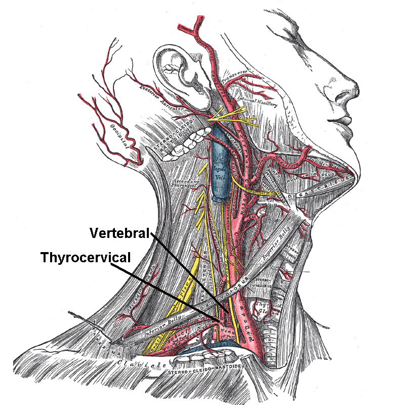
In human anatomy, the subclavian arteries are paired major arteries of the upper thorax, below the clavicle. They receive blood from the aortic arch. The left subclavian artery supplies blood to the left arm and the right subclavian artery supplies blood to the right arm, with some branches supplying the head and thorax. On the left side of the body, the subclavian comes directly off the aortic arch, while on the right side it arises from the relatively short brachiocephalic artery when it bifurcates into the subclavian and the right common carotid artery. The usual branches of the subclavian on both sides of the body are the vertebral artery, the internal thoracic artery, the thyrocervical trunk, the costocervical trunk and the dorsal scapular artery, which may branch off the transverse cervical artery, which is a branch of the thyrocervical trunk. The subclavian becomes the axillary artery at the lateral border of the first rib. Structure From its origin, the subclavian art ... [...More Info...] [...Related Items...] OR: [Wikipedia] [Google] [Baidu] [Amazon] |
Internal Thoracic Artery
The internal thoracic artery (ITA), also known as the internal mammary artery, is an artery that supplies the anterior chest wall and the breasts. It is a paired artery, with one running along each side of the sternum, to continue after its bifurcation as the superior epigastric and musculophrenic arteries. Structure The internal thoracic artery arises from the anterior surface of the subclavian artery near its origin. It has a width of between 1-2 mm. It travels downward on the inside of the rib cage, approximately 1 cm from the sides of the sternum, and thus medial to the nipple. It is accompanied by the internal thoracic vein. It runs deep to the abdominal external oblique muscle, but superficial to the vagus nerve. In adults, the internal thoracic artery lies closest to the sternum at the first intercostal space. The gap between the artery and lateral border of the sternum increases when going downwards, up to 1.1 cm to 1.3 cm at the sixth intercost ... [...More Info...] [...Related Items...] OR: [Wikipedia] [Google] [Baidu] [Amazon] |
Vertebrae
Each vertebra (: vertebrae) is an irregular bone with a complex structure composed of bone and some hyaline cartilage, that make up the vertebral column or spine, of vertebrates. The proportions of the vertebrae differ according to their spinal segment and the particular species. The basic configuration of a vertebra varies; the vertebral body (also ''centrum'') is of bone and bears the load of the vertebral column. The upper and lower surfaces of the vertebra body give attachment to the intervertebral discs. The posterior part of a vertebra forms a vertebral arch, in eleven parts, consisting of two pedicles (pedicle of vertebral arch), two laminae, and seven process (anatomy), processes. The laminae give attachment to the ligamenta flava (ligaments of the spine). There are vertebral notches formed from the shape of the pedicles, which form the intervertebral foramina when the vertebrae articulation (anatomy), articulate. These foramina are the entry and exit conduits for the spi ... [...More Info...] [...Related Items...] OR: [Wikipedia] [Google] [Baidu] [Amazon] |
Esophagus
The esophagus (American English), oesophagus (British English), or œsophagus (Œ, archaic spelling) (American and British English spelling differences#ae and oe, see spelling difference) all ; : ((o)e)(œ)sophagi or ((o)e)(œ)sophaguses), colloquially known also as the food pipe, food tube, or gullet, is an Organ (anatomy), organ in vertebrates through which food passes, aided by Peristalsis, peristaltic contractions, from the Human pharynx, pharynx to the stomach. The esophagus is a :wiktionary:fibromuscular, fibromuscular tube, about long in adults, that travels behind the trachea and human heart, heart, passes through the Thoracic diaphragm, diaphragm, and empties into the uppermost region of the stomach. During swallowing, the epiglottis tilts backwards to prevent food from going down the larynx and lungs. The word ''esophagus'' is from Ancient Greek οἰσοφάγος (oisophágos), from οἴσω (oísō), future form of φέρω (phérō, "I carry") + ἔφαγον ( ... [...More Info...] [...Related Items...] OR: [Wikipedia] [Google] [Baidu] [Amazon] |
Thoracic Duct
In human anatomy, the thoracic duct (also known as the ''left lymphatic duct'', ''alimentary duct'', ''chyliferous duct'', and ''Van Hoorne's canal'') is the larger of the two lymph ducts of the lymphatic system (the other being the right lymphatic duct). The thoracic duct usually begins from the upper aspect of the cisterna chyli, passing out of the abdomen through the aortic hiatus into first the posterior mediastinum and then the superior mediastinum, extending as high up as the root of the neck before descending to drain into the systemic (blood) circulation at the venous angle. The thoracic duct carries chyle, a liquid containing both lymph and emulsified fats, rather than pure lymph. It also collects most of the lymph in the body other than from the right thorax, arm, head, and neck (which are drained by the right lymphatic duct). When the duct ruptures, the resulting flood of liquid into the pleural cavity is known as chylothorax. Structure In adults, the thoraci ... [...More Info...] [...Related Items...] OR: [Wikipedia] [Google] [Baidu] [Amazon] |
Azygos Vein
The azygos vein (from Ancient Greek ἄζυγος (ázugos), meaning 'unwedded' or 'unpaired') is a vein running up the right side of the thoracic vertebral column draining itself towards the superior vena cava. It connects the systems of superior vena cava and inferior vena cava and can provide an alternative path for blood to the right atrium when either of the venae cavae is blocked. Structure The azygos vein transports deoxygenated blood from the posterior walls of the thorax and abdomen into the superior vena cava. It is formed by the union of the ascending lumbar veins with the right subcostal veins at the level of the 12th thoracic vertebra, ascending to the right of the descending aorta and thoracic duct, passing behind the right crus of diaphragm, anterior to the vertebral bodies of T12 to T5 and right posterior intercostal arteries. At the level of T4 vertebrae, it arches over the root of the right lung from behind to the front to join the superior vena cava. The tra ... [...More Info...] [...Related Items...] OR: [Wikipedia] [Google] [Baidu] [Amazon] |
Lung
The lungs are the primary Organ (biology), organs of the respiratory system in many animals, including humans. In mammals and most other tetrapods, two lungs are located near the Vertebral column, backbone on either side of the heart. Their function in the respiratory system is to extract oxygen from the atmosphere and transfer it into the bloodstream, and to release carbon dioxide from the bloodstream into the atmosphere, in a process of gas exchange. Respiration is driven by different muscular systems in different species. Mammals, reptiles and birds use their musculoskeletal systems to support and foster breathing. In early tetrapods, air was driven into the lungs by the pharyngeal muscles via buccal pumping, a mechanism still seen in amphibians. In humans, the primary muscle that drives breathing is the Thoracic diaphragm, diaphragm. The lungs also provide airflow that makes Animal communication#Auditory, vocalisation including speech possible. Humans have two lungs, a ri ... [...More Info...] [...Related Items...] OR: [Wikipedia] [Google] [Baidu] [Amazon] |
Superior Intercostal Vein
The superior intercostal veins are two veins that drain the 2nd, 3rd, and 4th intercostal spaces, one vein for each side of the body. Right superior intercostal vein The right superior intercostal vein drains the 2nd, 3rd, and 4th posterior intercostal veins on the right side of the body. It flows into the azygos vein. Left superior intercostal vein The left superior intercostal vein drains the 2nd and 3rd posterior intercostal veins on the left side of the body. It usually drains into the left brachiocephalic vein. It may also communicate with the accessory hemiazygos vein. As it passes posteriorly above the aortic arch, it crosses deep to the phrenic nerve and the pericardiacophrenic vessels and then superficial to the vagus nerve. See also * Supreme intercostal vein The supreme intercostal vein (highest intercostal vein) is a paired vein that drains the first intercostal space on its corresponding side. It usually drains into the brachiocephalic vein. Alternatively, it ... [...More Info...] [...Related Items...] OR: [Wikipedia] [Google] [Baidu] [Amazon] |
Posterior Intercostal Vein
The posterior intercostal veins are veins that drain the intercostal spaces posteriorly. They run with their corresponding posterior intercostal artery on the underside of the rib, the vein superior to the artery. Each vein also gives off a dorsal branch that drains blood from the muscles of the back. There are eleven posterior intercostal veins on each side. Their patterns are variable, but they are commonly arranged as: * The 1st posterior intercostal vein, supreme intercostal vein, drains into the brachiocephalic vein or the vertebral vein. * The 2nd and 3rd (and often 4th) posterior intercostal veins drain into the superior intercostal vein. * The remaining posterior intercostal veins drain into the azygos vein on the right, or the hemiazygos and accessory hemiazygos vein The accessory hemiazygos vein, also called the superior hemiazygous vein, is a vein on the left side of the vertebral column that generally drains the fourth through eighth intercostal spaces on the left ... [...More Info...] [...Related Items...] OR: [Wikipedia] [Google] [Baidu] [Amazon] |


