|
Mouth Ulcers
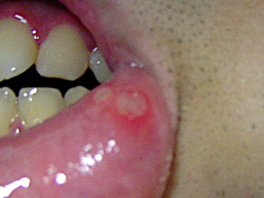
A mouth ulcer (aphtha), or sometimes called a canker sore or salt blister, is an ulcer that occurs on the mucous membrane of the oral cavity. Mouth ulcers are very common, occurring in association with many diseases and by many different mechanisms, but usually there is no serious underlying cause. Rarely, a mouth ulcer that does not heal may be a sign of oral cancer. These ulcers may form individually or multiple ulcers may appear at once (i.e., a "crop" of ulcers). Once formed, an ulcer may be maintained by inflammation and/or secondary infection. The two most common causes of oral ulceration are local trauma (e.g. rubbing from a sharp edge on a broken filling or braces, biting one's lip, etc.) and aphthous stomatitis ("canker sores"), a condition characterized by the recurrent formation of oral ulcers for largely unknown reasons. Mouth ulcers often cause pain and discomfort and may alter the person's choice of food while healing occurs (e.g. avoiding acidic, sugary, salty or sp ... [...More Info...] [...Related Items...] OR: [Wikipedia] [Google] [Baidu] |
Aphthous Stomatitis
Aphthous stomatitis, or recurrent aphthous stomatitis (RAS), commonly referred to as a canker sore or salt blister, is a common condition characterized by the repeated formation of benignity, benign and non-contagious disease, contagious mouth ulcers (aphthae) in otherwise healthy individuals. The cause is not completely understood but involves a T cell-mediated immune response triggered by a variety of factors which may include nutritional deficiencies, local Physical trauma, trauma, Stress (biology), stress, hormonal influences, allergies, genetic predisposition, certain foods, dehydration, some food additives, or some hygienic chemical additives like Sodium dodecyl sulfate#Mouth ulceration, SDS (common in toothpaste). These ulcers occur periodically and heal completely between attacks. In the majority of cases, the individual ulcers last about 7–10 days, and ulceration episodes occur 3–6 times per year. Most appear on the Stratified squamous epithelium#Non-keratinized, n ... [...More Info...] [...Related Items...] OR: [Wikipedia] [Google] [Baidu] |
Capillary
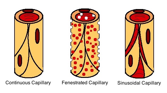
A capillary is a small blood vessel, from 5 to 10 micrometres in diameter, and is part of the microcirculation system. Capillaries are microvessels and the smallest blood vessels in the body. They are composed of only the tunica intima (the innermost layer of an artery or vein), consisting of a thin wall of simple squamous endothelial cells. They are the site of the exchange of many substances from the surrounding interstitial fluid, and they convey blood from the smallest branches of the arteries (arterioles) to those of the veins (venules). Other substances which cross capillaries include water, oxygen, carbon dioxide, urea, glucose, uric acid, lactic acid and creatinine. Lymph capillaries connect with larger lymph vessels to drain lymphatic fluid collected in microcirculation. Etymology ''Capillary'' comes from the Latin word , meaning "of or resembling hair", with use in English beginning in the mid-17th century. The meaning stems from the tiny, hairlike diameter of a capi ... [...More Info...] [...Related Items...] OR: [Wikipedia] [Google] [Baidu] |
Rete Pegs
Rete pegs (also known as rete processes or rete ridges) are the epithelial extensions that project into the underlying connective tissue in both skin and mucous membranes. In the epithelium of the mouth, the attached gingiva exhibit rete pegs, while the sulcularItoiz, ME; Carranza, FA: The Gingiva. In Newman, MG; Takei, HH; Carranza, FA; editors: ''Carranza’s Clinical Periodontology'', 9th Edition. Philadelphia: W.B. Saunders Company, 2002. pages 23. and junctional epithelia do not.Page, RC; Schroeder, HE. "Pathogenesis of Inflammatory Periodontal Disease: A Summary of Current Work." ''Lab Invest'' 1976;34(3):235-249 Scar tissue lacks rete pegs and scars tend to shear off more easily than normal tissue as a result. Also known as ''papillae'', they are downward thickenings of the epidermis The epidermis is the outermost of the three layers that comprise the skin, the inner layers being the dermis and Subcutaneous tissue, hypodermis. The epidermal layer provides a barrie ... [...More Info...] [...Related Items...] OR: [Wikipedia] [Google] [Baidu] |
Excoriation
A skin condition, also known as cutaneous condition, is any medical condition that affects the integumentary system—the organ system that encloses the body and includes skin, nails, and related muscle and glands. The major function of this system is as a barrier against the external environment. Conditions of the human integumentary system constitute a broad spectrum of diseases, also known as dermatoses, as well as many nonpathologic states (like, in certain circumstances, melanonychia and racquet nails). While only a small number of skin diseases account for most visits to the physician, thousands of skin conditions have been described. Classification of these conditions often presents many nosological challenges, since underlying causes and pathogenetics are often not known. Therefore, most current textbooks present a classification based on location (for example, conditions of the mucous membrane), morphology ( chronic blistering conditions), cause ( skin conditions res ... [...More Info...] [...Related Items...] OR: [Wikipedia] [Google] [Baidu] |
Basement Membrane
The basement membrane, also known as base membrane, is a thin, pliable sheet-like type of extracellular matrix that provides cell and tissue support and acts as a platform for complex signalling. The basement membrane sits between epithelial tissues including mesothelium and endothelium, and the underlying connective tissue. Structure As seen with the electron microscope, the basement membrane is composed of two layers, the basal lamina and the reticular lamina. The underlying connective tissue attaches to the basal lamina with collagen VII anchoring fibrils and fibrillin microfibrils. The basal lamina layer can further be subdivided into two layers based on their visual appearance in electron microscopy. The lighter-colored layer closer to the epithelium is called the lamina lucida, while the denser-colored layer closer to the connective tissue is called the lamina densa. The electron-dense lamina densa layer is about 30–70 nanometers thick and consists of an und ... [...More Info...] [...Related Items...] OR: [Wikipedia] [Google] [Baidu] |
Epidermis (skin)
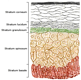
The epidermis is the outermost of the three layers that comprise the skin, the inner layers being the dermis and hypodermis. The epidermal layer provides a barrier to infection from environmental pathogens and regulates the amount of water released from the body into the atmosphere through transepidermal water loss. The epidermis is composed of multiple layers of flattened cells that overlie a base layer ( stratum basale) composed of columnar cells arranged perpendicularly. The layers of cells develop from stem cells in the basal layer. The thickness of the epidermis varies from 31.2μm for the penis to 596.6μm for the sole of the foot with most being roughly 90μm. Thickness does not vary between the sexes but becomes thinner with age. The human epidermis is an example of epithelium, particularly a stratified squamous epithelium. The word epidermis is derived through Latin , itself and . Something related to or part of the epidermis is termed epidermal. Structure ... [...More Info...] [...Related Items...] OR: [Wikipedia] [Google] [Baidu] |
Cutaneous Condition
A skin condition, also known as cutaneous condition, is any medical condition that affects the integumentary system—the organ system that encloses the body and includes skin, nails, and related muscle and glands. The major function of this system is as a barrier against the external environment. Conditions of the human integumentary system constitute a broad spectrum of diseases, also known as dermatoses, as well as many nonpathologic states (like, in certain circumstances, melanonychia and racquet nails). While only a small number of skin diseases account for most visits to the physician, thousands of skin conditions have been described. Classification of these conditions often presents many nosological challenges, since underlying causes and pathogenetics are often not known. Therefore, most current textbooks present a classification based on location (for example, conditions of the mucous membrane), morphology ( chronic blistering conditions), cause ( skin conditions res ... [...More Info...] [...Related Items...] OR: [Wikipedia] [Google] [Baidu] |
Lamina Propria
The lamina propria is a thin layer of connective tissue that forms part of the moist linings known as mucous membranes or mucosae, which line various tubes in the body, such as the respiratory tract, the gastrointestinal tract, and the urogenital tract. The lamina propria is a thin layer of loose (areolar) connective tissue, which lies beneath the epithelium, and together with the epithelium and basement membrane constitutes the mucosa. As its Latin name indicates, it is a characteristic component of the mucosa, or the mucosa's "own special layer." Thus, the term mucosa or mucous membrane refers to the combination of the epithelium and the lamina propria. The connective tissue of the lamina propria is loose and rich in cells. The cells of the lamina propria are variable and can include fibroblasts, lymphocytes, plasma cells, macrophages, eosinophilic leukocytes, and mast cells. It provides support and nutrition to the epithelium, as well as the means to bind to the underlying t ... [...More Info...] [...Related Items...] OR: [Wikipedia] [Google] [Baidu] |
Periosteum
The periosteum is a membrane that covers the outer surface of all bones, except at the articular surfaces (i.e. the parts within a joint space) of long bones. (At the joints of long bones the bone's outer surface is lined with "articular cartilage", a type of hyaline cartilage.) Endosteum lines the inner surface of the medullary cavity of all long bones. Structure The periosteum consists of an outer fibrous layer, and an inner ''cambium layer'' (or osteogenic layer). The fibrous layer is of dense irregular connective tissue, containing fibroblasts, while the cambium layer is highly cellular containing progenitor cells that develop into osteoblasts. These osteoblasts are responsible for increasing the width of a long bone (the length of a long bone is controlled by the epiphyseal plate) and the overall size of the other bone types. After a bone fracture A bone fracture (abbreviated FRX or Fx, Fx, or #) is a medical condition in which there is a partial or complete break ... [...More Info...] [...Related Items...] OR: [Wikipedia] [Google] [Baidu] |
Submucosa
The submucosa (or tela submucosa) is a thin layer of tissue in various organs of the gastrointestinal, respiratory, and genitourinary tracts. It is the layer of dense irregular connective tissue that supports the mucosa (mucous membrane) and joins it to the muscular layer, the bulk of overlying smooth muscle (fibers running circularly within layer of longitudinal muscle). The submucosa ('' sub-'' + ''mucosa'') is to a mucous membrane what the subserosa ('' sub-'' + ''serosa'') is to a serous membrane. Structure Blood vessels, lymphatic vessels, and nerves (all supplying the mucosa) will run through here. In the intestinal wall, tiny parasympathetic ganglia are scattered around forming the submucous plexus (or "Meissner's plexus") where preganglionic parasympathetic neurons synapse with postganglionic nerve fibers that supply the muscularis mucosae. Histologically, the wall of the alimentary canal shows four distinct layers (from the lumen moving out): mucosa, submuc ... [...More Info...] [...Related Items...] OR: [Wikipedia] [Google] [Baidu] |
Connective Tissue
Connective tissue is one of the four primary types of animal tissue, a group of cells that are similar in structure, along with epithelial tissue, muscle tissue, and nervous tissue. It develops mostly from the mesenchyme, derived from the mesoderm, the middle embryonic germ layer. Connective tissue is found in between other tissues everywhere in the body, including the nervous system. The three meninges, membranes that envelop the brain and spinal cord, are composed of connective tissue. Most types of connective tissue consists of three main components: elastic and collagen fibers, ground substance, and cells. Blood and lymph are classed as specialized fluid connective tissues that do not contain fiber. All are immersed in the body water. The cells of connective tissue include fibroblasts, adipocytes, macrophages, mast cells and leukocytes. The term "connective tissue" (in German, ) was introduced in 1830 by Johannes Peter Müller. The tissue was already recognized as ... [...More Info...] [...Related Items...] OR: [Wikipedia] [Google] [Baidu] |


