|
Monoderm
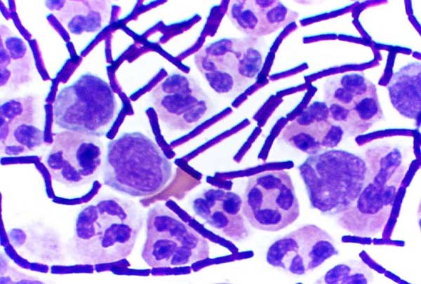
In bacteriology, gram-positive bacteria are bacteria that give a positive result in the Gram stain test, which is traditionally used to quickly classify bacteria into two broad categories according to their type of cell wall. Gram-positive bacteria take up the crystal violet stain used in the test, and then appear to be purple-coloured when seen through an optical microscope. This is because the thick peptidoglycan layer in the bacterial cell wall retains the stain after it is washed away from the rest of the sample, in the decolorization stage of the test. Conversely, gram-negative bacteria cannot retain the violet stain after the decolorization step; alcohol used in this stage degrades the outer membrane of gram-negative cells, making the cell wall more porous and incapable of retaining the crystal violet stain. Their peptidoglycan layer is much thinner and sandwiched between an inner cell membrane and a bacterial outer membrane, causing them to take up the counterstain (safran ... [...More Info...] [...Related Items...] OR: [Wikipedia] [Google] [Baidu] |
Gram-positive Cellwall-schematic
In bacteriology, gram-positive bacteria are bacteria that give a positive result in the Gram stain test, which is traditionally used to quickly classify bacteria into two broad categories according to their type of cell wall. Gram-positive bacteria take up the crystal violet stain used in the test, and then appear to be purple-coloured when seen through an optical microscope. This is because the thick peptidoglycan layer in the bacterial cell wall retains the stain after it is washed away from the rest of the sample, in the decolorization stage of the test. Conversely, gram-negative bacteria cannot retain the violet stain after the decolorization step; alcohol used in this stage degrades the outer membrane of gram-negative cells, making the cell wall more porous and incapable of retaining the crystal violet stain. Their peptidoglycan layer is much thinner and sandwiched between an inner cell membrane and a bacterial outer membrane, causing them to take up the counterstai ... [...More Info...] [...Related Items...] OR: [Wikipedia] [Google] [Baidu] |
Gram-negative Bacteria
Gram-negative bacteria are bacteria that do not retain the crystal violet stain used in the Gram staining method of bacterial differentiation. They are characterized by their cell envelopes, which are composed of a thin peptidoglycan cell wall sandwiched between an inner cytoplasmic cell membrane and a bacterial outer membrane. Gram-negative bacteria are found in virtually all environments on Earth that support life. The gram-negative bacteria include the model organism '' Escherichia coli'', as well as many pathogenic bacteria, such as '' Pseudomonas aeruginosa'', '' Chlamydia trachomatis'', and '' Yersinia pestis''. They are a significant medical challenge as their outer membrane protects them from many antibiotics (including penicillin), detergents that would normally damage the inner cell membrane, and lysozyme, an antimicrobial enzyme produced by animals that forms part of the innate immune system. Additionally, the outer leaflet of this membrane comprises a compl ... [...More Info...] [...Related Items...] OR: [Wikipedia] [Google] [Baidu] |
Periplasm
The periplasm is a concentrated gel-like matrix in the space between the inner cytoplasmic membrane and the bacterial outer membrane called the ''periplasmic space'' in gram-negative bacteria. Using cryo-electron microscopy it has been found that a much smaller periplasmic space is also present in gram-positive bacteria., Matias, V. R., and T. J. Beveridge. 2005. Cryo-electron microscopy reveals native polymeric cell wall structure in Bacillus subtilis 168 and the existence of a periplasmic space. Mol. Microbiol. 56:240-251. ., Zuber B, Haenni M, Ribeiro T, Minnig K, Lopes F, Moreillon P, Dubochet J. 2006. Granular layer in the periplasmic space of Gram-positive bacteria and fine structures of Enterococcus gallinarum and Streptococcus gordonii septa revealed by cryo-electron microscopy of vitreous sections. J Bacteriol. 188:6652-6660. The periplasm may constitute up to 40% of the total cell volume of gram-negative bacteria, but is a much smaller percentage in gram-positive bact ... [...More Info...] [...Related Items...] OR: [Wikipedia] [Google] [Baidu] |
Gram Stain
In microbiology and bacteriology, Gram stain (Gram staining or Gram's method), is a method of staining used to classify bacterial species into two large groups: gram-positive bacteria and gram-negative bacteria. The name comes from the Danish bacteriologist Hans Christian Gram, who developed the technique in 1884. Gram staining differentiates bacteria by the chemical and physical properties of their cell walls. Gram-positive cells have a thick layer of peptidoglycan in the cell wall that retains the primary stain, crystal violet. Gram-negative cells have a thinner peptidoglycan layer that allows the crystal violet to wash out on addition of ethanol. They are stained pink or red by the counterstain, commonly safranin or fuchsine. Lugol's iodine solution is always added after addition of crystal violet to strengthen the bonds of the stain with the cell membrane. Gram staining is almost always the first step in the preliminary identification of a bacterial organism. While Gra ... [...More Info...] [...Related Items...] OR: [Wikipedia] [Google] [Baidu] |
Gram Stain Anthrax
The gram (originally gramme; SI unit symbol g) is a unit of mass in the International System of Units (SI) equal to one one thousandth of a kilogram. Originally defined as of 1795 as "the absolute weight of a volume of pure water equal to the cube of the hundredth part of a metre the_temperature_of_Melting_point.html" "title="Melting_point_of_water.html" ;"title="Cubic_centimetre.html" ;"title=" Cubic centimetre">cm3], and at Melting point of water">the temperature of Melting point">melting ice", the defining temperature (~0 °C) was later changed to 4 °C, the temperature of maximum density of water. However, by the late 19th century, there was an effort to make the Base unit (measurement), base unit the kilogram and the gram a derived unit. In 1960, the new International System of Units defined a ''gram'' as one one-thousandth of a kilogram (i.e., one gram is Scientific notation, 1×10⁻³ kg). The kilogram, as of 2019, is defined by the International Bur ... [...More Info...] [...Related Items...] OR: [Wikipedia] [Google] [Baidu] |
S-layer
An S-layer (surface layer) is a part of the cell envelope found in almost all archaea, as well as in many types of bacteria. The S-layers of both archaea and bacteria consists of a monomolecular layer composed of only one (or, in a few cases, two) identical proteins or glycoproteins. This structure is built via self-assembly and encloses the whole cell surface. Thus, the S-layer protein can represent up to 15% of the whole protein content of a cell. S-layer proteins are poorly conserved or not conserved at all, and can differ markedly even between related species. Depending on species, the S-layers have a thickness between 5 and 25 nm and possess identical pores with 2–8 nm in diameter. The terminology “S-layer” was used the first time in 1976. The general use was accepted at the "First International Workshop on Crystalline Bacterial Cell Surface Layers, Vienna (Austria)" in 1984, and in the year 1987 S-layers were defined at the European Molecular Biology Organiz ... [...More Info...] [...Related Items...] OR: [Wikipedia] [Google] [Baidu] |
Basal Body
A basal body (synonymous with basal granule, kinetosome, and in older cytological literature with blepharoplast) is a protein structure found at the base of a eukaryotic undulipodium ( cilium or flagellum). The basal body was named by Theodor Wilhelm Engelmann in 1880 It is formed from a centriole and several additional protein structures, and is, essentially, a modified centriole. The basal body serves as a nucleation site for the growth of the axoneme microtubules. Centrioles, from which basal bodies are derived, act as anchoring sites for proteins that in turn anchor microtubules, and are known as the microtubule organizing center (MTOC). These microtubules provide structure and facilitate movement of vesicles and organelles within many eukaryotic cells. Assembly, structure Cilia and basal bodies form during quiescence or the G1 phase of the cell cycle. Before the cell enters G1 phase, i.e. before the formation of the cilium, the mother centriole serves as a compone ... [...More Info...] [...Related Items...] OR: [Wikipedia] [Google] [Baidu] |
Flagellum
A flagellum (; ) is a hairlike appendage that protrudes from certain plant and animal sperm cells, and from a wide range of microorganisms to provide motility. Many protists with flagella are termed as flagellates. A microorganism may have from one to many flagella. A gram-negative bacterium ''Helicobacter pylori'' for example uses its multiple flagella to propel itself through the mucus lining to reach the stomach epithelium, where it may cause a gastric ulcer to develop. In some bacteria the flagellum can also function as a sensory organelle, being sensitive to wetness outside the cell. Across the three domains of Bacteria, Archaea, and Eukaryota the flagellum has a different structure, protein composition, and mechanism of propulsion but shares the same function of providing motility. The Latin word means "whip" to describe its lash-like swimming motion. The flagellum in archaea is called the archaellum to note its difference from the bacterial flagellum. Eukary ... [...More Info...] [...Related Items...] OR: [Wikipedia] [Google] [Baidu] |
Flagellate
A flagellate is a cell or organism with one or more whip-like appendages called flagella. The word ''flagellate'' also describes a particular construction (or level of organization) characteristic of many prokaryotes and eukaryotes and their means of motion. The term presently does not imply any specific relationship or classification of the organisms that possess flagella. However, the term "flagellate" is included in other terms (such as " dinoflagellate" and " choanoflagellata") which are more formally characterized. Form and behavior Flagella in eukaryotes are supported by microtubules in a characteristic arrangement, with nine fused pairs surrounding two central singlets. These arise from a basal body. In some flagellates, flagella direct food into a cytostome or mouth, where food is ingested. Flagella often support hairs, called mastigonemes, or contain rods. Their ultrastructure plays an important role in classifying eukaryotes. Among protoctists and micro ... [...More Info...] [...Related Items...] OR: [Wikipedia] [Google] [Baidu] |
Polysaccharides
Polysaccharides (), or polycarbohydrates, are the most abundant carbohydrates found in food. They are long chain polymeric carbohydrates composed of monosaccharide units bound together by glycosidic linkages. This carbohydrate can react with water (hydrolysis) using amylase enzymes as catalyst, which produces constituent sugars ( monosaccharides, or oligosaccharides). They range in structure from linear to highly branched. Examples include storage polysaccharides such as starch, glycogen and galactogen and structural polysaccharides such as cellulose and chitin. Polysaccharides are often quite heterogeneous, containing slight modifications of the repeating unit. Depending on the structure, these macromolecules can have distinct properties from their monosaccharide building blocks. They may be amorphous or even insoluble in water. When all the monosaccharides in a polysaccharide are the same type, the polysaccharide is called a homopolysaccharide or homoglycan, but when more ... [...More Info...] [...Related Items...] OR: [Wikipedia] [Google] [Baidu] |
Bacterial Capsule
The bacteria capsule is a large structure common to many bacteria. It is a polysaccharide layer that lies outside the cell envelope, and is thus deemed part of the outer envelope of a bacterial cell. It is a well-organized layer, not easily washed off, and it can be the cause of various diseases. The capsule—which can be found in both gram negative and gram-positive bacteria—is different from the second lipid membrane – bacterial outer membrane, which contains lipopolysaccharides and lipoproteins and is found only in gram-negative bacteria. When the amorphous viscid secretion (that makes up the capsule) diffuses into the surrounding medium and remains as a loose undemarcated secretion, it is known as a slime layer. Capsule and slime layer are sometimes summarized under the term glycocalyx. Composition Most bacterial capsules are composed of polysaccharide, but some species use other materials, such as poly-D-glutamic acid in ''Bacillus anthracis''. Because most caps ... [...More Info...] [...Related Items...] OR: [Wikipedia] [Google] [Baidu] |


.jpg)

