|
Mesonephric Tubule
The mesonephros () is one of three excretory organs that develop in vertebrates. It serves as the main excretory organ of aquatic vertebrates and as a temporary kidney in reptiles, birds, and mammals. The mesonephros is included in the Wolffian body after Caspar Friedrich Wolff who described it in 1759. (The Wolffian body is composed of: mesonephros + paramesonephrotic blastema) Structure The mesonephros acts as a structure similar to the kidney that, in humans, functions between the sixth and tenth weeks of embryological life. Despite the similarity in structure, function, and terminology, however, the mesonephric nephrons do not form any part of the mature kidney or nephrons. In humans, the mesonephros consists of units which are similar in structure and function to nephrons of the adult kidney. Each of these consists of a glomerulus, a tuft of capillaries which arises from lateral branches of dorsal aorta and drains into the inferior cardinal vein; a Bowman's capsule, a f ... [...More Info...] [...Related Items...] OR: [Wikipedia] [Google] [Baidu] |
Embryo
An embryo ( ) is the initial stage of development for a multicellular organism. In organisms that reproduce sexually, embryonic development is the part of the life cycle that begins just after fertilization of the female egg cell by the male sperm cell. The resulting fusion of these two cells produces a single-celled zygote that undergoes many cell divisions that produce cells known as blastomeres. The blastomeres (4-cell stage) are arranged as a solid ball that when reaching a certain size, called a morula, (16-cell stage) takes in fluid to create a cavity called a blastocoel. The structure is then termed a blastula, or a blastocyst in mammals. The mammalian blastocyst hatches before implantating into the endometrial lining of the womb. Once implanted the embryo will continue its development through the next stages of gastrulation, neurulation, and organogenesis. Gastrulation is the formation of the three germ layers that will form all of the different parts of t ... [...More Info...] [...Related Items...] OR: [Wikipedia] [Google] [Baidu] |
Epoophoron
The epoophoron or epoöphoron (also called organ of RosenmüllerJ. C. Rosenmüller. De ovariis embryonum et foetuum humanorum. 1802. or the parovarium; : epoophora) is a remnant of the mesonephric duct that can be found next to the ovary and fallopian tube. Anatomy It may contain 10–15 transverse small ducts or tubules that lead to the Gartner's duct (also longitudinal duct of epoophoron) that represents the caudal remnant of the mesonephric ducts and passes through the broad ligament and the lateral wall of the cervix and vagina. The epoophoron is a homologue to the epididymis in the male. While the epoophoron is located in the lateral portion of the mesosalpinx and mesovarium, the paroophoron (residual remnant of that part of the mesonephric duct that forms the paradidymis in the male) lies more medially in the mesosalpinx. Histology It has a unique histological profile. Clinical significance Clinically the organ may give rise to a local paraovarian cyst or adenoma A ... [...More Info...] [...Related Items...] OR: [Wikipedia] [Google] [Baidu] |
Paramesonephric Ducts
The paramesonephric ducts (or Müllerian ducts) are paired ducts of the embryo in the reproductive system of humans and other mammals that run down the lateral sides of the genital ridge and terminate at the sinus tubercle in the primitive urogenital sinus. They form in both sexes during 6th week of fetal development. In the female, go on to form the fallopian tubes/oviducts, uterus, cervix, and the upper one-third of the vagina. In males fetuses, they are normally made to regress by anti-Müllerian hormone which begins to be secreted by the testes during 8th week of fetal development. Each maramesonephric duct is situated just lateral to the mesonephric ducts (Wolffian duct) of the same side. Development The female reproductive system is composed of two embryological segments: the urogenital sinus and the paramesonephric ducts. The two are conjoined at the sinus tubercle. Paramesonephric ducts are present on the embryo of both sexes. Only in females do they develop into repr ... [...More Info...] [...Related Items...] OR: [Wikipedia] [Google] [Baidu] |
Pronephros
Pronephros is the most basic of the three excretory organs that develop in vertebrates, corresponding to the first stage of kidney development. It is succeeded by the mesonephros, which in fish and amphibians remains as the adult kidney. In amniotes, the mesonephros is the embryonic kidney and a more complex metanephros acts as the adult kidney. Once a more advanced kidney forms, the previous version typically degenerates by apoptosis or becomes part of the male reproductive system. The pronephros develops from the intermediate mesoderm, as do the later kidneys. It is a paired organ, consisting of a single giant nephron that processes blood filtrate produced from glomeruli or glomera- large embryonic glomeruli. The filtrate is deposited into the coelom. It then passes through thin ciliated tubules into the pronephric nephron where it is processed for solute recovery. The organ is active in adult forms of some primitive fish, like lampreys or hagfish. It is present at the embry ... [...More Info...] [...Related Items...] OR: [Wikipedia] [Google] [Baidu] |
Metanephros
Kidney development, or nephrogenesis, describes the embryologic origins of the kidney, a major organ in the urinary system. This article covers a 3 part developmental process that is observed in most reptiles, birds and mammals, including humans. Nephrogenesis is often considered in the broader context of the development of the urinary and reproductive organs. Phases The development of the kidney proceeds through a series of successive phases, each marked by the development of a more advanced kidney: the archinephros, pronephros, mesonephros, and metanephros. The pronephros is the most immature form of kidney, while the metanephros is most developed. The metanephros persists as the definitive adult kidney. Archinephros The archinephros is considered as hypothetical or primitive kidney. Pronephros The pronephros develops in the cervical region of the embryo. During approximately day 22 of human gestation, the paired pronephri appears towards the cranial end of the intermediat ... [...More Info...] [...Related Items...] OR: [Wikipedia] [Google] [Baidu] |
Paradidymis
The term paradidymis (: paradidymides; organ of Giraldés) is applied to a small collection of convoluted tubules, situated in front of the lower part of the spermatic cord, above the head of the epididymis. These tubes are lined with columnar ciliated epithelium, and probably represent the remains of a part of the Wolffian body, like the epididymis, but are functionless and vestigial. The Wolffian body operates as a kidney (mesonephros The mesonephros () is one of three excretory system, excretory organs that develop in vertebrates. It serves as the main excretory organ of aquatic vertebrates and as a temporary kidney in reptiles, birds, and mammals. The mesonephros is included ...) in fishes and amphibians, but the corresponding tissue is co-opted to form parts of the male reproductive system in other classes of vertebrate. The paradidymis represents a remnant of an unused, atrophied part of the Wolffian body. The paradidymis is homologous to the female paroophoron, as th ... [...More Info...] [...Related Items...] OR: [Wikipedia] [Google] [Baidu] |
Appendix Epididymis
The appendix of the epididymis (or pedunculated hydatid) is a small stalked appendage (sometimes duplicated) on the head of the epididymis. It is usually regarded as a detached efferent duct. This structure is derived from the Wolffian duct (mesonephric duct) as opposed to the appendix testis which is derived from the Müllerian duct (paramesonephric duct) remnant. See also * Appendix testis The appendix testis (or hydatid of Morgagni) is a vestigial remnant of the Müllerian duct, present on the upper pole of the testis and attached to the tunica vaginalis. It is present about 90% of the time. Clinical significance Torsion The a ... References External links * - Torsion of Appendix Epididymis * * () Mammal male reproductive system {{Portal bar, Anatomy ... [...More Info...] [...Related Items...] OR: [Wikipedia] [Google] [Baidu] |
Appendix Testis
The appendix testis (or hydatid of Morgagni) is a vestigial remnant of the Müllerian duct, present on the upper pole of the testis and attached to the tunica vaginalis. It is present about 90% of the time. Clinical significance Torsion The appendix of testis can occasionally become twisted, causing acute one-sided testicular pain and may require surgical excision to achieve relief. One third of patients present with a palpable "blue dot" discoloration on the scrotum. This is nearly diagnostic of this condition. If clinical suspicion is high for the serious differential diagnosis In healthcare, a differential diagnosis (DDx) is a method of analysis that distinguishes a particular disease or condition from others that present with similar clinical features. Differential diagnostic procedures are used by clinicians to di ... of testicular torsion, a surgical exploration of the scrotum is warranted. Torsion of the appendix of testis occurs at ages 0–15 years, with a mean ... [...More Info...] [...Related Items...] OR: [Wikipedia] [Google] [Baidu] |
Seminal Vesicle
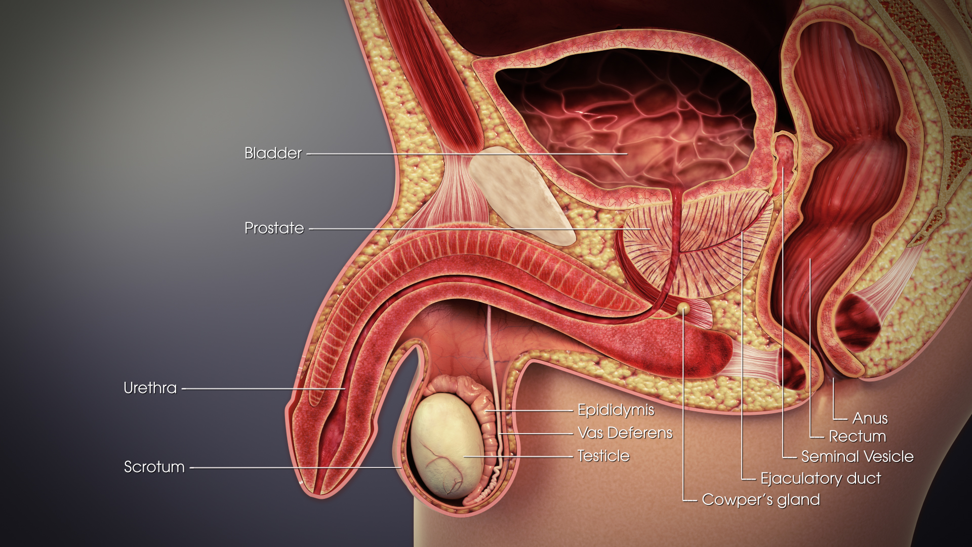
The seminal vesicles (also called vesicular glands or seminal glands) are a pair of convoluted tubular accessory glands that lie behind the urinary bladder of male mammals. They secrete fluid that largely composes the semen. The vesicles are 5–10 cm in size, 3–5 cm in diameter, and are located between the bladder and the rectum. They have multiple outpouchings, which contain secretory glands, which join together with the vasa deferentia at the ejaculatory ducts. They receive blood from the vesiculodeferential artery, and drain into the vesiculodeferential veins. The glands are lined with column-shaped and cuboidal cells. The vesicles are present in many groups of mammals, but not marsupials, monotremes or carnivores. Inflammation of the seminal vesicles is called seminal vesiculitis and most often is due to bacterial infection as a result of a sexually transmitted infection or following a surgical procedure. Seminal vesiculitis can cause pain in the lower abdo ... [...More Info...] [...Related Items...] OR: [Wikipedia] [Google] [Baidu] |
Vas Deferens
The vas deferens (: vasa deferentia), ductus deferens (: ductūs deferentes), or sperm duct is part of the male reproductive system of many vertebrates. In mammals, spermatozoa are produced in the seminiferous tubules and flow into the epididymal duct. The end of the epididymis is connected to the vas deferens. The vas deferens ends with an opening into the ejaculatory duct at a point where the duct of the seminal vesicle also joins the ejaculatory duct. The vas deferens is a partially coiled tube which exits the abdominal cavity through the inguinal canal. Etymology ''Vas deferens'' is Latin, meaning "carrying-away vessel" while ''ductus deferens'', also Latin, means "carrying-away duct". Structure The human vas deferens measures 30–35 cm in length, and 2–3 mm in diameter. It is continuous proximally with the tail of the epididymis, and exhibits a tortuous, convoluted initial/proximal section (which measures 2–3 cm in length). Distally, it forms a dilated ... [...More Info...] [...Related Items...] OR: [Wikipedia] [Google] [Baidu] |
Epididymis
The epididymis (; : epididymides or ) is an elongated tubular genital organ attached to the posterior side of each one of the two male reproductive glands, the testicles. It is a single, narrow, tightly coiled tube in adult humans, in length; uncoiled the tube would be approximately 6 m (20 feet) long. It connects the testicle to the vas deferens in the male reproductive system. The epididymis serves as an interconnection between the multiple efferent ducts at the rear of a testicle (proximally), and the vas deferens (distally). Its primary function is the storage, maturation and transport of sperm cells. Structure The human epididymis is situated posterior and somewhat lateral to the testis. The epididymis is invested completely by the tunica vaginalis (which is continuous with the tunica vaginalis covering the testis). The epididymis can be divided into three main regions: * The head (). The head of the epididymis receives spermatozoa via the efferent ducts of the medi ... [...More Info...] [...Related Items...] OR: [Wikipedia] [Google] [Baidu] |


