|
Mediastinum
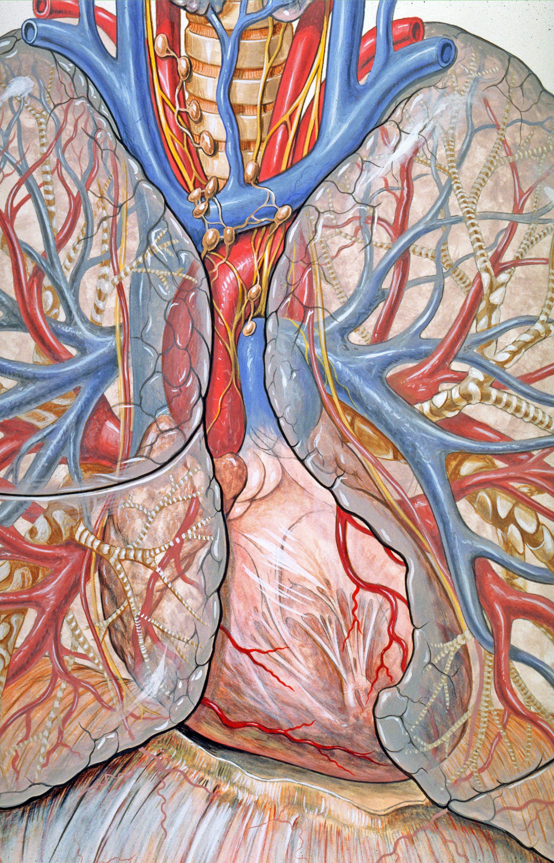
The mediastinum (from ;: mediastina) is the central compartment of the thoracic cavity. Surrounded by loose connective tissue, it is a region that contains vital organs and structures within the thorax, mainly the heart and its vessels, the esophagus, the trachea, the vagus nerve, vagus, phrenic nerve, phrenic and cardiac nerves, the thoracic duct, the thymus and the lymph nodes of the central chest. Anatomy The mediastinum lies within the thorax and is enclosed on the right and left by pulmonary pleurae, pleurae. It is surrounded by the chest wall in front, the lungs to the sides and the Spine (anatomy), spine at the back. It extends from the sternum in front to the vertebral column behind. It contains all the organs of the thorax except the lungs. It is continuous with the loose connective tissue of the neck. The mediastinum can be divided into an upper (or superior) and lower (or inferior) part: * The superior mediastinum starts at the superior thoracic aperture and ends ... [...More Info...] [...Related Items...] OR: [Wikipedia] [Google] [Baidu] |
Thoracic Plane
The mediastinum (from ;: mediastina) is the central compartment of the thoracic cavity. Surrounded by loose connective tissue, it is a region that contains vital organs and structures within the thorax, mainly the heart and its vessels, the esophagus, the trachea, the vagus nerve, vagus, phrenic nerve, phrenic and cardiac nerves, the thoracic duct, the thymus and the lymph nodes of the central chest. Anatomy The mediastinum lies within the thorax and is enclosed on the right and left by pulmonary pleurae, pleurae. It is surrounded by the chest wall in front, the lungs to the sides and the Spine (anatomy), spine at the back. It extends from the sternum in front to the vertebral column behind. It contains all the organs of the thorax except the lungs. It is continuous with the loose connective tissue of the neck. The mediastinum can be divided into an upper (or superior) and lower (or inferior) part: * The superior mediastinum starts at the superior thoracic aperture and end ... [...More Info...] [...Related Items...] OR: [Wikipedia] [Google] [Baidu] |
Thoracic Duct
In human anatomy, the thoracic duct (also known as the ''left lymphatic duct'', ''alimentary duct'', ''chyliferous duct'', and ''Van Hoorne's canal'') is the larger of the two lymph ducts of the lymphatic system (the other being the right lymphatic duct). The thoracic duct usually begins from the upper aspect of the cisterna chyli, passing out of the abdomen through the aortic hiatus into first the posterior mediastinum and then the superior mediastinum, extending as high up as the root of the neck before descending to drain into the systemic (blood) circulation at the venous angle. The thoracic duct carries chyle, a liquid containing both lymph and emulsified fats, rather than pure lymph. It also collects most of the lymph in the body other than from the right thorax, arm, head, and neck (which are drained by the right lymphatic duct). When the duct ruptures, the resulting flood of liquid into the pleural cavity is known as chylothorax. Structure In adults, the thoraci ... [...More Info...] [...Related Items...] OR: [Wikipedia] [Google] [Baidu] |
Pulmonary Pleurae
The pleurae (: pleura) are the two flattened closed sacs filled with pleural fluid, each ensheathing each lung and lining their surrounding tissues, locally appearing as two opposing layers of serous membrane separating the lungs from the mediastinum, the inside surfaces of the surrounding chest walls and the thoracic diaphragm, diaphragm. Although wrapped onto itself resulting in an apparent double layer, each lung is surrounded by a single, continuous pleural membrane. The portion of the pleura that covers the surface of each lung is often called the visceral pleura. This can lead to some confusion, as the lung is not the only visceral organ covered by the pleura. The pleura typically dips between the lobes of the lung as ''fissures'', and is formed by the invagination of lung buds into each coelomic cavity, thoracic sac during embryonic development. The portion of the pleura seen as the outer layer covers the chest wall, the diaphragm and the mediastinum and is often also mis ... [...More Info...] [...Related Items...] OR: [Wikipedia] [Google] [Baidu] |
Esophagus
The esophagus (American English), oesophagus (British English), or œsophagus (Œ, archaic spelling) (American and British English spelling differences#ae and oe, see spelling difference) all ; : ((o)e)(œ)sophagi or ((o)e)(œ)sophaguses), colloquially known also as the food pipe, food tube, or gullet, is an Organ (anatomy), organ in vertebrates through which food passes, aided by Peristalsis, peristaltic contractions, from the Human pharynx, pharynx to the stomach. The esophagus is a :wiktionary:fibromuscular, fibromuscular tube, about long in adults, that travels behind the trachea and human heart, heart, passes through the Thoracic diaphragm, diaphragm, and empties into the uppermost region of the stomach. During swallowing, the epiglottis tilts backwards to prevent food from going down the larynx and lungs. The word ''esophagus'' is from Ancient Greek οἰσοφάγος (oisophágos), from οἴσω (oísō), future form of φέρω (phérō, "I carry") + ἔφαγον ( ... [...More Info...] [...Related Items...] OR: [Wikipedia] [Google] [Baidu] |
Sternal Angle
The sternal angle (also known as the angle of Lewis, angle of Louis, angle of Ludovic, or manubriosternal junction) is the projecting angle formed between the manubrium and body of a sternum at their junction at the manubriosternal joint. The sternal angle is a palpable and visible landmark in surface anatomy, presenting as either a slight body ridge or depression upon the upper chest wall which corresponds to the underlying manubriosternal joint. The sternal angle is palpable and often visible in young people. The sternal angle corresponds to the level of the 2nd costal cartilage on either side, and the level between the fourth and fifth thoracic vertebra. The sternal angle is used to define the Thoracic plane, transverse thoracic plane which represents the imaginary boundary between the superior mediastinum, superior and inferior mediastinum. It is also used to identify the second rib during physical examination and then the rest of the ribs by counting. Anatomy The sternal angl ... [...More Info...] [...Related Items...] OR: [Wikipedia] [Google] [Baidu] |
Heart
The heart is a muscular Organ (biology), organ found in humans and other animals. This organ pumps blood through the blood vessels. The heart and blood vessels together make the circulatory system. The pumped blood carries oxygen and nutrients to the tissue, while carrying metabolic waste such as carbon dioxide to the lungs. In humans, the heart is approximately the size of a closed fist and is located between the lungs, in the middle compartment of the thorax, chest, called the mediastinum. In humans, the heart is divided into four chambers: upper left and right Atrium (heart), atria and lower left and right Ventricle (heart), ventricles. Commonly, the right atrium and ventricle are referred together as the right heart and their left counterparts as the left heart. In a healthy heart, blood flows one way through the heart due to heart valves, which prevent cardiac regurgitation, backflow. The heart is enclosed in a protective sac, the pericardium, which also contains a sma ... [...More Info...] [...Related Items...] OR: [Wikipedia] [Google] [Baidu] |
Thymus
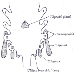
The thymus (: thymuses or thymi) is a specialized primary lymphoid organ of the immune system. Within the thymus, T cells mature. T cells are critical to the adaptive immune system, where the body adapts to specific foreign invaders. The thymus is located in the upper front part of the chest, in the anterior superior mediastinum, behind the sternum, and in front of the heart. It is made up of two lobes, each consisting of a central medulla and an outer cortex, surrounded by a capsule. The thymus is made up of immature T cells called thymocytes, as well as lining cells called epithelial cells which help the thymocytes develop. T cells that successfully develop react appropriately with Major histocompatibility complex, MHC immune receptors of the body (called ''positive selection'') and not against proteins of the body (called ''negative selection''). The thymus is the largest and most active during the neonatal and pre-adolescent periods. By the early teens, the Thymic involuti ... [...More Info...] [...Related Items...] OR: [Wikipedia] [Google] [Baidu] |
Pericardium
The pericardium (: pericardia), also called pericardial sac, is a double-walled sac containing the heart and the roots of the great vessels. It has two layers, an outer layer made of strong inelastic connective tissue (fibrous pericardium), and an inner layer made of serous membrane (serous pericardium). It encloses the pericardial cavity, which contains pericardial fluid, and defines the middle mediastinum. It separates the heart from interference of other structures, protects it against infection and blunt trauma, and lubricates the heart's movements. The English name originates from the Ancient Greek prefix ''peri-'' (περί) 'around' and the suffix ''-cardion'' (κάρδιον) 'heart'. Anatomy The pericardium is a tough fibroelastic sac which covers the heart from all sides except at the cardiac root (where the great vessels join the heart) and the bottom (where only the serous pericardium exists to cover the upper surface of the central tendon of diaphragm). ... [...More Info...] [...Related Items...] OR: [Wikipedia] [Google] [Baidu] |
Pericardial Cavity
The pericardium (: pericardia), also called pericardial sac, is a double-walled sac containing the heart and the roots of the great vessels. It has two layers, an outer layer made of strong inelastic connective tissue (fibrous pericardium), and an inner layer made of serous membrane (serous pericardium). It encloses the pericardial cavity, which contains pericardial fluid, and defines the middle mediastinum. It separates the heart from interference of other structures, protects it against infection and blunt trauma, and lubricates the heart's movements. The English name originates from the Ancient Greek prefix ''peri-'' (περί) 'around' and the suffix ''-cardion'' (κάρδιον) 'heart'. Anatomy The pericardium is a tough fibroelastic sac which covers the heart from all sides except at the cardiac root (where the great vessels join the heart) and the bottom (where only the serous pericardium exists to cover the upper surface of the central tendon of diaphragm). The ... [...More Info...] [...Related Items...] OR: [Wikipedia] [Google] [Baidu] |
Thoracic Cavity
The thoracic cavity (or chest cavity) is the chamber of the body of vertebrates that is protected by the thoracic wall (rib cage and associated skin, muscle, and fascia). The central compartment of the thoracic cavity is the mediastinum. There are two openings of the thoracic cavity, a superior thoracic aperture known as the thoracic inlet and a lower inferior thoracic aperture known as the thoracic outlet. The thoracic cavity includes the tendons as well as the cardiovascular system which could be damaged from injury to the back, spine or the neck. Structure Structures within the thoracic cavity include: * structures of the cardiovascular system, including the heart and great vessels, which include the thoracic aorta, the pulmonary artery and all its branches, the superior and inferior vena cava, the pulmonary veins, and the azygos vein * structures of the respiratory system, including the diaphragm, trachea, bronchi and lungs * structures of the digestive system, incl ... [...More Info...] [...Related Items...] OR: [Wikipedia] [Google] [Baidu] |
Superior Thoracic Aperture
The superior thoracic aperture, also known as the thoracic outlet, or thoracic inlet refers to the opening at the top of the thoracic cavity. It is also clinically referred to as the thoracic outlet, in the case of thoracic outlet syndrome. A lower thoracic opening is the ''inferior thoracic aperture''. Structure The superior thoracic aperture is essentially a hole surrounded by a bony ring, through which several vital structures pass. It is bounded by: the first thoracic vertebra (T1) ''posteriorly''; the first pair of ribs ''laterally'', forming lateral C-shaped curves posterior to anterior; and the costal cartilage of the first rib and the superior border of the manubrium ''anteriorly''. Dimensions The adult thoracic outlet is around 6.5 cm antero-posteriorly and 11 cm transversely. Because of the obliquity of the first pair of ribs, the aperture slopes antero-inferiorly. Relations The clavicle articulates with the manubrium to form the anterior border of the thoracic outle ... [...More Info...] [...Related Items...] OR: [Wikipedia] [Google] [Baidu] |
Phrenic Nerve
The phrenic nerve is a mixed nerve that originates from the C3–C5 spinal nerves in the neck. The nerve is important for breathing because it provides exclusive motor control of the diaphragm, the primary muscle of respiration. In humans, the right and left phrenic nerves are primarily supplied by the C4 spinal nerve, but there is also a contribution from the C3 and C5 spinal nerves. From its origin in the neck, the nerve travels downward into the chest to pass between the heart and lungs towards the diaphragm. In addition to motor fibers, the phrenic nerve contains sensory fibers, which receive input from the central tendon of the diaphragm and the mediastinal pleura, as well as some sympathetic nerve fibers. Although the nerve receives contributions from nerve roots of the cervical plexus and the brachial plexus, it is usually considered separate from either plexus. The name of the nerve comes from Ancient Greek ''phren'' 'diaphragm'. Structure The phrenic nerve or ... [...More Info...] [...Related Items...] OR: [Wikipedia] [Google] [Baidu] |


