|
Median Sacral Artery
The median sacral artery (or middle sacral artery) is a small artery that arises posterior to the abdominal aorta and superior to its bifurcation. Structure The median sacral artery arises from the abdominal aorta at the level of the bottom quarter of the third lumbar vertebra. It descends in the middle line in front of the fourth and fifth lumbar vertebrae, the sacrum and coccyx, ending in the glomus coccygeum (coccygeal gland). Minute branches pass from it, to the posterior surface of the rectum. On the last lumbar vertebra it anastomoses with the lumbar branch of the iliolumbar artery; in front of the sacrum it anastomoses with the lateral sacral arteries, sending offshoots into the anterior sacral foramina. It is crossed by the left common iliac vein and accompanied by a pair of venae comitantes; these unite to form a single vessel that opens into the left common iliac vein. Development The median sacral artery is morphologically the direct continuation of the abdo ... [...More Info...] [...Related Items...] OR: [Wikipedia] [Google] [Baidu] |
Abdominal Aorta
In human anatomy, the abdominal aorta is the largest artery in the abdominal cavity. As part of the aorta, it is a direct continuation of the descending aorta (of the thorax). Structure The abdominal aorta begins at the level of the diaphragm, crossing it via the aortic hiatus, technically behind the diaphragm, at the vertebral level of T12. It travels down the posterior wall of the abdomen, anterior to the vertebral column. It thus follows the curvature of the lumbar vertebrae, that is, convex anteriorly. The peak of this convexity is at the level of the third lumbar vertebra (L3). It runs parallel to the inferior vena cava, which is located just to the right of the abdominal aorta, and becomes smaller in diameter as it gives off branches. This is thought to be due to the large size of its principal branches. At the 11th rib, the diameter is 122mm long and 55mm wide and this is because of the constant pressure. The abdominal aorta is clinically divided into 2 segments: # Th ... [...More Info...] [...Related Items...] OR: [Wikipedia] [Google] [Baidu] |
Anastomoses
An anastomosis (, : anastomoses) is a connection or opening between two things (especially cavities or passages) that are normally diverging or branching, such as between blood vessels, leaf#Veins, leaf veins, or streams. Such a connection may be normal (such as the foramen ovale (heart), foramen ovale in a fetus' heart) or abnormal (such as the atrial septal defect#Patent foramen ovale, patent foramen ovale in an adult's heart); it may be acquired (such as an arteriovenous fistula) or innate (such as the arteriovenous shunt of a metarteriole); and it may be natural (such as the aforementioned examples) or artificial (such as a surgical anastomosis). The reestablishment of an anastomosis that had become blocked is called a reanastomosis. Anastomoses that are abnormal, whether congenital disorder, congenital or acquired, are often called fistulas. The term is used in medicine, biology, mycology, geology, and geography. Etymology Anastomosis: medical or Modern Latin, from Greek ἀ ... [...More Info...] [...Related Items...] OR: [Wikipedia] [Google] [Baidu] |
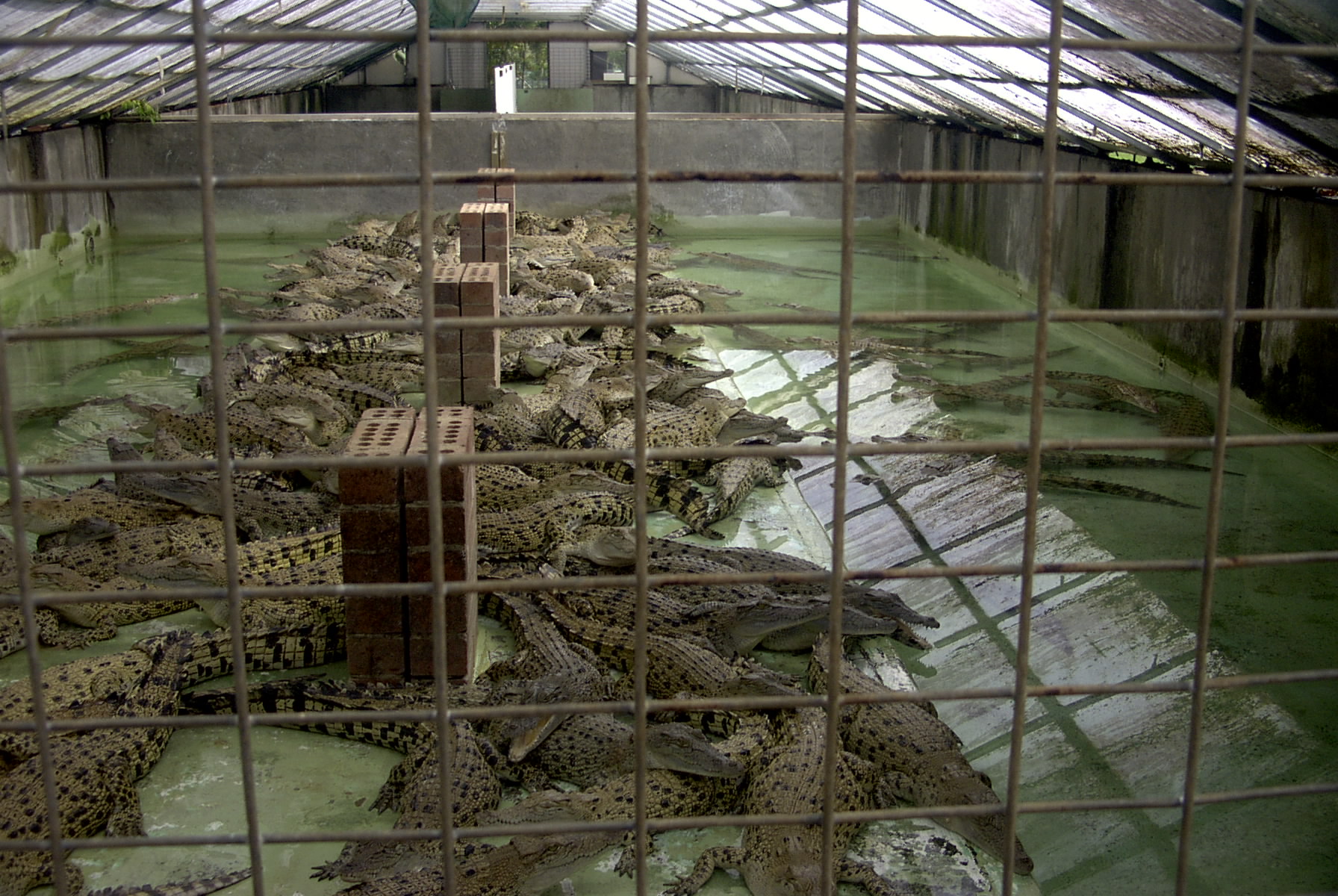
Crocodile
Crocodiles (family (biology), family Crocodylidae) or true crocodiles are large, semiaquatic reptiles that live throughout the tropics in Africa, Asia, the Americas and Australia. The term "crocodile" is sometimes used more loosely to include all extant taxon, extant members of the order (biology), order Crocodilia, which includes the alligators and caimans (both members of the family Alligatoridae), the gharial and false gharial (both members of the family Gavialidae) as well as other extinct Taxon, taxa. Crocodile Measurement, size, Morphology (biology), morphology, behaviour and ecology differ among species. However, they have many similarities in these areas as well. All crocodiles are semiaquatic and tend to congregate in freshwater habitats such as rivers, lakes, wetlands and sometimes in brackish water and Seawater, saltwater. They are carnivorous animals, feeding mostly on vertebrates such as fish, reptiles, birds and mammals, and sometimes on invertebrates such as mol ... [...More Info...] [...Related Items...] OR: [Wikipedia] [Google] [Baidu] |
Vestigiality
Vestigiality is the retention, during the process of evolution, of genetically determined structures or attributes that have lost some or all of the ancestral function in a given species. Assessment of the vestigiality must generally rely on comparison with homology (biology), homologous features in related species. The emergence of vestigiality occurs by normal evolutionary processes, typically by loss of function of a feature that is no longer subject to Directional selection, positive Evolutionary pressure, selection pressures when it loses its value in a changing environment. The feature may be Negative selection (natural selection), selected against more urgently when its function becomes definitively harmful, but if the lack of the feature provides no advantage, and its presence provides no disadvantage, the feature may not be phased out by natural selection and persist across species. Examples of vestigial structures (also called degenerate, atrophied, or rudimentary orga ... [...More Info...] [...Related Items...] OR: [Wikipedia] [Google] [Baidu] |
Venae Comitantes
Vena comitans (Latin for accompanying vein, also known as a satellite vein) refers to a vein that is usually paired, with both veins lying on the sides of an artery. Because they are generally found in pairs, they are often referred to by their plural form: venae comitantes. Venae comitantes are usually found with certain smaller arteries, especially those in the extremities. Larger arteries, on the other hand, generally do not have venae comitantes. They usually have a single, similarly sized vein which is not as intimately associated with the artery. Function As the vein is found in close proximity to an artery the pulsations of the artery aid venous return. Claude Bernard suggested the interchange of heat between the arteries and adjacent veins might moderate cooling of the arterial blood, for which there is experimental evidence. Examples Examples of arteries and their venae comitantes: * Radial artery and radial veins * Ulnar artery and ulnar veins * Brachial artery a ... [...More Info...] [...Related Items...] OR: [Wikipedia] [Google] [Baidu] |
Common Iliac Vein
In human anatomy, the common iliac veins are formed by the external iliac veins and internal iliac veins. The left and right common iliac veins come together in the abdomen at the level of the lumbar vertebrae, fifth lumbar vertebra, forming the inferior vena cava. They drain blood from the pelvis and lower limbs. Both common iliac veins are accompanied along their course by common iliac artery, common iliac arteries. Structure The external iliac vein and internal iliac vein unite in front of the sacroiliac joint to form the common iliac veins. Both common iliac veins ascend to form the inferior vena cava behind the right common iliac artery at the level of the fifth lumbar vertebra. The vena cava is to the right of the midline and therefore the left common iliac vein is longer than the right. The left common iliac vein occasionally travels upwards to the left of the aorta to the level of the kidney, where it receives the left renal vein and crosses in front of the aorta to jo ... [...More Info...] [...Related Items...] OR: [Wikipedia] [Google] [Baidu] |
Anterior Sacral Foramina
The sacrum (: sacra or sacrums), in human anatomy, is a triangular bone at the base of the spine that forms by the fusing of the sacral vertebrae (S1S5) between ages 18 and 30. The sacrum situates at the upper, back part of the pelvic cavity, between the two wings of the pelvis. It forms joints with four other bones. The two projections at the sides of the sacrum are called the alae (wings), and articulate with the ilium at the L-shaped sacroiliac joints. The upper part of the sacrum connects with the last lumbar vertebra (L5), and its lower part with the coccyx (tailbone) via the sacral and coccygeal cornua. The sacrum has three different surfaces which are shaped to accommodate surrounding pelvic structures. Overall, it is concave (curved upon itself). The base of the sacrum, the broadest and uppermost part, is tilted forward as the sacral promontory internally. The central part is curved outward toward the posterior, allowing greater room for the pelvic cavity. In all ot ... [...More Info...] [...Related Items...] OR: [Wikipedia] [Google] [Baidu] |
Lateral Sacral Arteries
The lateral sacral arteries is an artery in the pelvis that arises from the posterior division of the internal iliac artery. It later splits into two smaller branches, a superior and an inferior. Structure The lateral sacral artery is the second branch of the posterior division of the internal iliac artery. It is a parietal branch. Superior The superior, of large size, passes medialward, and, after anastomosing with branches from the middle sacral, enters the first or second anterior sacral foramen, supplies branches to the contents of the sacral canal, and, escaping by the corresponding posterior sacral foramen, is distributed to the skin and muscles on the dorsum of the sacrum, anastomosing with the superior gluteal. Inferior The inferior runs obliquely across the front of the piriformis and the sacral nerves to the medial side of the anterior sacral foramina, descends on the front of the sacrum, and anastomoses over the coccyx with the middle sacral and opposite late ... [...More Info...] [...Related Items...] OR: [Wikipedia] [Google] [Baidu] |
Iliolumbar Artery
The iliolumbar artery is the first branch of the posterior trunk of the internal iliac artery. Structure The iliolumbar artery is the first branch of the posterior trunk of the internal iliac artery. It turns upward behind the obturator nerve and the external iliac artery and vein, to the medial border of the psoas major muscle The psoas major ( or ; from ) is a long fusiform muscle located in the lateral lumbar region between the vertebral column and the brim of the lesser pelvis. It joins the iliacus muscle to form the iliopsoas. In other animals, this muscle is equ ..., behind which it divides into: * Lumbar branch of iliolumbar artery * Iliac branch of iliolumbar artery Anastomoses *1. Last lumbar→iliolumbar *2. Lateral sacral↔lateral sacral *3. Middle sacral→lateral sacral *4. Superior hemorrhoidal→middle hemorrhoidal *5. Medial femoral circumflex→inferior gluteal *6. Medial femoral circumflex↔obturator *7. Lateral femoral circumflex→superior gluteal *8. ... [...More Info...] [...Related Items...] OR: [Wikipedia] [Google] [Baidu] |
Rectum
The rectum (: rectums or recta) is the final straight portion of the large intestine in humans and some other mammals, and the gut in others. Before expulsion through the anus or cloaca, the rectum stores the feces temporarily. The adult human rectum is about long, and begins at the rectosigmoid junction (the end of the sigmoid colon) at the level of the third sacral vertebra or the sacral promontory depending upon what definition is used. Its diameter is similar to that of the sigmoid colon at its commencement, but it is dilated near its termination, forming the rectal ampulla. It terminates at the level of the anorectal ring (the level of the puborectalis sling) or the dentate line, again depending upon which definition is used. In humans, the rectum is followed by the anal canal, which is about long, before the gastrointestinal tract terminates at the anal verge. The word rectum comes from the Latin '' rēctum intestīnum'', meaning ''straight intestine''. Struc ... [...More Info...] [...Related Items...] OR: [Wikipedia] [Google] [Baidu] |
Abdominal Aorta
In human anatomy, the abdominal aorta is the largest artery in the abdominal cavity. As part of the aorta, it is a direct continuation of the descending aorta (of the thorax). Structure The abdominal aorta begins at the level of the diaphragm, crossing it via the aortic hiatus, technically behind the diaphragm, at the vertebral level of T12. It travels down the posterior wall of the abdomen, anterior to the vertebral column. It thus follows the curvature of the lumbar vertebrae, that is, convex anteriorly. The peak of this convexity is at the level of the third lumbar vertebra (L3). It runs parallel to the inferior vena cava, which is located just to the right of the abdominal aorta, and becomes smaller in diameter as it gives off branches. This is thought to be due to the large size of its principal branches. At the 11th rib, the diameter is 122mm long and 55mm wide and this is because of the constant pressure. The abdominal aorta is clinically divided into 2 segments: # Th ... [...More Info...] [...Related Items...] OR: [Wikipedia] [Google] [Baidu] |
Glomus Coccygeum
The coccygeal glomus (coccygeal gland or body; Luschka’s gland) is a vestigial structure placed in front of, or immediately below, the tip of the coccyx. Anatomy It is about 2.5 mm. in diameter and is irregularly oval in shape; several smaller nodules are found around or near the main mass. It consists of irregular masses of round or polyhedral cells epitheloid cells, which are grouped around a dilated sinusoidal capillary vessel. Each cell contains a large round or oval nucleus, the protoplasm surrounding which is clear, and is not stained by chromic salts. Since it is not stained by chromic salts, it is not truly a part of Chromafin system; viz. the system which includes cells stained by chromic salts, consisting of renal medulla, para ganglia, and para aortic bodies. It is situated near the ganglion impar in pelvis, and also at the termination of median sacral artery. Clinical significance It may appear similar to a glomus tumor :''Glomus tumor was also the name for ... [...More Info...] [...Related Items...] OR: [Wikipedia] [Google] [Baidu] |


