|
Mammography
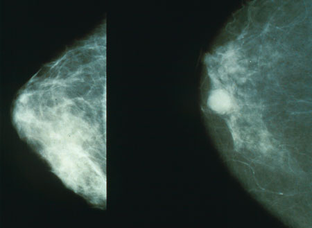
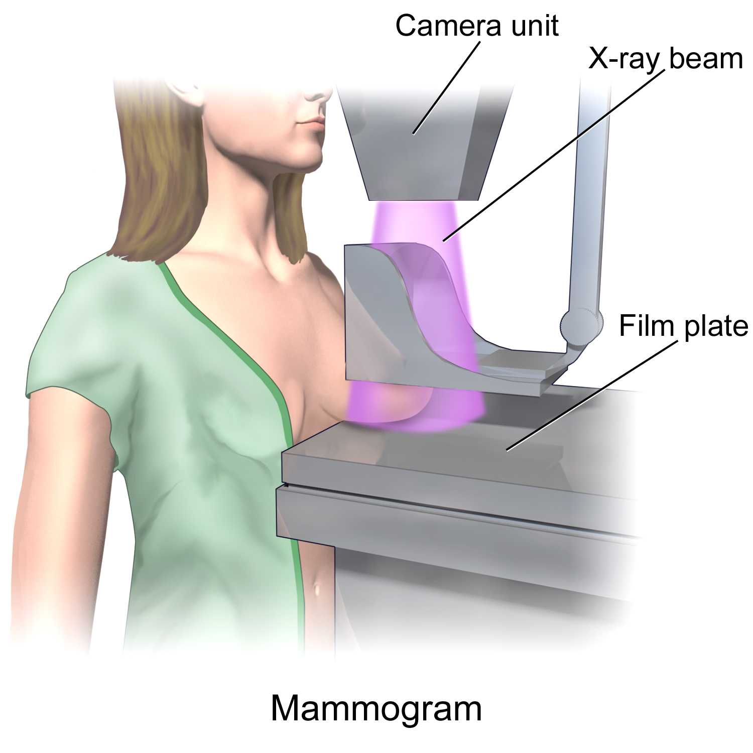
Mammography (also called mastography; DICOM modality: MG) is the process of using low-energy X-rays (usually around 30 kVp) to examine the human breast for diagnosis and screening. The goal of mammography is the early detection of breast cancer, typically through detection of characteristic masses, microcalcifications, asymmetries, and distortions. As with all X-rays, mammograms use doses of ionizing radiation to create images. These images are then analyzed for abnormal findings. It is usual to employ lower-energy X-rays, typically Mo (K-shell X-ray energies of 17.5 and 19.6 keV) and Rh (20.2 and 22.7 keV) than those used for radiography of bones. Mammography may be 2D or 3D ( tomosynthesis), depending on the available equipment or purpose of the examination. Ultrasound, ductography, positron emission mammography (PEM), and magnetic resonance imaging (MRI) are adjuncts to mammography. Ultrasound is typically used for further evaluation of masses found on mammography or palp ... [...More Info...] [...Related Items...] OR: [Wikipedia] [Google] [Baidu] |
Three-dimensional Mammography
Mammography (also called mastography; DICOM modality: MG) is the process of using low-energy X-rays (usually around 30 Peak kilovoltage, kVp) to examine the human breast for diagnosis and screening. The goal of mammography is the early detection of breast cancer, typically through detection of characteristic masses, microcalcifications, asymmetries, and distortions. As with all X-rays, mammograms use doses of ionizing radiation to create images. These images are then analyzed for abnormal findings. It is usual to employ lower-energy X-rays, typically Mo (K-shell X-ray energies of 17.5 and 19.6 keV) and Rh (20.2 and 22.7 keV) than those used for radiography of bones. Mammography may be 2D or 3D (tomosynthesis), depending on the available equipment or purpose of the examination. Ultrasound, ductography, positron emission mammography (PEM), and magnetic resonance imaging (MRI) are adjuncts to mammography. Ultrasound is typically used for further evaluation of masses found on mammogr ... [...More Info...] [...Related Items...] OR: [Wikipedia] [Google] [Baidu] |
Spectral Imaging (radiography)
Spectral imaging is an umbrella term for energy-resolved X-ray imaging in medicine. The technique makes use of the energy dependence of X-ray attenuation to either increase the contrast-to-noise ratio, or to provide quantitative image data and reduce image artefacts by so-called material decomposition. Dual-energy imaging, i.e. imaging at two energy levels, is a special case of spectral imaging and is still the most widely used terminology, but the terms "spectral imaging" and "spectral CT" have been coined to acknowledge the fact that photon-counting detectors have the potential for measurements at a larger number of energy levels. Background The first medical application of spectral imaging appeared in 1953 when B. Jacobson at the Karolinska University Hospital, inspired by X-ray absorption spectroscopy, presented a method called "dichromography" to measure the concentration of iodine in X-ray images. In the 70's, spectral computed tomography (CT) with exposures at two different ... [...More Info...] [...Related Items...] OR: [Wikipedia] [Google] [Baidu] |
Breast Cancer
Breast cancer is a cancer that develops from breast tissue. Signs of breast cancer may include a Breast lump, lump in the breast, a change in breast shape, dimpling of the skin, Milk-rejection sign, milk rejection, fluid coming from the nipple, a newly inverted nipple, or a red or scaly patch of skin. In those with Metastatic breast cancer, distant spread of the disease, there may be bone pain, swollen lymph nodes, shortness of breath, or yellow skin. Risk factors for developing breast cancer include obesity, a Sedentary lifestyle, lack of physical exercise, alcohol consumption, hormone replacement therapy during menopause, ionizing radiation, an early age at Menarche, first menstruation, having children late in life (or not at all), older age, having a prior history of breast cancer, and a family history of breast cancer. About five to ten percent of cases are the result of an inherited genetic predisposition, including BRCA mutation, ''BRCA'' mutations among others. Breast ... [...More Info...] [...Related Items...] OR: [Wikipedia] [Google] [Baidu] |
Positron Emission Mammography
Positron emission mammography (PEM) is a nuclear medicine imaging modality used to detect or characterise breast cancer. Mammography typically refers to x-ray imaging of the breast, while PEM uses an injected positron emitting isotope and a dedicated scanner to locate breast tumors. Scintimammography is another nuclear medicine breast imaging technique, however it is performed using a gamma camera. Breasts can be imaged on standard whole-body PET scanners, however dedicated PEM scanners offer advantages including improved resolution. PEM is not recommended for routine use or for breast cancer screening, in part due to higher radiation dose compared to other modalities. Compared to breast MRI, PEM offers higher specificity. Specific indications can include "high-risk patients with masses > 2 cm or aggressive malignancy and serum tumor marker elevation". 18F-FDG is the most common radiopharmaceutical used for PEM. Equipment PEM uses a specialised scanning system. Though some s ... [...More Info...] [...Related Items...] OR: [Wikipedia] [Google] [Baidu] |
Tomosynthesis
Tomosynthesis, also digital tomosynthesis (DTS), is a method for performing high-resolution limited-angle tomography at radiation dose levels comparable with projectional radiography. It has been studied for a variety of clinical applications, including vascular imaging, dental imaging, orthopedic imaging, mammographic imaging, musculoskeletal imaging, and chest imaging. History The concept of tomosynthesis was derived from the work of Ziedses des Plantes, who developed methods of reconstructing an arbitrary number of planes from a set of projections. Though this idea was displaced by the advent of computed tomography, tomosynthesis later gained interest as a low-dose tomographic alternative to CT. Reconstruction Tomosynthesis reconstruction algorithms are similar to CT reconstructions, in that they are based on performing an inverse Radon transform. Due to partial data sampling with very few projections, approximation algorithms have to be used. Filtered back projection and ite ... [...More Info...] [...Related Items...] OR: [Wikipedia] [Google] [Baidu] |
Microcalcification
Microcalcifications are tiny Calculus (medicine), deposits of calcium salts that are too small to be felt but can be detected by medical imaging, imaging. They can be scattered throughout the mammary gland, or occur in clusters. Microcalcifications can be an early sign of breast cancer. Based on morphology, it is possible to classify by radiography how likely microcalcifications are to indicate cancer. In breast Microcalcifications in the breast are made up of calcium phosphate or calcium oxalate. When consisting of calcium phosphate, they are usually dystrophic calcifications (occurring in degenerated or necrotic tissue). Yet, the mechanism of their formation is not fully known. Calcium oxalate crystals in the breast may be seen on mammography and are usually benign, but can be associated with lobular carcinoma in situ. Last author update: 1 June 2010 Microcalcification was first described in 1913 by surgeon Albert Salomon (surgeon), Albert Salomon. File:Histopathology of ... [...More Info...] [...Related Items...] OR: [Wikipedia] [Google] [Baidu] |
Radiography
Radiography is an imaging technology, imaging technique using X-rays, gamma rays, or similar ionizing radiation and non-ionizing radiation to view the internal form of an object. Applications of radiography include medical ("diagnostic" radiography and "therapeutic radiography") and industrial radiography. Similar techniques are used in airport security, (where "body scanners" generally use backscatter X-ray). To create an image in conventional radiography, a beam of X-rays is produced by an X-ray generator and it is projected towards the object. A certain amount of the X-rays or other radiation are absorbed by the object, dependent on the object's density and structural composition. The X-rays that pass through the object are captured behind the object by a X-ray detector, detector (either photographic film or a digital detector). The generation of flat two-dimensional images by this technique is called Projection radiography, projectional radiography. In computed tomography (C ... [...More Info...] [...Related Items...] OR: [Wikipedia] [Google] [Baidu] |
DICOM
Digital Imaging and Communications in Medicine (DICOM) is a technical standard for the digital storage and Medical image sharing, transmission of medical images and related information. It includes a file format definition, which specifies the structure of a DICOM file, as well as a network communication protocol that uses Internet protocol suite, TCP/IP to communicate between systems. The primary purpose of the standard is to facilitate communication between the software and Computer hardware, hardware entities involved in medical imaging, especially those that are created by different manufacturers. Entities that utilize DICOM files include components of Picture archiving and communication system, picture archiving and communication systems (PACS), such as Modality (medical imaging), imaging machines (modalities), Radiological information system, radiological information systems (RIS), Image scanner, scanners, Printer (computing), printers, Server (computing), computing servers, a ... [...More Info...] [...Related Items...] OR: [Wikipedia] [Google] [Baidu] |
Fujifilm
, trading as , or simply Fuji, is a Japanese Multinational corporation, multinational Conglomerate (company), conglomerate headquartered in Tokyo, Japan, operating in the areas of photography, optics, Office supplies, office and Biomedical engineering, medical electronics, biotechnology, and Chemical substance, chemicals. The company started as a manufacturer of photographic films, which it still produces. Fujifilm products include document solutions, medical imaging and diagnostics equipment, cosmetics, Medication, pharmaceutical drugs, regenerative medicine, stem cells, Contract manufacturing organization, biologics manufacturing, magnetic tape data storage, Optical coating, optical films for flat-panel displays, Optical instrument, optical devices, photocopiers, printers, digital cameras, Color photography, color films, color paper, Photographic processing, photofinishing and graphic arts equipment and materials. Fujifilm is part of the Sumitomo Mitsui Financial Group financia ... [...More Info...] [...Related Items...] OR: [Wikipedia] [Google] [Baidu] |
Radiologist
Radiology ( ) is the medical specialty that uses medical imaging to diagnose diseases and guide treatment within the bodies of humans and other animals. It began with radiography (which is why its name has a root referring to radiation), but today it includes all imaging modalities. This includes technologies that use no ionizing electromagnetic radiation, such as ultrasonography and magnetic resonance imaging (MRI), as well as others that do use radiation, such as computed tomography (CT), fluoroscopy, and nuclear medicine including positron emission tomography (PET). Interventional radiology is the performance of usually minimally invasive medical procedures with the guidance of imaging technologies such as those mentioned above. The modern practice of radiology involves a team of several different healthcare professionals. A radiologist, who is a medical doctor with specialized post-graduate training, interprets medical images, communicates these findings to other physic ... [...More Info...] [...Related Items...] OR: [Wikipedia] [Google] [Baidu] |
X-ray
An X-ray (also known in many languages as Röntgen radiation) is a form of high-energy electromagnetic radiation with a wavelength shorter than those of ultraviolet rays and longer than those of gamma rays. Roughly, X-rays have a wavelength ranging from 10 Nanometre, nanometers to 10 Picometre, picometers, corresponding to frequency, frequencies in the range of 30 Hertz, petahertz to 30 Hertz, exahertz ( to ) and photon energies in the range of 100 electronvolt, eV to 100 keV, respectively. X-rays were discovered in 1895 in science, 1895 by the German scientist Wilhelm Röntgen, Wilhelm Conrad Röntgen, who named it ''X-radiation'' to signify an unknown type of radiation.Novelline, Robert (1997). ''Squire's Fundamentals of Radiology''. Harvard University Press. 5th edition. . X-rays can penetrate many solid substances such as construction materials and living tissue, so X-ray radiography is widely used in medical diagnostics (e.g., checking for Bo ... [...More Info...] [...Related Items...] OR: [Wikipedia] [Google] [Baidu] |
United States Preventive Services Task Force
The United States Preventive Services Task Force (USPSTF) is "an independent panel of experts in primary care and prevention that systematically reviews the evidence of effectiveness and develops recommendations for clinical preventive services". The task force, a volunteer panel of primary care clinicians (including those from internal medicine, pediatrics, family medicine, obstetrics and gynecology, nursing, and psychology) with methodology experience including epidemiology, biostatistics, health services research, decision sciences, and health economics, is funded, staffed, and appointed by the U.S. Department of Health and Human Services' Agency for Healthcare Research and Quality. Intent The USPSTF evaluates scientific evidence to determine whether medical screenings, counseling, and preventive medications work for adults and children who have no symptoms. Methods The methods of evidence synthesis used by the Task Force have been described in detail. In 2007, their metho ... [...More Info...] [...Related Items...] OR: [Wikipedia] [Google] [Baidu] |


