|
Intracardiac Echocardiography
Intracardiac echocardiography (ICE) is a specialized form of echocardiography that utilizes an ultrasound-tipped catheter to perform imaging of the heart from within the heart. Unlike transthoracic echocardiography (TTE), ICE is not limited by body habitus. An ICE catheter is inserted into the body, typically, through the femoral vein and advanced into the heart. Uses The use of ICE is specialized and not intended for general echocardiography due to its cost and invasiveness. It is used as a part of a larger heart procedure. A typical use of ICE is for performing a transseptal puncture across the interatrial septum; in other words, pushing a catheter from the right atrium to the left atrium. Next to the septum is the aorta, and puncturing from the right atrium to the aorta is dangerous, and ICE visualization increases the confidence of performing this procedure safely. Risks The use of an ICE catheter has the same risks of use and advancing any other catheter into the heart, namel ... [...More Info...] [...Related Items...] OR: [Wikipedia] [Google] [Baidu] |
Echocardiography
Echocardiography, also known as cardiac ultrasound, is the use of ultrasound to examine the heart. It is a type of medical imaging, using standard ultrasound or Doppler ultrasound. The visual image formed using this technique is called an echocardiogram, a cardiac echo, or simply an echo. Echocardiography is routinely used in the diagnosis, management, and follow-up of patients with any suspected or known heart diseases. It is one of the most widely used diagnostic imaging modalities in cardiology. It can provide a wealth of helpful information, including the size and shape of the heart (internal chamber size quantification), pumping capacity, location and extent of any tissue damage, and assessment of valves. An echocardiogram can also give physicians other estimates of heart function, such as a calculation of the cardiac output, ejection fraction, and diastolic function (how well the heart relaxes). Echocardiography is an important tool in assessing wall motion abnorma ... [...More Info...] [...Related Items...] OR: [Wikipedia] [Google] [Baidu] |
Atrial Septal Defect
Atrial septal defect (ASD) is a congenital heart defect in which blood flows between the atrium (heart), atria (upper chambers) of the heart. Some flow is a normal condition both pre-birth and immediately post-birth via the Foramen ovale (heart), foramen ovale; however, when this does not naturally close after birth it is referred to as a patent (open) foramen ovale (PFO). It is common in patients with a congenital interatrial septum, atrial septal aneurysm (ASA). After PFO closure the atria normally are separated by a dividing wall, the interatrial septum. If this septum is defective or absent, then oxygen-rich blood can flow directly from the left side of the heart to mix with the oxygen-poor blood in the right side of the heart; or the opposite, depending on whether the left or right atrium has the higher blood pressure. In the absence of other heart defects, the left atrium has the higher pressure. This can lead to lower-than-normal oxygen levels in the arterial blood that su ... [...More Info...] [...Related Items...] OR: [Wikipedia] [Google] [Baidu] |
Transesophageal Echocardiography
A transesophageal echocardiogram (TEE; also spelled transoesophageal echocardiogram; TOE in British English) is an alternative way to perform an echocardiogram. A specialized probe containing an ultrasound transducer at its tip is passed into the patient's esophagus. This allows image and Doppler evaluation which can be recorded. It is commonly used during cardiac surgery and is an excellent modality for assessing the aorta, although there are some limitations. It has several advantages and some disadvantages compared with a transthoracic echocardiogram (TTE). Details TEE is a semi-invasive procedure in that the probe must enter the body but does not require surgical (i.e., invasive) cutting for this procedure. Before inserting the probe, mild to moderate sedation is induced in the patient to ease the discomfort and to decrease the gag reflex. Usually a local anesthetic spray (e.g., lidocaine, benzocaine, xylocaine) is used for the back of the throat or as a jell ... [...More Info...] [...Related Items...] OR: [Wikipedia] [Google] [Baidu] |
Transthoracic Echocardiography
Echocardiography, also known as cardiac ultrasound, is the use of ultrasound to examine the heart. It is a type of medical imaging, using standard ultrasound or Doppler ultrasound. The visual image formed using this technique is called an echocardiogram, a cardiac echo, or simply an echo. Echocardiography is routinely used in the diagnosis, management, and follow-up of patients with any suspected or known heart diseases. It is one of the most widely used diagnostic imaging modalities in cardiology. It can provide a wealth of helpful information, including the size and shape of the heart (internal chamber size quantification), pumping capacity, location and extent of any tissue damage, and assessment of valves. An echocardiogram can also give physicians other estimates of heart function, such as a calculation of the cardiac output, ejection fraction, and diastolic function (how well the heart relaxes). Echocardiography is an important tool in assessing wall motion abnormality in ... [...More Info...] [...Related Items...] OR: [Wikipedia] [Google] [Baidu] |
Percutaneous Aortic Valve Replacement
Transcatheter aortic valve replacement (TAVR) is the implantation of the aortic valve of the heart through the blood vessels without actual removal of the native valve (as opposed to the aortic valve replacement by open heart surgery, surgical aortic valve replacement, AVR). The first TAVI was performed on 16 April 2002 by Alain Cribier, which became a new alternative in the management of high-risk patients with severe aortic stenosis. The implantated valve is delivered via one of several access methods: transfemoral (in the upper leg), transapical (through the wall of the heart), subclavian (beneath the collar bone), direct aortic (through a minimally invasive surgical incision into the aorta), and transcaval (from a temporary hole in the aorta near the navel through a vein in the upper leg), among others. Severe symptomatic aortic stenosis carries a poor prognosis. At present, there is no treatment via medication, making the timing of aortic valve replacement the most imp ... [...More Info...] [...Related Items...] OR: [Wikipedia] [Google] [Baidu] |
MitraClip
MitraClip (mitral clip) is a medical device used to treat mitral valve regurgitation for individuals who should not have open-heart surgery. It is implanted via a tri-axial transcatheter technique and involves suturing together the anterior and posterior mitral valve leaflets. Medical use and indications MitraClip is used for patients with severe secondary mitral valve regurgitation that is refractory to medical therapy. Primary mitral regurgitation is usually due to an organic cause whereas secondary mitral regurgitation is due to a secondary ischemia or cardiomyopathy. Open-heart surgery remains the preferred treatment option when possible for primary mitral regurgitation, due to the effectiveness and long-term record of the procedure in reducing mitral valve regurgitation. Secondary Mitral regurgitation however can have different options in management as surgery has not been proven in clinical trials to be superior The indications for using MitraClip are as follows: # Those w ... [...More Info...] [...Related Items...] OR: [Wikipedia] [Google] [Baidu] |
Mitral Valve
The mitral valve ( ), also known as the bicuspid valve or left atrioventricular valve, is one of the four heart valves. It has two Cusps of heart valves, cusps or flaps and lies between the atrium (heart), left atrium and the ventricle (heart), left ventricle of the heart. The heart valves are all one-way valves allowing blood flow in just one direction. The mitral valve and the tricuspid valve are known as the Heart valve#Atrioventricular valves, atrioventricular valves because they lie between the atria and the ventricles. In normal conditions, blood flows through an open mitral valve during diastole with contraction of the left atrium, and the mitral valve closes during systole with contraction of the left ventricle. The valve opens and closes because of pressure differences, opening when there is greater pressure in the left atrium than ventricle and closing when there is greater pressure in the left ventricle than atrium. In abnormal conditions, blood may flow backward thro ... [...More Info...] [...Related Items...] OR: [Wikipedia] [Google] [Baidu] |
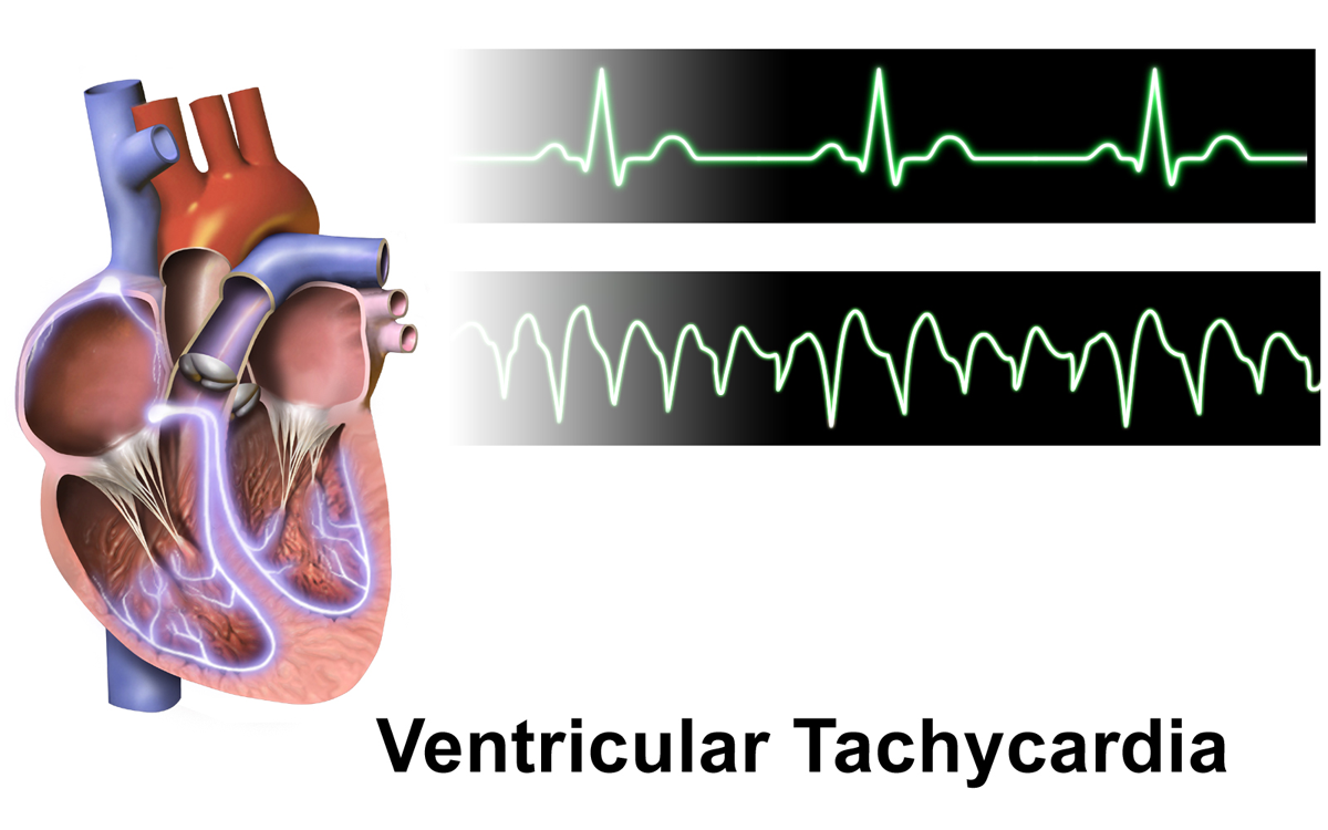
Ventricular Tachycardia
Ventricular tachycardia (V-tach or VT) is a cardiovascular disorder in which fast heart rate occurs in the ventricles of the heart. Although a few seconds of VT may not result in permanent problems, longer periods are dangerous; and multiple episodes over a short period of time are referred to as an electrical storm. Short periods may occur without symptoms, or present with lightheadedness, palpitations, shortness of breath, chest pain, and decreased level of consciousness. Ventricular tachycardia may lead to coma and persistent vegetative state due to lack of blood and oxygen to the brain. Ventricular tachycardia may result in ventricular fibrillation (VF) and turn into cardiac arrest. This conversion of the VT into VF is called the degeneration of the VT. It is found initially in about 7% of people in cardiac arrest. Ventricular tachycardia can occur due to coronary heart disease, aortic stenosis, cardiomyopathy, electrolyte imbalance, or a heart attack. Diagnosis is ... [...More Info...] [...Related Items...] OR: [Wikipedia] [Google] [Baidu] |
Atrial Flutter
Atrial flutter (AFL) is a common abnormal heart rhythm that starts in the atrial chambers of the heart. When it first occurs, it is usually associated with a fast heart rate and is classified as a type of supraventricular tachycardia (SVT). Atrial flutter is characterized by a sudden-onset (usually) regular abnormal heart rhythm on an electrocardiogram (ECG) in which the heart rate is fast. Symptoms may include a feeling of the heart beating too fast, too hard, or skipping beats, chest discomfort, difficulty breathing, a feeling as if one's stomach has dropped, a feeling of being light-headed, or loss of consciousness. Although this abnormal heart rhythm typically occurs in individuals with cardiovascular disease (e.g., high blood pressure, coronary artery disease, and cardiomyopathy) and diabetes mellitus, it may occur spontaneously in people with otherwise normal hearts. It is typically not a stable rhythm and often degenerates into atrial fibrillation (AF). But rar ... [...More Info...] [...Related Items...] OR: [Wikipedia] [Google] [Baidu] |
Atrial Fibrillation
Atrial fibrillation (AF, AFib or A-fib) is an Heart arrhythmia, abnormal heart rhythm (arrhythmia) characterized by fibrillation, rapid and irregular beating of the Atrium (heart), atrial chambers of the heart. It often begins as short periods of abnormal cardiac cycle, beating, which become longer or continuous over time. It may also start as other forms of arrhythmia such as atrial flutter that then transform into AF. Episodes can be asymptomatic. Symptomatic episodes may involve heart palpitations, syncope (medicine), fainting, Presyncope, lightheadedness, Unconsciousness, loss of consciousness, or shortness of breath. Atrial fibrillation is associated with an increased risk of heart failure, dementia, and stroke. It is a type of supraventricular tachycardia. Atrial fibrillation frequently results from bursts of tachycardia that originate in muscle bundles extending from the Atrium (heart), atrium to the pulmonary veins. Pulmonary vein isolation by catheter ablation, trans ... [...More Info...] [...Related Items...] OR: [Wikipedia] [Google] [Baidu] |
Electroanatomic Mapping
Electroanatomic mapping is a method of creating a three dimensional model of the human heart during clinical cardiac electrophysiology procedures. Technology The fundamental concept of electroanatomic mapping systems is to localize catheters within the heart in three dimensional space (a sort of "GPS" within the heart). Building a 3-D model of the heart with real-time visualization permits reduction in fluoroscopy use. In addition to 3-D structure, the voltage and timing of signals at each point of the heart is recorded to generate different maps to understand and treat different rhythm disturbances. Each of the three systems utilizes different techniques to localize catheters: * Carto uses a low-intensity magnetic field (5-50 μT) with tri-axial inductors in the tip of the catheters to triangulate the tip based on the sense magnetic field relative to sensors placed on the front and back of the chest. Carto 3 adds emission of electric fields from each unique electrode on a cat ... [...More Info...] [...Related Items...] OR: [Wikipedia] [Google] [Baidu] |


