|
Interstitial Nucleus Of Cajal
The interstitial nucleus of Cajal is a collection of neurons in the mesencephalon (midbrain) which are involved in integrating eye position-velocity information in order to coordinate head-eye movements - especially those related to vertical and torsional conjugate eye movements (gaze). It also mediates vertical gaze holding. Bilateral projections to the oculomotor (cranial nerve III) and trochlear (cranial nerve IV) nuclei represent its principal outputs. It forms reciprocal connections with vestibular nuclei. It also has additional afferents and efferents. Some of the nucleus' connections pass through the medial longitudinal fasciculus, and the posterior commissure. It is one of the accessory oculomotor nuclei. Anatomy The interstitial nucleus of Cajal is a diffuse collection of mid-sized, parvalbumin-containing premotor neurons of the midbrain reticular formation. Connections The nucleus forms reciprocal connections with the vestibular nuclei (through the MLF). It f ... [...More Info...] [...Related Items...] OR: [Wikipedia] [Google] [Baidu] |
Midbrain
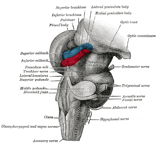
The midbrain or mesencephalon is the uppermost portion of the brainstem connecting the diencephalon and cerebrum with the pons. It consists of the cerebral peduncles, tegmentum, and tectum. It is functionally associated with vision, hearing, motor control, sleep and wakefulness, arousal (alertness), and temperature regulation.Breedlove, Watson, & Rosenzweig. Biological Psychology, 6th Edition, 2010, pp. 45-46 The name ''mesencephalon'' comes from the Greek ''mesos'', "middle", and ''enkephalos'', "brain". Structure The midbrain is the shortest segment of the brainstem, measuring less than 2cm in length. It is situated mostly in the posterior cranial fossa, with its superior part extending above the tentorial notch. The principal regions of the midbrain are the tectum, the cerebral aqueduct, tegmentum, and the cerebral peduncles. Rostral and caudal, Rostrally the midbrain adjoins the diencephalon (thalamus, hypothalamus, etc.), while Rostral and caudal, cau ... [...More Info...] [...Related Items...] OR: [Wikipedia] [Google] [Baidu] |
Rostral Interstitial Nucleus Of Medial Longitudinal Fasciculus
The rostral interstitial nucleus of medial longitudinal fasciculus (riMLF) is a collection of neurons in the medial longitudinal fasciculus in the midbrain. It is responsible for mediating vertical conjugate eye movements (vertical gaze (physiology), gaze) and vertical saccades. It mostly projects efferents to the ipsilateral oculomotor and trochlear nuclei. To mediate downgaze, it projects efferents to the ipsilateral oculomotor nucleus and trochlear nucleus; mediate upgaze, it projects efferents to the contralateral aforementioned nuclei through the posterior commissure. It is one of the accessory oculomotor nuclei. Anatomy Structure The riMLF is a wing-shaped nucleus. The riMLF contains two populations of neurons: excitatory burst neurons mediating vertical gaze/saccades, as well as omnipause neurons which are functionally similar to those mediating horizontal gaze. Relations It is situated at the caudal extremity of the mesencephalon at its junction with the telenceph ... [...More Info...] [...Related Items...] OR: [Wikipedia] [Google] [Baidu] |
Medial Longitudinal Fasciculus
The medial longitudinal fasciculus (MLF) is a prominent bundle of nerve fibres which pass within the ventral/anterior portion of periaqueductal gray of the mesencephalon (midbrain). It contains the interstitial nucleus of Cajal, responsible for oculomotor control, head posture, and vertical eye movement. The MLF interconnects interneurons of each abducens nucleus with motor neurons of the contralateral oculomotor nucleus; thus, the MLF mediates coordination of horizontal (side to side) eye movements, ensuring the two eyes move in unison (thus also enabling saccadic eye movements). The MLF also contains fibers projecting from the vestibular nuclei to the oculomotor and trochlear nuclei as well as the interstitial nucleus of Cajal; these connections ensure that eye movements are coordinated with head movements (as sensed by the vestibular system). The medial longitudinal fasciculus is the main central connection for the oculomotor nerve, trochlear nerve, and abduce ... [...More Info...] [...Related Items...] OR: [Wikipedia] [Google] [Baidu] |
Nucleus Of Darkschewitsch
The nucleus of Darkschewitsch is an accessory oculomotor nucleus situated in the ventrolateral portion of the periaqueductal gray of the mesencephalon (midbrain) near its junction with the diencephalon. It is involved in mediating vertical eye movements. It projects to the trochlear nucleus, receives afferents from the visual cortex, and forms a reciprocal (looping) connection with the cerebellum by way of the inferior olive. Anatomy Connections It receives afferents from the visual association areas (via the corticotectal tract), vestibular nuclei (via the medial longitudinal fasciculus), and from the spinomesencephalic tract. It gives rise to the medial tegmental tract to project efferents to the (rostral portion of) medial accessory olivary nucleus → ((decussation) inferior cerebellar peduncle → (contralateral) globose nucleus of cerebellum → superior cerebellar peduncle (decussation) → (rostral part of) medial accessory olivary nucleus → (ipsilateral) nu ... [...More Info...] [...Related Items...] OR: [Wikipedia] [Google] [Baidu] |
Oculomotor Nucleus
The fibers of the oculomotor nerve arise from a nucleus in the midbrain, which lies in the gray substance of the floor of the cerebral aqueduct and extends in front of the aqueduct for a short distance into the floor of the third ventricle. From this nucleus the fibers pass forward through the tegmentum, the red nucleus, and the medial part of the substantia nigra, forming a series of curves with a lateral convexity, and emerge from the oculomotor sulcus on the medial side of the cerebral peduncle. The nucleus of the oculomotor nerve does not consist of a continuous column of cells, but is broken up into a number of smaller nuclei, which are arranged in two groups, anterior and posterior. Those of the posterior group are six in number, five of which are symmetrical on the two sides of the middle line, while the sixth is centrally placed and is common to the nerves of both sides. The anterior group consists of two nuclei, an antero-medial and an antero-lateral. The nucleus ... [...More Info...] [...Related Items...] OR: [Wikipedia] [Google] [Baidu] |
Red Nucleus
The red nucleus or nucleus ruber is a structure in the rostral midbrain involved in motor coordination. The red nucleus is pale pink, which is believed to be due to the presence of iron in at least two different forms: hemoglobin and ferritin. The structure is located in the midbrain tegmentum next to the substantia nigra and comprises caudal magnocellular and rostral parvocellular components. The red nucleus and substantia nigra are subcortical centers of the extrapyramidal motor system. Function In a vertebrate without a significant corticospinal tract, gait is mainly controlled by the red nucleus. However, in primates, where the corticospinal tract is dominant, the rubrospinal tract may be regarded as vestigial in motor function. Therefore, the red nucleus is less important in primates than in many other mammals. Nevertheless, the crawling of babies is controlled by the red nucleus, as is arm swinging in typical walking. The red nucleus may play an additional role ... [...More Info...] [...Related Items...] OR: [Wikipedia] [Google] [Baidu] |
Periaqueductal Gray
The periaqueductal gray (PAG), also known as the central gray, is a brain region that plays a critical role in autonomic function, motivated behavior and behavioural responses to threatening stimuli. PAG is also the primary control center for descending pain modulation. It has enkephalin-producing cells that suppress pain. The periaqueductal gray is the gray matter located around the cerebral aqueduct within the tegmentum of the midbrain. It projects to the nucleus raphe magnus, and also contains descending autonomic tracts. The ascending pain and temperature fibers of the spinothalamic tract send information to the PAG via the spinomesencephalic pathway (so-named because the fibers originate in the spine and terminate in the PAG, in the mesencephalon or midbrain). This region has been used as the target for brain-stimulating implants in patients with chronic pain. Role in analgesia Stimulation of the periaqueductal gray matter of the midbrain activates enkephalin-r ... [...More Info...] [...Related Items...] OR: [Wikipedia] [Google] [Baidu] |
Diencephalon
In the human brain, the diencephalon (or interbrain) is a division of the forebrain (embryonic ''prosencephalon''). It is situated between the telencephalon and the midbrain (embryonic ''mesencephalon''). The diencephalon has also been known as the tweenbrain in older literature. It consists of structures that are on either side of the third ventricle, including the thalamus, the hypothalamus, the epithalamus and the subthalamus. The diencephalon is one of the main brain vesicle, vesicles of the brain formed during human embryonic development, embryonic development. During the third week of development a neural tube is created from the ectoderm, one of the three primary germ layers, and forms three main vesicles: the prosencephalon, the Midbrain, mesencephalon and the Hindbrain, rhombencephalon. The prosencephalon gradually divides into the telencephalon (the cerebrum) and the diencephalon. Structure The diencephalon consists of the following structures: * Thalamus * Hypothalamus ... [...More Info...] [...Related Items...] OR: [Wikipedia] [Google] [Baidu] |
Mesencephalic Tegmentum
The midbrain is anatomically delineated into the tectum (roof) and the tegmentum (floor). The midbrain tegmentum extends from the substantia nigra to the cerebral aqueduct in a horizontal section of the midbrain. It forms the floor of the midbrain that surrounds below the cerebral aqueduct as well as the floor of the fourth ventricle while the midbrain tectum forms the roof of the fourth ventricle. The tegmentum contains a collection of tracts and nuclei with movement-related, species-specific, and pain-perception functions. The general structures of midbrain tegmentum include red nucleus and the periaqueductal grey matter. Unlike the midbrain tectum (which is a sensory structure located posteriorly), the midbrain tegmentum, which locates anteriorly, is related to a number of motor functions. Within the tegmentum, the red nucleus is in charge of motor coordination (specifically for limb movements) and the periaqueductal gray matter (PAG) contains critical circuits for modulating ... [...More Info...] [...Related Items...] OR: [Wikipedia] [Google] [Baidu] |
Mesencephalon
The midbrain or mesencephalon is the uppermost portion of the brainstem connecting the diencephalon and cerebrum with the pons. It consists of the cerebral peduncles, tegmentum, and tectum. It is functionally associated with vision, hearing, motor control, sleep and wakefulness, arousal (alertness), and temperature regulation.Breedlove, Watson, & Rosenzweig. Biological Psychology, 6th Edition, 2010, pp. 45-46 The name ''mesencephalon'' comes from the Greek ''mesos'', "middle", and ''enkephalos'', "brain". Structure The midbrain is the shortest segment of the brainstem, measuring less than 2cm in length. It is situated mostly in the posterior cranial fossa, with its superior part extending above the tentorial notch. The principal regions of the midbrain are the tectum, the cerebral aqueduct, tegmentum, and the cerebral peduncles. Rostral and caudal, Rostrally the midbrain adjoins the diencephalon (thalamus, hypothalamus, etc.), while Rostral and caudal, cau ... [...More Info...] [...Related Items...] OR: [Wikipedia] [Google] [Baidu] |
Medial Vestibular Nucleus
The medial vestibular nucleus (Schwalbe nucleus) is one of the vestibular nuclei. It is located in the medulla oblongata. Lateral vestibulo-spinal tract (lateral vestibular nucleus “Deiters”)- via ventrolateral medulla and spinal cord to ventral funiculus (lumbo-sacral segments). ..Ipsilaterally for posture Medial vestibulo-spinal tract (medial, lateral, inferior, vestibular nuclei), bilateral projection via descending medial longitudinal fasciculus to cervical segments. DESCENDING MLF..Bilaterally for head/neck/eye movements It is one of the nuclei that corresponds to CN VIII, corresponding to the vestibular nerve, which joins with the cochlear nerve. It receives its blood supply from the Posterior Inferior Cerebellar Artery, which is compromised in the lateral medullary syndrome. See also * Vestibular nerve * Vestibular system The vestibular system, in vertebrates, is a sensory system that creates the sense of balance and spatial orientation for the purpose of coordin ... [...More Info...] [...Related Items...] OR: [Wikipedia] [Google] [Baidu] |
