|
Internal Carotid Nerve Plexus
The internal carotid plexus is a nerve plexus situated upon the lateral side of the internal carotid artery. It is composed of post-ganglionic sympathetic fibres which have synapsed at (i.e. have their nerve cell bodies at) the superior cervical ganglion. The plexus gives rise to the deep petrosal nerve. Anatomy Postganglionic sympathetic fibres ascend from the superior cervical ganglion, along the walls of the internal carotid artery, to enter the internal carotid plexus. These fibres are then distributed to deep structures, including the superior tarsal muscle and pupillary dilator muscle. It includes fibres destined for the pupillary dilator muscle as part of a neural circuit regulating pupillary dilatation component of the pupillary reflex. Some fibres of the plexus converge to form the deep petrosal nerve.Richard L. Drake, Wayne Vogel & Adam W M Mitchell, "Gray's Anatomy for Students", Elsevier inc., 2005 The internal carotid plexus communicates with the trigeminal gangli ... [...More Info...] [...Related Items...] OR: [Wikipedia] [Google] [Baidu] |
Nerve Plexus
A nerve plexus is a plexus (branching network) of intersecting nerves. A nerve plexus is composed of afferent and efferent fibers that arise from the merging of the anterior rami of spinal nerves and blood vessels. There are five spinal nerve plexuses, except in the thoracic region, as well as other forms of autonomic nervous system, autonomic plexuses, many of which are a part of the enteric nervous system. The nerves that arise from the plexuses have both sensory and motor functions. These functions include muscle contraction, the maintenance of body coordination and control, and the reaction to sensations such as heat, cold, pain, and pressure. There are several plexuses in the body, including: *Spinal plexuses **Cervical plexusserves the head, neck and shoulders **Brachial plexusserves the chest, shoulders, arms and hands **Lumbosacral plexus ***Lumbar plexusserves the back, abdomen, groin, thighs, knees, and calves ****Subsartorial plexusbelow the sartorius muscle of thigh *** ... [...More Info...] [...Related Items...] OR: [Wikipedia] [Google] [Baidu] |
Internal Carotid Artery
The internal carotid artery is an artery in the neck which supplies the anterior cerebral artery, anterior and middle cerebral artery, middle cerebral circulation. In human anatomy, the internal and external carotid artery, external carotid arise from the common carotid artery, where it bifurcates at cervical vertebrae C3 or C4. The internal carotid artery supplies the brain, including the eyes, while the external carotid nourishes other portions of the head, such as the face, scalp, skull, and meninges. Classification Terminologia Anatomica in 1998 subdivided the artery into four parts: "cervical", "petrous", "cavernous", and "cerebral". In clinical settings, however, usually the classification system of the internal carotid artery follows the 1996 recommendations by Bouthillier, describing seven anatomical segments of the internal carotid artery, each with a corresponding alphanumeric identifier: C1 cervical; C2 petrous; C3 lacerum; C4 cavernous; C5 clinoid; C6 ophthalmic; ... [...More Info...] [...Related Items...] OR: [Wikipedia] [Google] [Baidu] |
Superior Cervical Ganglion
The superior cervical ganglion (SCG) is the upper-most and largest of the cervical sympathetic ganglia of the sympathetic trunk. It probably formed by the union of four sympathetic ganglia of the cervical spinal nerves C1–C4. It is the only ganglion of the sympathetic nervous system that innervates the head and neck. The SCG innervates numerous structures of the head and neck. Structure The superior cervical ganglion is reddish-gray color, and usually shaped like a spindle with tapering ends. It measures about 3 cm in length. Sometimes the SCG is broad and flattened, and occasionally constricted at intervals. It formed by the coalescence of four ganglia, corresponding to the four upper-most cervical nerves C1–C4. The bodies of its preganglionic sympathetic afferent neurons are located in the lateral horn of the spinal cord. Their axons enter the SCG to synapse with postganglionic neurons whose axons then exit the rostral end of the SCG and proceed to innervate their targ ... [...More Info...] [...Related Items...] OR: [Wikipedia] [Google] [Baidu] |
Deep Petrosal Nerve
The deep petrosal nerve is a post-ganglionic branch of the ( sympathetic) internal carotid (nervous) plexus (which is in turn derived from the superior cervical ganglion, a part of the cervical sympathetic trunk) that enters the cranial cavity through the carotid canal, then passes perpendicular to the carotid canal in the cartilaginous substance which fills the foramen lacerum to unite with the (parasympathetic) greater petrosal nerve to form the nerve of pterygoid canal (Vidian nerve). Anatomy intermediate grey column (of spinal cord at around the level of T1) → white rami communicantes (of cervical part of sympathetic chain) → superior cervical ganglion (synapse) → gray rami communicantes → internal carotid plexus → deep petrosal nerve → nerve of pterygoid canal → pterygopalatine ganglion (fibres pass through without synapsing) → zygomatic nerve → zygomaticotemporal nerve → lacrimal nerve Origin The cell bodies of pre-ganglionic sympathetic axons th ... [...More Info...] [...Related Items...] OR: [Wikipedia] [Google] [Baidu] |
Superior Tarsal Muscle
The superior tarsal muscle is a smooth muscle adjoining the levator palpebrae superioris muscle muscle that helps to raise the upper eyelid. Structure The superior tarsal muscle originates on the underside of levator palpebrae superioris muscle and inserts on the superior tarsal plate of the eyelid. Nerve supply The superior tarsal muscle receives its innervation from the sympathetic nervous system. Postganglionic sympathetic fibers originate in the superior cervical ganglion, and travel via the internal carotid plexus, where small branches communicate with the oculomotor nerve as it passes through the cavernous sinus. The sympathetic fibres continue to the superior division of the oculomotor nerve The oculomotor nerve, also known as the third cranial nerve, cranial nerve III, or simply CN III, is a cranial nerve that enters the orbit through the superior orbital fissure and innervates extraocular muscles that enable most movements o ..., where they enter th ... [...More Info...] [...Related Items...] OR: [Wikipedia] [Google] [Baidu] |
Pupillary Dilator Muscle
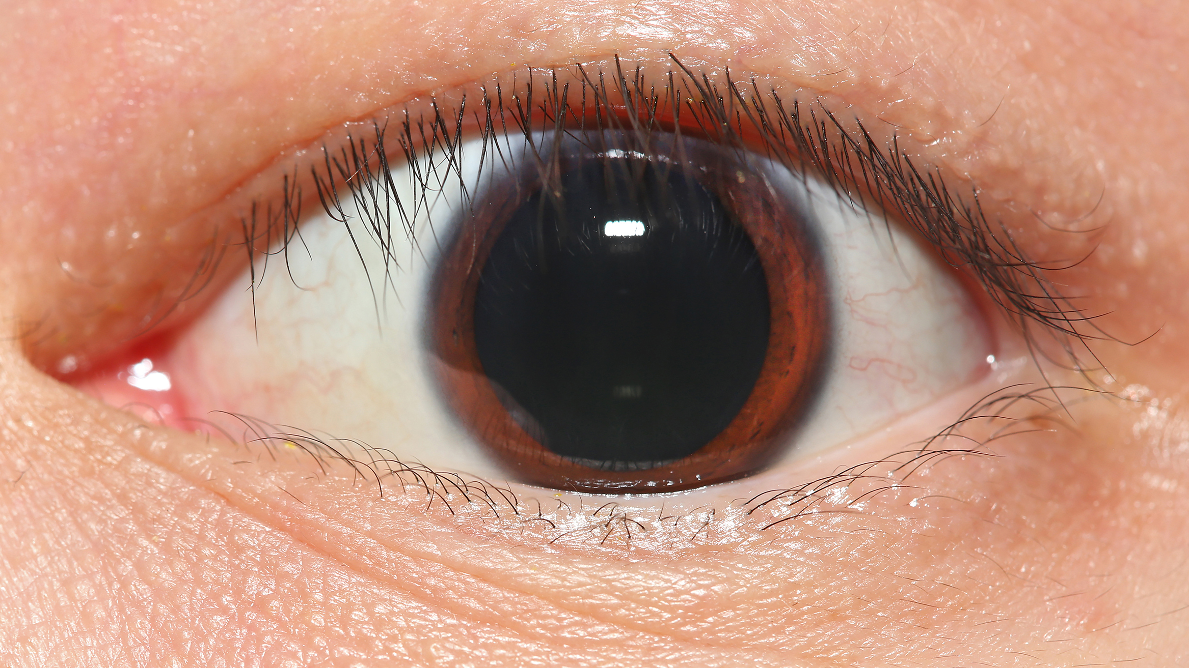
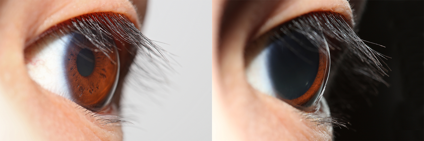
The iris dilator muscle (pupil dilator muscle, pupillary dilator, radial muscle of iris, radiating fibers), is a smooth muscle of the eye, running radially in the iris and therefore fit as a dilator. The pupillary dilator consists of a spokelike arrangement of modified contractile cells called myoepithelial cells. These cells are stimulated by the sympathetic nervous system. When stimulated, the cells contract, widening the pupil and allowing more light to enter the eye. The ciliary muscle, pupillary sphincter muscle and pupillary dilator muscle sometimes are called intrinsic ocular muscles or intraocular muscles. Structure Innervation It is innervated by the sympathetic system, which acts by releasing noradrenaline, which acts on α1-receptors. Thus, when presented with a threatening stimulus that activates the fight-or-flight response, this innervation contracts the muscle and dilates the pupil, thus temporarily letting more light reach the retina. The dilator muscle is inne ... [...More Info...] [...Related Items...] OR: [Wikipedia] [Google] [Baidu] |
Hal Blumenfeld
Hal Blumenfeld (born March 28, 1962) is a professor of neurology, neuroscience, and neurosurgery at Yale University. His focus is on brain mechanisms of consciousness and on altered consciousness in epilepsy. As director of the Yale Clinical Neuroscience Imaging Center, he leads multi-disciplinary research and is also well known for his teaching contributions in neuroanatomy and clinical neuroscience. Biography Blumenfeld was born in California, grew up in New York and began his career in Bio-electrical Engineering at Harvard University (1984).Curriculum Vitae. Blumenfeld, H. (Accessed 20 May 2021). https://prod.admin.profile.yale.edu/Profiles/Cv/user/hal_blumenfeld His passion for science would lead him to Columbia University where, working with Eric Kandel and Steven Siegelbaum, he obtained his PhD (1990) in Physiology and Cellular Biophysics and his MD (1992). He completed his internal medicine internship at Columbia Presbyterian Medical Center(1993) and entered the field of ... [...More Info...] [...Related Items...] OR: [Wikipedia] [Google] [Baidu] |
Pupillary Dilatation
Mydriasis is the dilation of the pupil, usually having a non-physiological cause, or sometimes a physiological pupillary response. Non-physiological causes of mydriasis include disease, trauma, or the use of certain types of drugs. It may also be of unknown cause. Normally, as part of the pupillary light reflex, the pupil dilates in the dark and constricts in the light to respectively improve vividity at night and to protect the retina from sunlight damage during the day. A ''mydriatic'' pupil will remain excessively large even in a bright environment. The excitation of the radial fibres of the iris which increases the pupillary aperture is referred to as a mydriasis. More generally, mydriasis also refers to the natural dilation of pupils, for instance in low light conditions or under sympathetic stimulation. Mydriasis is frequently induced by drugs for certain ophthalmic examinations and procedures, particularly those requiring visual access to the retina. Fixed, unilateral ... [...More Info...] [...Related Items...] OR: [Wikipedia] [Google] [Baidu] |
Pupillary Reflex
Pupillary reflex refers to one of the reflexes associated with pupillary function. These include the pupillary light reflex and accommodation reflex. Although the pupillary response, in which the pupil dilates or constricts due to light is not usually called a "reflex", it is still usually considered a part of this topic. Adjustment to close-range vision is known as "the near response", while relaxation of the ciliary muscle to view distant objects is known as the "far response". In "the near response" there are three processes that occur to focus an image on the retina. Convergence of the eyes, or the orientation of the visual axis of each eye towards an object in order to focus its image on each fovea, is the first of the three responses. This can be observed by the cross-eyed movement of the eyes when a finger is held up in front of a face and moved towards the face. Next, constriction of the pupil occurs. Because the lens cannot refract In physics, refraction is the r ... [...More Info...] [...Related Items...] OR: [Wikipedia] [Google] [Baidu] |
Trigeminal Ganglion
The trigeminal ganglion (also known as: Gasserian ganglion, semilunar ganglion, or Gasser's ganglion) is the sensory ganglion of each trigeminal nerve (CN V). The trigeminal ganglion is located within the trigeminal cave (Meckel's cave), a cavity formed by dura mater. Anatomy The trigeminal ganglion contains cell bodies of the pseudo-unipolar sensory neurons of the trigeminal nerve which extend their axons both distally/peripherally into the three divisions of the trigeminal nerve on the one end, and proximally/centrally to the brainstem on the other end; the trigeminal root extends from the trigeminal ganglion to the ventrolateral aspect of the pons. The trigeminal ganglion is situated within the trigeminal cave (or Meckel's cave), a cerebrospinal fluid-filled cavity formed by a double layer of dura mater overlying the trigeminal impression near the apex of the petrous part of the temporal bone. Structure The trigeminal ganglion is somewhat crescent-shaped, with its co ... [...More Info...] [...Related Items...] OR: [Wikipedia] [Google] [Baidu] |
Abducent Nerve
The abducens nerve or abducent nerve, also known as the sixth cranial nerve, cranial nerve VI, or simply CN VI, is a cranial nerve in humans and various other animals that controls the movement of the lateral rectus muscle, one of the extraocular muscles responsible for outward gaze. It is a somatic efferent nerve. Structure Nucleus The abducens nucleus is located in the pons, on the floor of the fourth ventricle, at the level of the facial colliculus. Axons from the facial nerve loop around the abducens nucleus, creating a slight bulge (the facial colliculus) that is visible on the dorsal surface of the floor of the fourth ventricle. The abducens nucleus is close to the midline, like the other motor nuclei that control eye movements (the oculomotor and trochlear nuclei). Motor axons leaving the abducens nucleus run ventrally and caudally through the pons. They pass lateral to the corticospinal tract (which runs longitudinally through the pons at this level) before exiting t ... [...More Info...] [...Related Items...] OR: [Wikipedia] [Google] [Baidu] |


