|
Inferior Orbital
The inferior orbital fissure is a gap between the greater wing of sphenoid bone, and the maxilla. It connects the orbit (anteriorly) with the infratemporal fossa and pterygopalatine fossa (posteriorly). Anatomy The medial end of the inferior orbital fissure diverges laterally from the medial end of the superior orbital fissure. It is situated between the lateral wall of the orbit and the floor of the orbit. Contents The fissure gives passage to multiple structures, including: * Infraorbital nerve, artery and vein * Inferior ophthalmic vein * Zygomatic nerve * Orbital branches of the pharyngeal nerve * Maxillary nerve Additional images File:Gray189.png, Left infratemporal fossa. File:Gray191.png, Horizontal section of nasal and orbital cavities. File:Gray787.png, Dissection showing origins of right ocular muscles, and nerves entering by the superior orbital fissure. File:Slide2rome.JPG, Inferior orbital fissure. See also *Foramina of skull *Superior orbital fissur ... [...More Info...] [...Related Items...] OR: [Wikipedia] [Google] [Baidu] |
Infraorbital Groove
The infraorbital groove (or sulcus) is located in the middle of the posterior part of the orbital surface of the maxilla. Its function is to act as the passage of the infraorbital artery, the infraorbital vein, and the infraorbital nerve. Structure The infraorbital groove begins at the middle of the posterior border of the maxilla (with which it is continuous). This is near the upper edge of the infratemporal surface of the maxilla. It passes forward, and ends in a canal which subdivides into two branches. The infraorbital groove has an average length of 16.7 mm, with a small amount of variation between people. It is similar in men and women. Function The infraorbital groove creates space that allows for passage of the infraorbital artery, the infraorbital vein, and the infraorbital nerve. Clinical significance The infraorbital groove is an important Landmark, surgical landmark for Local anesthesia, local anaesthesia of the infraorbital nerve. See also * Infraorbital f ... [...More Info...] [...Related Items...] OR: [Wikipedia] [Google] [Baidu] |
Infratemporal Fossa
The infratemporal fossa is an irregularly shaped cavity that is a part of the skull. It is situated below and medial to the zygomatic arch. It is not fully enclosed by bone in all directions. It contains superficial muscles, including the lower part of the temporalis muscle, the lateral pterygoid muscle, and the medial pterygoid muscle. It also contains important blood vessels such as the middle meningeal artery, the pterygoid plexus, and the retromandibular vein, and nerves such as the mandibular nerve (CN V3) and its branches. Structure Boundaries The boundaries of the infratemporal fossa occur: * ''anteriorly'', by the infratemporal surface of the maxilla, and the ridge which descends from its zygomatic process. This contains the alveolar canal. * ''posteriorly'', by the tympanic part of the temporal bone, and the spina angularis of the sphenoid. * ''superiorly'', by the greater wing of the sphenoid below the infratemporal crest, and by the under surface of the ... [...More Info...] [...Related Items...] OR: [Wikipedia] [Google] [Baidu] |
Foramina Of Skull
This article lists foramina that occur in the human body. __TOC__ Skull The human skull has numerous openings (foramina), through which cranial nerves, arteries, veins, and other structures pass. These foramina vary in size and number, with age. Gray193.png , Base of the skull, upper surface Gray187.png , Base of the skull, inferior surface, attachment of muscles marked in red Spine Within the vertebral column (spine) of vertebrates, including the human spine, each bone has an opening at both its top and bottom to allow nerves, arteries, veins, etc. to pass through. Other * Apical foramen, the opening at the tip of the root of a tooth * Foramen ovale (heart), an opening between the venous and arterial sides of the fetal heart * Foramen transversarium, one of a pair of openings in each cervical vertebra, in which the vertebral artery travels * Greater sciatic foramen, a major foramen of the pelvis * Interventricular foramen, channels connecting ventricles in ... [...More Info...] [...Related Items...] OR: [Wikipedia] [Google] [Baidu] |
Pharyngeal Nerve
The pharyngeal nerve is a small branch of the maxillary nerve (CN V2), arising at the posterior part of the pterygopalatine ganglion. It passes through the palatovaginal canal with the pharyngeal branch of the maxillary artery. It is distributed to the mucous membrane of the nasopharynx (its posterior wall, posterior to the pharyngotympanic tube). It also issues some minute orbital branches which pass through the inferior orbital fissure to enter the orbit and innervate the periosteum of the floor of the orbit, and the mucosa of the sphenoid sinus and ethmoid sinus The ethmoid sinuses or ethmoid air cells of the ethmoid bone are one of the four paired paranasal sinuses. Unlike the other three pairs of paranasal sinuses which consist of one or two large cavities, the ethmoidal sinuses entail a number of small .... See also * Pharyngeal branch of vagus nerve References External links Trigeminal nerve {{Neuroanatomy-stub ... [...More Info...] [...Related Items...] OR: [Wikipedia] [Google] [Baidu] |
Zygomatic Nerve
The zygomatic nerve is a branch of the maxillary nerve (itself a branch of the trigeminal nerve (CN V)). It arises in the pterygopalatine fossa and enters the orbit through the inferior orbital fissure before dividing into its two terminal branches: the zygomaticotemporal nerve and zygomaticofacial nerve. Through its branches, the zygomatic nerve provides sensory invervation to skin over the zygomatic bone and the temporal bone. It also carries post-ganglionic parasympathetic axons to the lacrimal gland. It may be blocked by anaesthetising the maxillary nerve. Structure Origin The zygomatic nerve is a branch of the maxillary nerve (CN V2). It arises at the pterygopalatine ganglion. Course It exits from the pterygopalatine fossa through the inferior orbital fissure to enter the orbit. In the orbit, it travels anteriorly along its lateral wall. Branches Soon after the zygomatic nerve enters the orbit, it divides into its branches. These include: * Zygomaticotemporal ... [...More Info...] [...Related Items...] OR: [Wikipedia] [Google] [Baidu] |
Inferior Ophthalmic Vein
The inferior ophthalmic vein is a vein of the orbit that - together with the superior ophthalmic vein - represents the principal drainage system of the orbit. It begins from a venous network in the front of the orbit, then passes backwards through the lower orbit. It drains several structures of the orbit. It may end by splitting into two branches, one draining into the pterygoid venous plexus and the other ultimately (i.e. directly or indirectly) into the cavernous sinus. Structure The inferior ophthalmic vein - together with the superior ophthalmic vein - represents the principal drainage system of the orbit. It forms/represents a connection between facial veins, and intracranial veins. It is valveless. Origin The inferior ophthalmic vein originates from a venous network at the anterior part of the floor and anterior part of the medial wall of the orbit. Course The inferior ophthalmic vein passes posterior-ward through the inferior orbit upon the inferior rectus muscle. ... [...More Info...] [...Related Items...] OR: [Wikipedia] [Google] [Baidu] |
Infraorbital Vein
The infraorbital vein is a vein that drains structures of the floor of the orbit. It arises on the face and passes backwards through the orbit alongside infraorbital artery and nerve, exiting the orbit through the inferior orbital fissure to drain into the pterygoid venous plexus. Anatomy Origin The infraorbital vein arises on the face by the union of several tributaries. Course and relations Accompanied by the infraorbital artery and the infraorbital nerve, it passes posteriorly through the infraorbital foramen, infraorbital canal, and infraorbital groove. It exits the orbit through the inferior orbital fissure to drain into the pterygoid venous plexus. Distribution The infraorbital vein drains structures of the floor of the orbit; receives tributaries from structures that lie close to the floor of the orbit. Anastomoses The infraorbital vein communicates with the inferior ophthalmic vein The inferior ophthalmic vein is a vein of the orbit that - together with t ... [...More Info...] [...Related Items...] OR: [Wikipedia] [Google] [Baidu] |
Infraorbital Artery
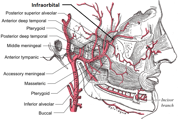
The infraorbital artery is a small artery in the head that arises from the maxillary artery and passes through the inferior orbital fissure to enter the orbit, then passes forward along the floor of the orbit, finally exiting the orbit through the infraorbital foramen to reach the face. Anatomy Origin The infraorbital artery arises from the maxillary artery; it often arises in conjunction with the posterior superior alveolar artery. It may be considered a continuation of the third part of the maxillary artery and continues the direction of the maxillary artery. Course It passes anterior-ward to enter the orbit through the inferior orbital fissure. In the orbit, it courses along the floor of the orbit with the infraorbital nerve first along the infraorbital groove and then the infraorbital canal. It exits the orbit (with the infraorbital nerve) through infraorbital foramen to reach the face, beneath the infraorbital head of the levator labii superioris muscle. Branches Wh ... [...More Info...] [...Related Items...] OR: [Wikipedia] [Google] [Baidu] |
Infraorbital Nerve
The infraorbital nerve is a branch of the maxillary nerve (itself a branch of the trigeminal nerve (CN V)). It arises in the pterygopalatine fossa. It passes through the inferior orbital fissure to enter the orbit. It travels through the orbit, then enters and traverses the infraorbital canal, exiting the canal at the infraorbital foramen to reach the face. It provides sensory innervation to the skin and mucous membranes around the middle of the face. Structure Origin The infraorbital nerve is a branch of the maxillary nerve (CN V2), itself a branch of the trigeminal nerve (CN V); it may be considered as the terminal branch of the maxillary nerve. It arises from the maxillary nerve in the pterygopalatine fossa. Course It travels through the inferior orbital fissure to enter the orbit. It runs anteriorly along the floor of the orbit in the infraorbital groove to the infraorbital canal of the maxilla. Within the infraorbital canal it has three branches, the posterior ... [...More Info...] [...Related Items...] OR: [Wikipedia] [Google] [Baidu] |
Superior Orbital Fissure
The superior orbital fissure is a foramen or cleft of the skull between the lesser and greater wings of the sphenoid bone. It gives passage to multiple structures, including the oculomotor nerve, trochlear nerve, ophthalmic nerve, abducens nerve, ophthalmic veins, and sympathetic fibres from the cavernous plexus. Structure The superior orbital fissure is usually 22 mm wide in adults, and is much larger medially. Its boundaries are formed by the (caudal surface of the) lesser wing of the sphenoid bone, and (medial border of the) greater wing of the sphenoid bone. Contents The superior orbital fissure is traversed by the following structures: * (superior and inferior divisions of the) oculomotor nerve (CN III) * trochlear nerve (CN IV) * lacrimal, frontal, and nasociliary branches of ophthalmic nerve (CN V1) * abducens nerve (CN VI) * superior ophthalmic vein and superior division of the inferior ophthalmic vein * sympathetic fibres from the cavernous nerve plex ... [...More Info...] [...Related Items...] OR: [Wikipedia] [Google] [Baidu] |
Pterygopalatine Fossa
In human anatomy, the pterygopalatine fossa (sphenopalatine fossa) is a fossa in the skull. A human skull contains two pterygopalatine fossae—one on the left side, and another on the right side. Each fossa is a cone-shaped paired depression deep to the infratemporal fossa and posterior to the maxilla on each side of the skull, located between the pterygoid process and the maxillary tuberosity close to the apex of the orbit. It is the indented area medial to the pterygomaxillary fissure leading into the sphenopalatine foramen. It communicates with the nasal and oral cavities, infratemporal fossa, orbit, pharynx, and middle cranial fossa through eight foramina. Structure Boundaries It has the following boundaries: * ''anterior'': superomedial part of the infratemporal surface of maxilla * ''posterior'': root of the pterygoid process and adjoining anterior surface of the greater wing of sphenoid bone * ''medial'': perpendicular plate of the palatine bone and its orbital an ... [...More Info...] [...Related Items...] OR: [Wikipedia] [Google] [Baidu] |
Orbit (anatomy)
In anatomy Anatomy () is the branch of morphology concerned with the study of the internal structure of organisms and their parts. Anatomy is a branch of natural science that deals with the structural organization of living things. It is an old scien ..., the orbit is the Body cavity, cavity or socket/hole of the skull in which the eye and Accessory visual structures, its appendages are situated. "Orbit" can refer to the bony socket, or it can also be used to imply the contents. In the adult human, the volume of the orbit is about , of which the eye occupies . The orbital contents comprise the eye, the Orbital fascia, orbital and retrobulbar fascia, extraocular muscles, cranial nerves optic nerve, II, oculomotor nerve, III, trochlear nerve, IV, trigeminal nerve, V, and abducens nerve, VI, blood vessels, fat, the lacrimal gland with its Lacrimal sac, sac and nasolacrimal duct, duct, the eyelids, Medial palpebral ligament, medial and Lateral palpebral raphe, lateral palpebr ... [...More Info...] [...Related Items...] OR: [Wikipedia] [Google] [Baidu] |


