|
Inferior Colliculus
The inferior colliculus (IC) (Latin for ''lower hill'') is the principal midbrain nucleus of the Auditory system, auditory pathway and receives input from several peripheral brainstem nuclei in the auditory pathway, as well as inputs from the auditory cortex.Shore, S. E.: ''Auditory/Somatosensory Interactions''. In: Squire (Ed.): ''Encyclopedia of Neuroscience'', Academic Press, 2009, pp. 691–695 The inferior colliculus has three subdivisions: the central nucleus, a dorsal cortex by which it is surrounded, and an external cortex which is located laterally. Its bimodal neurons are implicated in auditory-Somatosensory system, somatosensory interaction, receiving projections from Somatosensory system, somatosensory nuclei. This multisensory integration may underlie a filtering of self-effected sounds from vocalization, chewing, or respiration activities. The inferior colliculi together with the superior colliculi form the eminences of the corpora quadrigemina, and also part of the ... [...More Info...] [...Related Items...] OR: [Wikipedia] [Google] [Baidu] |
Sagittal Section
The sagittal plane (; also known as the longitudinal plane) is an anatomical plane that divides the body into right and left sections. It is perpendicular to the transverse and coronal planes. The plane may be in the center of the body and divide it into two equal parts ( mid-sagittal), or away from the midline and divide it into unequal parts (para-sagittal). The term ''sagittal'' was coined by Gerard of Cremona. Variations in terminology Examples of sagittal planes include: * The terms '' median plane'' or ''mid-sagittal plane'' are sometimes used to describe the sagittal plane running through the midline. This plane cuts the body into halves (assuming bilateral symmetry), passing through midline structures such as the navel and spine. It is one of the planes which, combined with the umbilical plane, defines the four quadrants of the human abdomen. * The term ''parasagittal'' is used to describe any plane parallel or adjacent to a given sagittal plane. Specific named par ... [...More Info...] [...Related Items...] OR: [Wikipedia] [Google] [Baidu] |
Lateral Lemniscus
The lateral lemniscus is a tract of axons in the brainstem that carries information about sound from the cochlear nucleus to various brainstem nuclei and ultimately the contralateral inferior colliculus of the midbrain. Three distinct, primarily inhibitory, cellular groups are located interspersed within these fibers, and are thus named the nuclei of the lateral lemniscus. Connections There are three small nuclei on each of the lateral lemnisci: * the intermediate nucleus of the lateral lemniscus (INLL) * the ventral nucleus of the lateral lemniscus (VNLL) * the dorsal nucleus of the lateral lemniscus (DNLL) Fibers leaving these brainstem nuclei ascending to the inferior colliculus rejoin the lateral lemniscus. In that sense, this is not a ' lemniscus' in the true sense of the word (second order, decussated sensory axons), as there is third (and out of the lateral superior olive, fourth) order information coming out of some of these brainstem nuclei. The lateral lemniscus is ... [...More Info...] [...Related Items...] OR: [Wikipedia] [Google] [Baidu] |
Neuroscience Information Framework
The Neuroscience Information Framework is a repository of global neuroscience web resources, including experimental, clinical, and translational neuroscience databases, knowledge bases, atlases, and genetic/ genomic resources and provides many authoritative links throughout the neuroscience portal of Wikipedia. Description The Neuroscience Information Framework (NIF) is an initiative of the NIH Blueprint for Neuroscience Research, which was established in 2004 by the National Institutes of Health. Development of the NIF started in 2008, when the University of California, San Diego School of Medicine obtained an NIH contract to create and maintain "a dynamic inventory of web-based neurosciences data, resources, and tools that scientists and students can access via any computer connected to the Internet". The project is headed by Maryann Martone, co-director of the National Center for Microscopy and Imaging Research (NCMIR), part of the multi-disciplinary Center for Research in Bi ... [...More Info...] [...Related Items...] OR: [Wikipedia] [Google] [Baidu] |
List Of Regions In The Human Brain
The human brain anatomical regions are ordered following standard neuroanatomy hierarchies. Functional, connective, and developmental regions are listed in parentheses where appropriate. Hindbrain (rhombencephalon) Myelencephalon * Medulla oblongata ** Medullary pyramids ** Arcuate nucleus ** Olivary body *** Inferior olivary nucleus ** Rostral ventrolateral medulla ** Caudal ventrolateral medulla ** Solitary nucleus (Nucleus of the solitary tract) **Respiratory center- Respiratory groups *** Dorsal respiratory group *** Ventral respiratory group or Apneustic centre **** Pre-Bötzinger complex **** Botzinger complex **** Retrotrapezoid nucleus **** Nucleus retrofacialis **** Nucleus retroambiguus **** Nucleus para-ambiguus ** Paramedian reticular nucleus ** Gigantocellular reticular nucleus ** Parafacial zone ** Cuneate nucleus ** Gracile nucleus ** Perihypoglossal nuclei *** Intercalated nucleus *** Prepositus nucleus *** Sublingual nucleus ** Area postrema **Medul ... [...More Info...] [...Related Items...] OR: [Wikipedia] [Google] [Baidu] |
Population Code
Neural coding (or neural representation) is a neuroscience field concerned with characterising the hypothetical relationship between the Stimulus (physiology), stimulus and the neuronal responses, and the relationship among the Electrophysiology, electrical activities of the neurons in the Neuronal ensemble, ensemble. Based on the theory that sensory and other information is represented in the brain by Biological neural network, networks of neurons, it is believed that neurons can encode both Digital data, digital and analog signal, analog information. Overview Neurons have an ability uncommon among the cells of the body to propagate signals rapidly over large distances by generating characteristic electrical pulses called action potentials: voltage spikes that can travel down axons. Sensory neurons change their activities by firing sequences of action potentials in various temporal patterns, with the presence of external sensory stimuli, such as light, sound, taste, Olfaction, sm ... [...More Info...] [...Related Items...] OR: [Wikipedia] [Google] [Baidu] |
Just-noticeable Difference
In the branch of experimental psychology focused on sense, sensation, and perception, which is called psychophysics, a just-noticeable difference or JND is the amount something must be changed in order for a difference to be noticeable, detectable at least half the time. This limen is also known as the difference limen, difference threshold, or least perceptible difference. Quantification For many sensory modalities, over a wide range of stimulus magnitudes sufficiently far from the upper and lower limits of perception, the 'JND' is a fixed proportion of the reference sensory level, and so the ratio of the JND/reference is roughly constant (that is the JND is a constant proportion/percentage of the reference level). Measured in physical units, we have: \frac = k, where I\! is the original intensity of the particular stimulation, \Delta I\! is the addition to it required for the change to be perceived (the JND), and ''k'' is a constant. This rule was first discovered by Erns ... [...More Info...] [...Related Items...] OR: [Wikipedia] [Google] [Baidu] |
Interaural Time Difference
The interaural time difference (or ITD) when concerning humans or animals, is the difference in arrival time of a sound between two ears. It is important in the Sound localization, localization of sounds, as it provides a cue to the direction or angle of the sound source from the head. If a signal arrives at the head from one side, the signal has further to travel to reach the far ear than the near ear. This pathlength difference results in a time difference between the sound's arrivals at the ears, which is detected and aids the process of identifying the direction of sound source. When a signal is produced in the horizontal plane, its angle in relation to the head is referred to as its azimuth, with 0 degrees (0°) azimuth being directly in front of the listener, 90° to the right, and 180° being directly behind. Different methods for measuring ITDs * For an abrupt stimulus such as a click, onset ITDs are measured. An onset ITD is the time difference between the onset of the s ... [...More Info...] [...Related Items...] OR: [Wikipedia] [Google] [Baidu] |
Vestibulo-ocular Reflex
The vestibulo-ocular reflex (VOR) is a reflex that acts to stabilize Gaze (physiology), gaze during head movement, with eye movement due to activation of the vestibular system, it is also known as the cervico-ocular reflex. The reflex acts to image stabilization, stabilize images on the retinas of the eye during head movement. Gaze is held steadily on a location by producing eye movements in the direction opposite that of head movement. For example, when the head moves to the right, the eyes move to the left, meaning the image a person sees stays the same even though the head has turned. Since slight head movement is present all the time, VOR is necessary for stabilizing vision: people with an impaired reflex find it difficult to read using print, because the eyes do not stabilise during small head tremors, and also because damage to reflex can cause nystagmus. The VOR does not depend on what is seen. It can also be activated by hot or cold stimulation of the inner ear, where the ... [...More Info...] [...Related Items...] OR: [Wikipedia] [Google] [Baidu] |
Startle Response
In animals, including humans, the startle response is a largely unconscious defensive response to sudden or threatening Stimulus (physiology), stimuli, such as sudden noise or sharp movement, and is associated with negative Affect (psychology), affect.Rammirez-Moreno, David. "A computational model for the modulation of the prepulse inhibition of the acoustic startle reflex". ''Biological Cybernetics'', 2012, p. 169 Usually the onset of the startle response is a startle reflex reaction. The startle reflex is a brainstem reflectory reaction (reflex) that serves to protect vulnerable parts, such as the back of the neck (whole-body startle) and the eyes (eyeblink) and Fight-or-flight response, facilitates escape from sudden stimuli. It is found across many different species, throughout all stages of life. A variety of responses may occur depending on the affected individual's emotional state, body posture, preparation for execution of a motor task, or other activities. The startle respo ... [...More Info...] [...Related Items...] OR: [Wikipedia] [Google] [Baidu] |
Thalamus
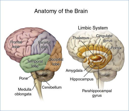
The thalamus (: thalami; from Greek language, Greek Wikt:θάλαμος, θάλαμος, "chamber") is a large mass of gray matter on the lateral wall of the third ventricle forming the wikt:dorsal, dorsal part of the diencephalon (a division of the forebrain). Nerve fibers project out of the thalamus to the cerebral cortex in all directions, known as the thalamocortical radiations, allowing hub (network science), hub-like exchanges of information. It has several functions, such as the relaying of sensory neuron, sensory and motor neuron, motor signals to the cerebral cortex and the regulation of consciousness, sleep, and alertness. Anatomically, the thalami are paramedian symmetrical structures (left and right), within the vertebrate brain, situated between the cerebral cortex and the midbrain. It forms during embryonic development as the main product of the diencephalon, as first recognized by the Swiss embryologist and anatomist Wilhelm His Sr. in 1893. Anatomy The thalami ar ... [...More Info...] [...Related Items...] OR: [Wikipedia] [Google] [Baidu] |
Lateralization
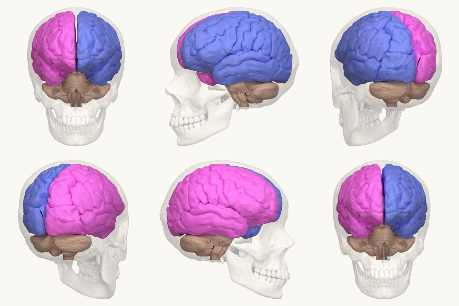
The lateralization of brain function (or hemispheric dominance/ lateralization) is the tendency for some neural functions or cognitive processes to be specialized to one side of the brain or the other. The median longitudinal fissure separates the human brain into two distinct cerebral hemispheres connected by the corpus callosum. Both hemispheres exhibit brain asymmetries in both structure and neuronal network composition associated with specialized function. Lateralization of brain structures has been studied using both healthy and split-brain patients. However, there are numerous counterexamples to each generalization and each human's brain develops differently, leading to unique lateralization in individuals. This is different from specialization, as lateralization refers only to the function of one structure divided between two hemispheres. Specialization is much easier to observe as a trend, since it has a stronger anthropological history. The best example of an establi ... [...More Info...] [...Related Items...] OR: [Wikipedia] [Google] [Baidu] |


