|
Iliotibial Band Syndrome
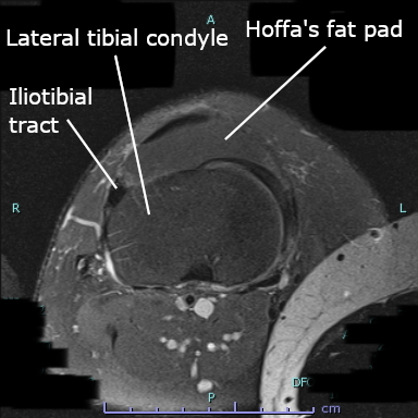
Iliotibial band syndrome (ITBS) is the second most common knee injury, and is caused by inflammation located on the lateral aspect of the knee due to friction between the iliotibial band and the lateral epicondyle of the femur. Pain is felt most commonly on the lateral aspect of the knee and is most intensive at 30 degrees of knee flexion. Risk factors in women include increased hip adduction and knee internal rotation. Risk factors seen in men are increased hip internal rotation and knee adduction. ITB syndrome is most associated with long-distance running, cycling, weight-lifting, and with military training. Signs and symptoms ITBS symptoms range from a stinging sensation just above the knee and outside of the knee (lateral side of the knee) joint, to swelling or thickening of the tissue in the area where the band moves over the femur. The stinging sensation just above the knee joint is felt on the outside of the knee or along the entire length of the iliotibial band. At ini ... [...More Info...] [...Related Items...] OR: [Wikipedia] [Google] [Baidu] |
Sports Medicine
Sports medicine is a branch of medicine that deals with physical fitness and the treatment and prevention of injuries related to sports and exercise. Although most sports teams have employed team physicians for many years, it is only since the late 20th century that sports medicine emerged as a distinct field of health care. In many countries, now over 50, sports medicine (or sport and exercise medicine) is a recognized medical specialty (with similar training and standards to other medical specialties or sub-specialties). In the majority of countries where sports medicine is recognized and practiced, it is a physician (non-surgical) specialty, but in some (such as the USA), it can equally be a surgical or non-surgical medical specialty, and also a specialty field within primary care. In other contexts, the field of sports medicine encompasses the scope of both medical specialists as well as Allied health professions, allied health practitioners who work in the field of sport, su ... [...More Info...] [...Related Items...] OR: [Wikipedia] [Google] [Baidu] |
Iliotibial Tract
The iliotibial tract or iliotibial band (ITB; also known as Maissiat's band or the IT band) is a longitudinal fibrous reinforcement of the fascia lata. The action of the muscles associated with the ITB ( tensor fasciae latae and some fibers of gluteus maximus) flex, extend, abduct, and laterally and medially rotate the hip. The ITB contributes to lateral knee stabilization. During knee extension the ITB moves anterior to the lateral condyle of the femur, while ~30 degrees knee flexion, the ITB moves posterior to the lateral condyle. However, it has been suggested that this is only an illusion due to the changing tension in the anterior and posterior fibers during movement. It originates at the anterolateral iliac tubercle portion of the external lip of the iliac crest and inserts at the lateral condyle of the tibia at Gerdy's tubercle. The figure shows only the proximal part of the iliotibial tract. The part of the iliotibial band which lies beneath the tensor fasciae latae ... [...More Info...] [...Related Items...] OR: [Wikipedia] [Google] [Baidu] |
Fibular Collateral Ligament
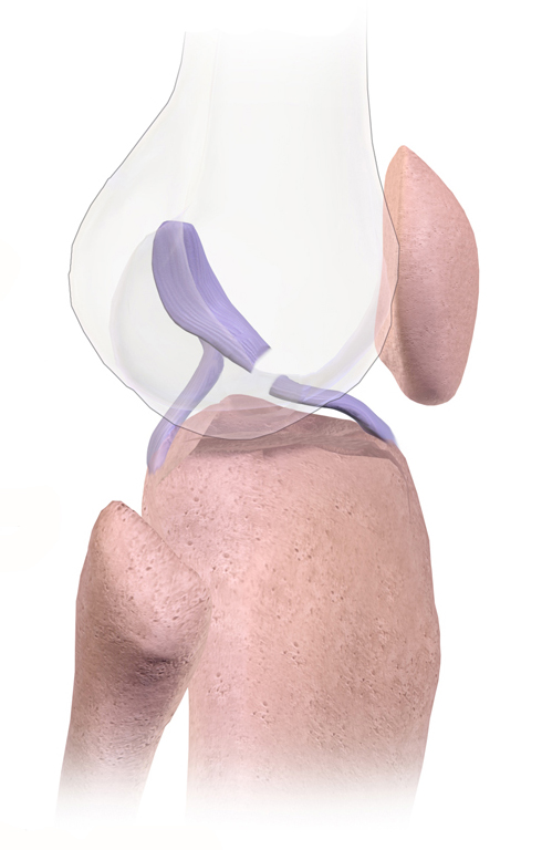
The lateral collateral ligament (LCL, long external lateral ligament or fibular collateral ligament) is an extrinsic ligament of the knee located on the lateral side of the knee. Its superior attachment is at the lateral epicondyle of the femur (superoposterior to the popliteal groove); its inferior attachment is at the lateral aspect of the head of fibula (anterior to the apex). The LCL is not fused with the joint capsule. Inferiorly, the LCL splits the tendon of insertion of the biceps femoris muscle. Structure The LCL measures some 5 cm in length. It is rounded, and is more narrow and less broad compared to the medial collateral ligament. It extends obliquely inferoposteriorly from its superior attachment to its inferior attachment. In contrast to the medial collateral ligament, it is not fused with either the capsular ligament nor the lateral meniscus. Because of this, the LCL is more flexible than its medial counterpart, and is therefore less susceptible to injury. ... [...More Info...] [...Related Items...] OR: [Wikipedia] [Google] [Baidu] |
Patellofemoral Pain Syndrome
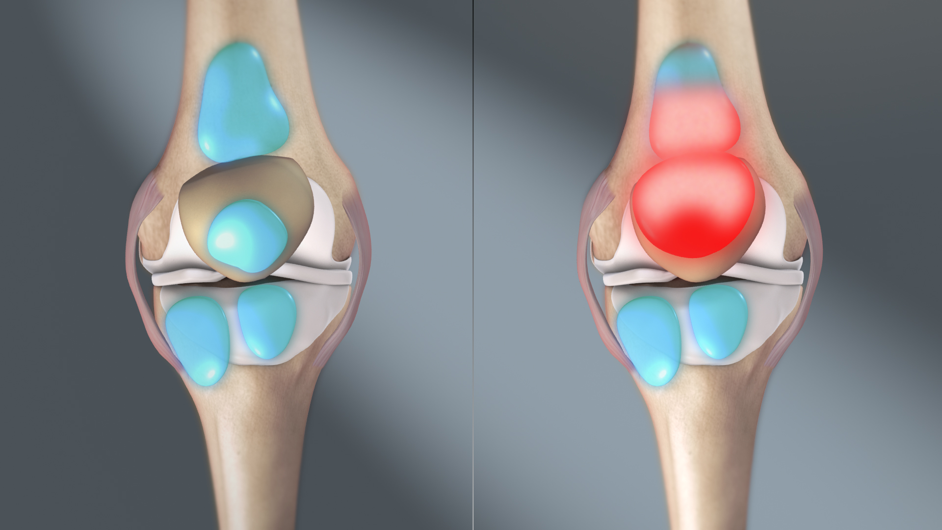
Patellofemoral pain syndrome (PFPS; not to be confused with jumper's knee) is knee pain as a result of problems between the kneecap and the femur. The pain is generally in the front of the knee and comes on gradually. Pain may worsen with sitting down with a bent knee for long periods of time, excessive use, or climbing and descending stairs. While the exact cause is unclear, it is believed to be due to overuse. Risk factors include trauma, increased training, and a weak quadriceps muscle. It is particularly common among runners. The diagnosis is generally based on the symptoms and examination. If pushing the kneecap into the femur increases the pain, the diagnosis is more likely. Treatment typically involves rest and rehabilitation with a physical therapist. Runners may need to switch to activities such as cycling or swimming. Insoles may help some people. Symptoms may last for years despite treatment. Patellofemoral pain syndrome is the most common cause of knee pain, aff ... [...More Info...] [...Related Items...] OR: [Wikipedia] [Google] [Baidu] |
Stress Fracture
A stress fracture is a fatigue-induced bone fracture caused by repeated stress over time. Instead of resulting from a single severe impact, stress fractures are the result of accumulated injury from repeated submaximal loading, such as running or jumping. Because of this mechanism, stress fractures are common overuse injuries in athletes. Stress fractures can be described as small cracks in the bone, or hairline fractures. Stress fractures of the foot are sometimes called " march fractures" because of the injury's prevalence among heavily marching soldiers. Stress fractures most frequently occur in weight-bearing bones of the lower extremities, such as the tibia and fibula (bones of the lower leg), calcaneus (heel bone), metatarsal and navicular bones (bones of the foot). Less common are stress fractures to the femur, pelvis, sacrum, lumbar spine (lower back), hips, hands, and wrists. Stress fractures make up about 20% of overall sports injuries. Treatment usually consists o ... [...More Info...] [...Related Items...] OR: [Wikipedia] [Google] [Baidu] |
Biceps Femoris Muscle
The biceps femoris () is a muscle of the thigh located to the posterior, or back. As its name implies, it consists of two heads; the long head is considered part of the hamstring muscle group, while the short head is sometimes excluded from this characterization, as it only causes knee flexion (but not hip extension) and is activated by a separate nerve (the peroneal, as opposed to the tibial branch of the sciatic nerve). Structure It has two heads of origin: *the ''long head'' arises from the lower and inner impression on the posterior part of the tuberosity of the ischium. This is a common tendon origin with the semitendinosus muscle, and from the lower part of the sacrotuberous ligament. *the ''short head'', arises from the lateral lip of the linea aspera, between the adductor magnus and vastus lateralis extending up almost as high as the insertion of the gluteus maximus, from the lateral prolongation of the linea aspera to within 5 cm. of the lateral condyle; and fr ... [...More Info...] [...Related Items...] OR: [Wikipedia] [Google] [Baidu] |
Tendinopathy
Tendinopathy is a type of tendon disorder that results in pain, swelling, and impaired function. The pain is typically worse with movement. It most commonly occurs around the shoulder ( rotator cuff tendinitis, biceps tendinitis), elbow ( tennis elbow, golfer's elbow), wrist, hip, knee ( jumper's knee, popliteus tendinopathy), or ankle ( Achilles tendinitis). Causes may include an injury or repetitive activities. Less common causes include infection, arthritis, gout, thyroid disease, diabetes and the use of quinolone antibiotic medicines. Groups at risk include people who do manual labor, musicians, and athletes. Diagnosis is typically based on symptoms, examination, and occasionally medical imaging. A few weeks following an injury little inflammation remains, with the underlying problem related to weak or disrupted tendon fibrils. Treatment may include rest, NSAIDs, splinting, and physiotherapy. Less commonly steroid injections or surgery may be done. About 80% of over ... [...More Info...] [...Related Items...] OR: [Wikipedia] [Google] [Baidu] |
Lateral Meniscus
The lateral meniscus (external semilunar fibrocartilage) is a fibrocartilaginous band that spans the lateral side of the interior of the knee joint. It is one of two menisci of the knee, the other being the medial meniscus. It is nearly circular and covers a larger portion of the articular surface than the medial. It can occasionally be injured or torn by twisting the knee or applying direct force, as seen in contact sports. Structure The lateral meniscus is grooved laterally for the tendon of the popliteus, which separates it from the fibular collateral ligament. Its anterior end is attached in front of the intercondyloid eminence of the tibia, lateral to, and behind, the anterior cruciate ligament, with which it blends; the posterior end is attached behind the intercondyloid eminence of the tibia and in front of the posterior end of the medial meniscus. The anterior attachment of the lateral meniscus is twisted on itself so that its free margin looks backward and upward, its ... [...More Info...] [...Related Items...] OR: [Wikipedia] [Google] [Baidu] |
Bursitis
Bursitis is the inflammation of one or more bursae (synovial sacs) of synovial fluid in the body. They are lined with a synovial membrane that secretes a lubricating synovial fluid. There are more than 150 bursae in the human body. The bursae (bur-see) rest at the points where internal functionaries, such as muscles and tendons, slide across bone. Healthy bursae create a smooth, almost frictionless functional gliding surface making normal movement painless. When bursitis occurs, however, movement relying on the inflamed bursa becomes difficult and painful. Moreover, movement of tendons and muscles over the inflamed bursa aggravates its inflammation, perpetuating the problem. Muscle can also be stiffened. Signs and symptoms Bursitis commonly affects superficial bursae. These include the subacromial, prepatellar, retrocalcaneal, and ''pes anserinus'' bursae of the shoulder, knee, heel and shin, etc. (see below). Symptoms vary from localized warmth and erythema (redness) to joi ... [...More Info...] [...Related Items...] OR: [Wikipedia] [Google] [Baidu] |
Flexion
Motion, the process of movement, is described using specific anatomical terminology, anatomical terms. Motion includes movement of Organ (anatomy), organs, joints, Limb (anatomy), limbs, and specific sections of the body. The terminology used describes this motion according to its direction relative to the anatomical position of the body parts involved. Anatomy, Anatomists and others use a unified set of terms to describe most of the movements, although other, more specialized terms are necessary for describing unique movements such as those of the hands, feet, and eyes. In general, motion is classified according to the anatomical plane it occurs in. ''Flexion'' and ''extension'' are examples of ''angular'' motions, in which two axes of a joint are brought closer together or moved further apart. ''Rotational'' motion may occur at other joints, for example the shoulder, and are described as ''internal'' or ''external''. Other terms, such as ''elevation'' and ''depression'', descri ... [...More Info...] [...Related Items...] OR: [Wikipedia] [Google] [Baidu] |
Lateral Epicondyle Of The Femur .
The lateral epicondyle of the femur, smaller and less prominent than the medial epicondyle, gives attachment to the fibular collateral ligament of the knee-joint. Directly below it is a small depression from which a smooth well-marked groove curves obliquely upward and backward to the posterior extremity of the condyle A condyle (; in References External links * * Bones of the lower limb Femur {{musculoskeletal-stub ...[...More Info...] [...Related Items...] OR: [Wikipedia] [Google] [Baidu] |
Hip (anatomy)
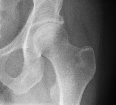
In vertebrate anatomy, the hip, or coxaLatin ''coxa'' was used by Celsus in the sense "hip", but by Pliny the Elder in the sense "hip bone" (Diab, p 77) (: ''coxae'') in medical terminology, refers to either an anatomical region or a joint on the outer (lateral) side of the pelvis. The hip region is located lateral and anterior to the gluteal region, inferior to the iliac crest, and lateral to the obturator foramen, with muscle tendons and soft tissues overlying the greater trochanter of the femur. In adults, the three pelvic bones ( ilium, ischium and pubis) have fused into one hip bone, which forms the superomedial/deep wall of the hip region. The hip joint, scientifically referred to as the acetabulofemoral joint (''art. coxae''), is the ball-and-socket joint between the pelvic acetabulum and the femoral head. Its primary function is to support the weight of the torso in both static (e.g. standing) and dynamic (e.g. walking or running) postures. The hip joints have very ... [...More Info...] [...Related Items...] OR: [Wikipedia] [Google] [Baidu] |
