|
Hallux
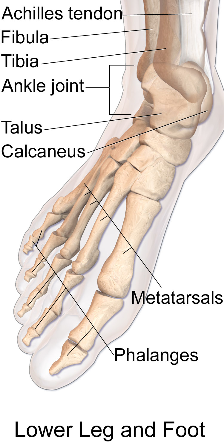
Toes are the digits of the foot of a tetrapod. Animal species such as cats that walk on their toes are described as being ''digitigrade''. Humans, and other animals that walk on the soles of their feet, are described as being ''plantigrade''; '' unguligrade'' animals are those that walk on hooves at the tips of their toes. Structure There are normally five toes present on each human foot. Each toe consists of three phalanx bones, the proximal, middle, and distal, with the exception of the big toe (). For a minority of people, the little toe also is missing a middle bone. The hallux only contains two phalanx bones, the proximal and distal. The joints between each phalanx are the interphalangeal joints. The proximal phalanx bone of each toe articulates with the metatarsal bone of the foot at the metatarsophalangeal joint. Each toe is surrounded by skin, and present on all five toes is a toenail. The toes are, from medial to lateral: * the first toe, also known as the h ... [...More Info...] [...Related Items...] OR: [Wikipedia] [Google] [Baidu] |
Foot On White Background (cropped)
The foot (: feet) is an anatomical structure found in many vertebrates. It is the terminal portion of a limb which bears weight and allows locomotion. In many animals with feet, the foot is an organ at the terminal part of the leg made up of one or more segments or bones, generally including claws and/or nails. Etymology The word "foot", in the sense of meaning the "terminal part of the leg of a vertebrate animal" comes from Old English ''fot'', from Proto-Germanic *''fot'' (source also of Old Frisian ''fot'', Old Saxon ''fot'', Old Norse ''fotr'', Danish ''fod'', Swedish ''fot'', Dutch ''voet'', Old High German ''fuoz'', German ''Fuß'', Gothic ''fotus'', all meaning "foot"), from PIE root *''ped-'' "foot". The plural form ''feet'' is an instance of i-mutation. Structure The human foot is a strong and complex mechanical structure containing 26 bones, 33 joints (20 of which are actively articulated), and more than a hundred muscles, tendons, and ligaments.Podiatry Channel, ... [...More Info...] [...Related Items...] OR: [Wikipedia] [Google] [Baidu] |
Foot
The foot (: feet) is an anatomical structure found in many vertebrates. It is the terminal portion of a limb which bears weight and allows locomotion. In many animals with feet, the foot is an organ at the terminal part of the leg made up of one or more segments or bones, generally including claws and/or nails. Etymology The word "foot", in the sense of meaning the "terminal part of the leg of a vertebrate animal" comes from Old English ''fot'', from Proto-Germanic *''fot'' (source also of Old Frisian ''fot'', Old Saxon ''fot'', Old Norse ''fotr'', Danish ''fod'', Swedish ''fot'', Dutch ''voet'', Old High German ''fuoz'', German ''Fuß'', Gothic ''fotus'', all meaning "foot"), from PIE root *''ped-'' "foot". The plural form ''feet'' is an instance of i-mutation. Structure The human foot is a strong and complex mechanical structure containing 26 bones, 33 joints (20 of which are actively articulated), and more than a hundred muscles, tendons, and ligaments.Podiatry Chan ... [...More Info...] [...Related Items...] OR: [Wikipedia] [Google] [Baidu] |
Digit (anatomy)
A digit is one of several most distal parts of a limb, such as fingers or toes, present in many vertebrates. Names Some languages have different names for hand and foot digits (English: respectively "finger" and " toe", German: "Finger" and "Zeh", French: "doigt" and "orteil"). In other languages, e.g. Arabic, Russian, Polish, Spanish, Portuguese, Italian, Czech, Tagalog, Turkish, Bulgarian, and Persian, there are no specific one-word names for fingers and toes; these are called "digit of the hand" or "digit of the foot" instead. In Japanese, yubi (指) can mean either, depending on context. Human digits Humans normally have five digits on each extremity. Each digit is formed by several bones called phalanges, surrounded by soft tissue. Human fingers normally have a nail at the distal phalanx. The phenomenon of polydactyly occurs when extra digits are present; fewer digits than normal are also possible, for instance in ectrodactyly. Whether such a mutation can ... [...More Info...] [...Related Items...] OR: [Wikipedia] [Google] [Baidu] |
Abductor Hallucis Muscle
The abductor hallucis muscle is an intrinsic muscle of the foot. It participates in the Anatomical terms of motion#Abduction and adduction, abduction and Anatomical terms of motion#Flexion and extension, flexion of the Toe#Hallux, great toe. Structure The abductor hallucis muscle is located in the medial border of the foot and contributes to form the prominence that is observed on the region. It is inserted behind on the tuberosity of the calcaneus, the Flexor retinaculum of foot, flexor retinaculum, and the plantar aponeurosis. Its muscle body, relatively thick behind, flattens as it goes forward. It ends in a common tendon with the medial head of the flexor hallucis brevis that inserts on the medial surface of the base of the first Phalanx bone, proximal phalanx and its related sesamoid bone. Its medial surface is superficial and covered with the muscle's fascia and the skin. Nerve supply Abductor hallucis is supplied by the medial plantar nerve. The nerves that supply it ente ... [...More Info...] [...Related Items...] OR: [Wikipedia] [Google] [Baidu] |
Flexor Hallucis Brevis
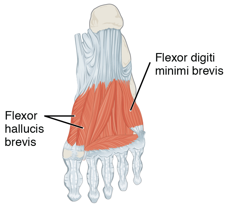
Flexor hallucis brevis muscle is a muscle of the foot that flexes the big toe. Structure Flexor hallucis brevis muscle arises, by a pointed tendinous process, from the medial part of the under surface of the cuboid bone, from the contiguous portion of the third cuneiform, and from the prolongation of the tendon of the tibialis posterior muscle which is attached to that bone. It divides in front into two portions, which are inserted into the medial and lateral sides of the base of the first phalanx of the great toe, a sesamoid bone being present in each tendon at its insertion. The medial portion is blended with the abductor hallucis muscle previous to its insertion; the lateral portion (sometimes described as the first plantar interosseus) with the adductor hallucis muscle. The tendon of the flexor hallucis longus muscle lies in a groove between the two. Its tendon usually contains two sesamoid bones at the point under the first metatarsophalangeal joint. Innervation The med ... [...More Info...] [...Related Items...] OR: [Wikipedia] [Google] [Baidu] |
Flexor Hallucis Longus
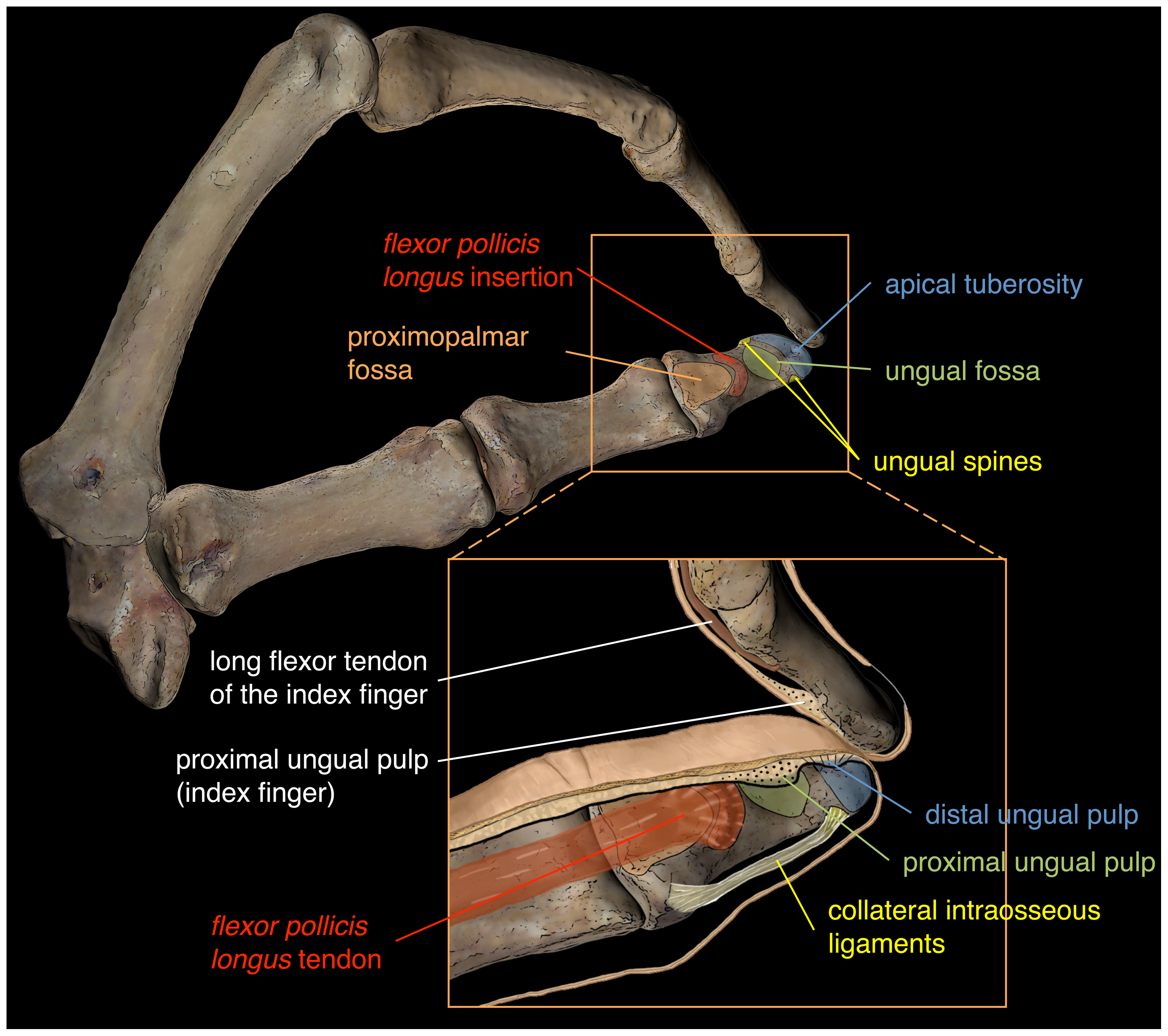
The flexor hallucis longus muscle (FHL) attaches to the plantar surface of phalanx of the great toe and is responsible for flexing that toe. The FHL is one of the three deep muscles of the posterior compartment of the leg, the others being the flexor digitorum longus and the tibialis posterior. The tibialis posterior is the most powerful of these deep muscles. All three muscles are innervated by the tibial nerve which comprises half of the sciatic nerve. Structure The flexor hallucis longus is situated on the fibular side of the leg. It arises from the inferior two-thirds of the posterior surface of the body of the fibula, with the exception of 2.5 cm at its lowest part; from the lower part of the interosseous membrane; from an intermuscular septum between it and the peroneus muscles, laterally, and from the fascia covering the tibialis posterior, medially. The fibers pass obliquely downward and backward, where it passes through the tarsal tunnel on the medial sid ... [...More Info...] [...Related Items...] OR: [Wikipedia] [Google] [Baidu] |
Phalanges Of The Foot
The phalanges (: phalanx ) are digit (anatomy), digital bones in the hands and foot, feet of most vertebrates. In primates, the Thumb, thumbs and Hallux, big toes have two phalanges while the other Digit (anatomy), digits have three phalanges. The phalanges are classed as long bones. Structure The phalanges are the bones that make up the fingers of the hand and the toes of the foot. There are 56 phalanges in the human body, with fourteen on each hand and foot. Three phalanges are present on each finger and toe, with the exception of the thumb and hallux, big toe, which possess only two. The middle and far phalanges of the fifth toes are often fused together (symphalangism). The phalanges of the hand are commonly known as the finger bones. The phalanges of the foot differ from the hand in that they are often shorter and more compressed, especially in the proximal phalanges, those closest to the torso. A phalanx is named according to whether it is Anatomical terms of locatio ... [...More Info...] [...Related Items...] OR: [Wikipedia] [Google] [Baidu] |
Interphalangeal Joints Of Foot
The interphalangeal joints of the foot are the joints between the phalanx bones of the toes in the foot, feet. Since the Toe#Hallux, great toe only has two phalanx bones (proximal and distal phalanges), it only has one interphalangeal joint, which is often abbreviated as the "IP joint". The rest of the toes each have three phalanx bones (proximal, middle, and distal phalanges), so they have two interphalangeal joints: the proximal interphalangeal joint between the proximal and middle phalanges (abbreviated "PIP joint") and the distal interphalangeal joint between the middle and distal phalanges (abbreviated "DIP joint"). All interphalangeal joints are ginglymoid (hinge) joints, and each has a plantar (underside) and two Collateral ligaments of interphalangeal articulations of foot, collateral ligaments. In the arrangement of these ligaments, Extension (kinesiology), extensor tendons supply the places of Anatomical terms of location#Dorsal and ventral, dorsal ligaments, which is ... [...More Info...] [...Related Items...] OR: [Wikipedia] [Google] [Baidu] |
Phalanx Bone
The phalanges (: phalanx ) are digital bones in the hands and feet of most vertebrates. In primates, the thumbs and big toes have two phalanges while the other digits have three phalanges. The phalanges are classed as long bones. Structure The phalanges are the bones that make up the fingers of the hand and the toes of the foot. There are 56 phalanges in the human body, with fourteen on each hand and foot. Three phalanges are present on each finger and toe, with the exception of the thumb and big toe, which possess only two. The middle and far phalanges of the fifth toes are often fused together (symphalangism). The phalanges of the hand are commonly known as the finger bones. The phalanges of the foot differ from the hand in that they are often shorter and more compressed, especially in the proximal phalanges, those closest to the torso. A phalanx is named according to whether it is proximal, middle, or distal and its associated finger or toe. The proximal phalange ... [...More Info...] [...Related Items...] OR: [Wikipedia] [Google] [Baidu] |
Metatarsophalangeal Joints
The metatarsophalangeal joints (MTP joints) are the joints between the metatarsal bones of the foot and the proximal bones (proximal phalanges) of the toes. They are analogous to the knuckles of the hand, and are consequently known as toe knuckles in common speech. They are condyloid joints, meaning that an elliptical or rounded surface (of the metatarsal bones) comes close to a shallow cavity (of the proximal phalanges). The region of skin directly below the joints forms the ball of the foot. The ligaments are the plantar and two collateral. Movements The movements permitted in the metatarsophalangeal joints are flexion, extension, abduction, adduction and circumduction. File:The feet of C. H. Unthan, the armless fiddler Wellcome L0034227.jpg, Left: toes adducted (pulled towards the center) and spread (abducted); right, both feet clenched (plantar flexed) File:Footgym rings1.jpg, The upper foot is clenching (plantarflexing) at the MTP joints and at the joints of the to ... [...More Info...] [...Related Items...] OR: [Wikipedia] [Google] [Baidu] |
Adductor Hallucis Muscle
The Adductor hallucis (adductor obliquus hallucis) arises by two heads—oblique and transverse and is responsible for adducting the big toe. It has two heads, both are innervated by the lateral plantar nerve. Structure Oblique head The ''oblique head'' is a large, thick, fleshy mass, crossing the foot obliquely and occupying the hollow space under the first, second, third and fourth metatarsal bones. It arises from the bases of the second, third, and fourth metatarsal bones, and from the sheath of the tendon of the Peroneus longus, and is inserted, together with the lateral portion of the flexor hallucis brevis, into the lateral side of the base of the first phalanx of the great toe. Transverse head The ''transverse head'' (''Transversus pedis'') is a narrow, flat fasciculus which arises from the plantar metatarsophalangeal ligaments of the third, fourth, and fifth toes (sometimes only from the third and fourth), and from the transverse ligament of the metatarsals. It is i ... [...More Info...] [...Related Items...] OR: [Wikipedia] [Google] [Baidu] |


