|
Granulomatous Meningoencephalitis
Granulomatous meningoencephalitis (GME) is an inflammatory disease of the central nervous system (CNS) of dogs and, rarely, cats. It is a form of meningoencephalitis. GME is likely second only to encephalitis caused by ''canine distemper virus'' as the most common cause of inflammatory disease of the canine CNS. The disease is more common in female dogs of young and middle age. It has a rapid onset. The lesions of GME exist mainly in the white matter of the cerebrum, brainstem, cerebellum, and spinal cord. The cause is only known to be noninfectious and is considered at this time to be idiopathic. Because lesions resemble those seen in allergic meningoencephalitis, GME is thought to have an immune-mediated cause, but it is also thought that the disease may be based on an abnormal response to an infectious agent. One study searched for viral DNA from '' canine herpesvirus'', '' canine adenovirus'', and ''canine parvovirus'' in brain tissue from dogs with GME, necrotizing mening ... [...More Info...] [...Related Items...] OR: [Wikipedia] [Google] [Baidu] |
Central Nervous System
The central nervous system (CNS) is the part of the nervous system consisting primarily of the brain, spinal cord and retina. The CNS is so named because the brain integrates the received information and coordinates and influences the activity of all parts of the bodies of bilateria, bilaterally symmetric and triploblastic animals—that is, all multicellular animals except sponges and Coelenterata, diploblasts. It is a structure composed of nervous tissue positioned along the Anatomical_terms_of_location#Rostral,_cranial,_and_caudal, rostral (nose end) to caudal (tail end) axis of the body and may have an enlarged section at the rostral end which is a brain. Only arthropods, cephalopods and vertebrates have a true brain, though precursor structures exist in onychophorans, gastropods and lancelets. The rest of this article exclusively discusses the vertebrate central nervous system, which is radically distinct from all other animals. Overview In vertebrates, the brain and spinal ... [...More Info...] [...Related Items...] OR: [Wikipedia] [Google] [Baidu] |
Pathologic Nystagmus
Nystagmus is a condition of involuntary (or voluntary, in some cases) eye movement. People can be born with it but more commonly acquire it in infancy or later in life. In many cases it may result in reduced or limited vision. In normal eyesight, while the head rotates about an axis, distant visual images are sustained by rotating eyes in the opposite direction of the respective axis. The semicircular canals in the vestibule of the ear sense angular acceleration, and send signals to the nuclei for eye movement in the brain. From here, a signal is relayed to the extraocular muscles to allow one's gaze to fix on an object as the head moves. Nystagmus occurs when the semicircular canals are stimulated (e.g., by means of the caloric test, or by disease) while the head is stationary. The direction of ocular movement is related to the semicircular canal that is being stimulated. There are two key forms of nystagmus: pathological and physiological, with variations within each type. ... [...More Info...] [...Related Items...] OR: [Wikipedia] [Google] [Baidu] |
Magnetic Resonance Imaging
Magnetic resonance imaging (MRI) is a medical imaging technique used in radiology to generate pictures of the anatomy and the physiological processes inside the body. MRI scanners use strong magnetic fields, magnetic field gradients, and radio waves to form images of the organs in the body. MRI does not involve X-rays or the use of ionizing radiation, which distinguishes it from computed tomography (CT) and positron emission tomography (PET) scans. MRI is a medical application of nuclear magnetic resonance (NMR) which can also be used for imaging in other NMR applications, such as NMR spectroscopy. MRI is widely used in hospitals and clinics for medical diagnosis, staging and follow-up of disease. Compared to CT, MRI provides better contrast in images of soft tissues, e.g. in the brain or abdomen. However, it may be perceived as less comfortable by patients, due to the usually longer and louder measurements with the subject in a long, confining tube, although ... [...More Info...] [...Related Items...] OR: [Wikipedia] [Google] [Baidu] |
Computed Tomography
A computed tomography scan (CT scan), formerly called computed axial tomography scan (CAT scan), is a medical imaging technique used to obtain detailed internal images of the body. The personnel that perform CT scans are called radiographers or radiology technologists. CT scanners use a rotating X-ray tube and a row of detectors placed in a gantry to measure X-ray attenuations by different tissues inside the body. The multiple X-ray measurements taken from different angles are then processed on a computer using tomographic reconstruction algorithms to produce tomographic (cross-sectional) images (virtual "slices") of a body. CT scans can be used in patients with metallic implants or pacemakers, for whom magnetic resonance imaging (MRI) is contraindicated. Since its development in the 1970s, CT scanning has proven to be a versatile imaging technique. While CT is most prominently used in medical diagnosis, it can also be used to form images of non-living objects. The 1979 N ... [...More Info...] [...Related Items...] OR: [Wikipedia] [Google] [Baidu] |
Monocytes
Monocytes are a type of leukocyte or white blood cell. They are the largest type of leukocyte in blood and can differentiate into macrophages and monocyte-derived dendritic cells. As a part of the vertebrate innate immune system monocytes also influence adaptive immune responses and exert tissue repair functions. There are at least three subclasses of monocytes in human blood based on their phenotypic receptors. Structure Monocytes are amoeboid in appearance, and have nongranulated cytoplasm. Thus they are classified as agranulocytes, although they might occasionally display some azurophil granules and/or vacuoles. With a diameter of 15–22 μm, monocytes are the largest cell type in peripheral blood. Monocytes are mononuclear cells and the ellipsoidal nucleus is often lobulated/indented, causing a bean-shaped or kidney-shaped appearance. Monocytes compose 2% to 10% of all leukocytes in the human body. Development Monocytes are produced by the bone marrow from precur ... [...More Info...] [...Related Items...] OR: [Wikipedia] [Google] [Baidu] |
Lymphocyte
A lymphocyte is a type of white blood cell (leukocyte) in the immune system of most vertebrates. Lymphocytes include T cells (for cell-mediated and cytotoxic adaptive immunity), B cells (for humoral, antibody-driven adaptive immunity), and innate lymphoid cells (ILCs; "innate T cell-like" cells involved in mucosal immunity and homeostasis), of which natural killer cells are an important subtype (which functions in cell-mediated, cytotoxic innate immunity). They are the main type of cell found in lymph, which prompted the name "lymphocyte" (with ''cyte'' meaning cell). Lymphocytes make up between 18% and 42% of circulating white blood cells. Types The three major types of lymphocyte are T cells, B cells and natural killer (NK) cells. They can also be classified as small lymphocytes and large lymphocytes based on their size and appearance. Lymphocytes can be identified by their large nucleus. T cells and B cells T cells (thymus cells) and B cells ( bone marrow- ... [...More Info...] [...Related Items...] OR: [Wikipedia] [Google] [Baidu] |
White Blood Cell
White blood cells (scientific name leukocytes), also called immune cells or immunocytes, are cells of the immune system that are involved in protecting the body against both infectious disease and foreign entities. White blood cells are generally larger than red blood cells. They include three main subtypes: granulocytes, lymphocytes and monocytes. All white blood cells are produced and derived from multipotent cells in the bone marrow known as hematopoietic stem cells. Leukocytes are found throughout the body, including the blood and lymphatic system. All white blood cells have nuclei, which distinguishes them from the other blood cells, the anucleated red blood cells (RBCs) and platelets. The different white blood cells are usually classified by cell lineage ( myeloid cells or lymphoid cells). White blood cells are part of the body's immune system. They help the body fight infection and other diseases. Types of white blood cells are granulocytes (neutrophils, eosino ... [...More Info...] [...Related Items...] OR: [Wikipedia] [Google] [Baidu] |
Cerebrospinal Fluid
Cerebrospinal fluid (CSF) is a clear, colorless Extracellular fluid#Transcellular fluid, transcellular body fluid found within the meninges, meningeal tissue that surrounds the vertebrate brain and spinal cord, and in the ventricular system, ventricles of the brain. CSF is mostly produced by specialized Ependyma, ependymal cells in the choroid plexuses of the ventricles of the brain, and absorbed in the arachnoid granulations. It is also produced by ependymal cells in the lining of the ventricles. In humans, there is about 125 mL of CSF at any one time, and about 500 mL is generated every day. CSF acts as a shock absorber, cushion or buffer, providing basic mechanical and immune system, immunological protection to the brain inside the Human skull, skull. CSF also serves a vital function in the cerebral autoregulation of cerebral blood flow. CSF occupies the subarachnoid space (between the arachnoid mater and the pia mater) and the ventricular system around and inside t ... [...More Info...] [...Related Items...] OR: [Wikipedia] [Google] [Baidu] |
Secondary Glaucoma
Secondary glaucoma is a collection of progressive optic nerve disorders associated with a rise in intraocular pressure (IOP) which results in the loss of vision. In clinical settings, it is defined as the occurrence of IOP above 21 mmHg requiring the prescription of IOP-managing drugs. It can be broadly divided into two subtypes: secondary open-angle glaucoma and secondary angle-closure glaucoma, depending on the closure of the angle between the cornea and the iris. Principal causes of secondary glaucoma include optic nerve trauma or damage, eye disease, surgery, neovascularization, tumours and use of steroid and sulfa drugs. Risk factors for secondary glaucoma include uveitis, cataract surgery and also intraocular tumours. Common treatments are designed according to the type (open-angle or angle-closure) and the underlying causative condition, in addition to the consequent rise in IOP. These include drug therapy, the use of miotics, surgery or laser therapy. Pathophysiology Sec ... [...More Info...] [...Related Items...] OR: [Wikipedia] [Google] [Baidu] |
Retinal Detachment
Retinal detachment is a condition where the retina pulls away from the tissue underneath it. It may start in a small area, but without quick treatment, it can spread across the entire retina, leading to serious vision loss and possibly blindness. Retinal detachment is a medical emergency that requires surgery. The retina is a thin layer at the back of the eye that processes visual information and sends it to the brain. When the retina detaches, common symptoms include seeing floaters, flashing lights, a dark shadow in vision, and sudden blurry vision. The most common type of retinal detachment is rhegmatogenous, which occurs when a tear or hole in the retina lets fluid from the center of the eye get behind it, causing the retina to pull away. Rhegmatogenous retinal detachment is most commonly caused by posterior vitreous detachment, a condition where the gel inside the eye breaks down and pulls on the retina. Risk factors include older age, nearsightedness ( myopia), eye injury, ... [...More Info...] [...Related Items...] OR: [Wikipedia] [Google] [Baidu] |
Uveitis
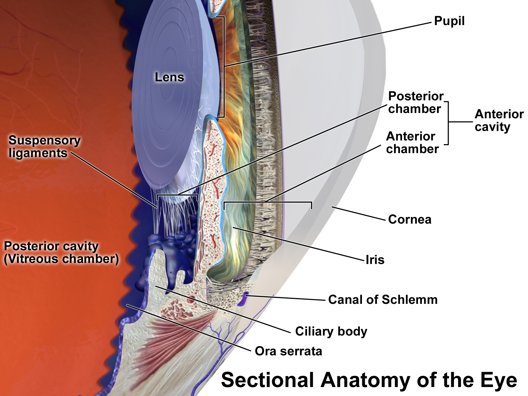
Uveitis () is inflammation of the uvea, the pigmented layer of the eye between the inner retina and the outer fibrous layer composed of the sclera and cornea. The uvea consists of the middle layer of pigmented vascular structures of the eye and includes the iris, ciliary body, and choroid. Uveitis is described anatomically, by the part of the eye affected, as anterior, intermediate or posterior, or panuveitic if all parts are involved. Anterior uveitis ( iridocyclitis) is the most common, with the incidence of uveitis overall affecting approximately 1:4500, most commonly those between the ages of 20–60. Symptoms include eye pain, eye redness, floaters and blurred vision, and ophthalmic examination may show dilated ciliary blood vessels and the presence of cells in the anterior chamber. Uveitis may arise spontaneously, have a genetic component, or be associated with an autoimmune disease or infection. While the eye is a relatively protected environment, its immune mecha ... [...More Info...] [...Related Items...] OR: [Wikipedia] [Google] [Baidu] |
Uvea
The uvea (; derived from meaning "grape"), also called the uveal layer, uveal coat, uveal tract, vascular tunic or vascular layer, is the pigmented middle layer of the three concentric layers that make up an eye, precisely between the inner retina and the outer fibrous layer composed of the sclera and cornea The cornea is the transparency (optics), transparent front part of the eyeball which covers the Iris (anatomy), iris, pupil, and Anterior chamber of eyeball, anterior chamber. Along with the anterior chamber and Lens (anatomy), lens, the cornea .... History and etymology The originally medieval Latin term comes from the Latin word ''uva'' ("grape") and is a reference to its grape-like appearance (reddish-blue or almost black colour, wrinkled appearance and grape-like size and shape when stripped intact from a cadaveric eye). In fact, it is a partial loan translation of the Ancient Greek term for the choroid, which literally means “covering resembling a grape ... [...More Info...] [...Related Items...] OR: [Wikipedia] [Google] [Baidu] |


