|
Gluteus Minimus
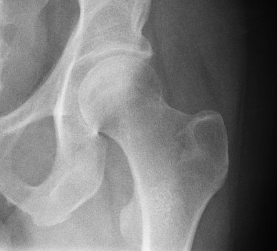
The gluteus minimus, or glutæus minimus, the smallest of the three gluteal muscles, is situated immediately beneath the gluteus medius. Structure It is fan-shaped, arising from the outer surface of the ilium, between the anterior and inferior gluteal lines, and behind, from the margin of the greater sciatic notch. The fibers converge to the deep surface of a radiated aponeurosis, and this ends in a tendon which is inserted into an impression on the anterior border of the greater trochanter, and gives an expansion to the capsule of the hip joint. Relations A bursa is interposed between the tendon and the greater trochanter. Between the gluteus medius and gluteus minimus are the deep branches of the superior gluteal vessels and the superior gluteal nerve. The deep surface of the gluteus minimus is in relation with the reflected tendon of the rectus femoris and the capsule of the hip joint. Variations The muscle may be divided into an anterior and a posterior pa ... [...More Info...] [...Related Items...] OR: [Wikipedia] [Google] [Baidu] |
Ilium (bone)
The ilium () (: ilia) is the uppermost and largest region of the coxal bone, and appears in most vertebrates including mammals and birds, but not bony fish. All reptiles have an ilium except snakes, with the exception of some snake species which have a tiny bone considered to be an ilium. The ilium of the human is divisible into two parts, the body and the wing; the separation is indicated on the top surface by a curved line, the arcuate line, and on the external surface by the margin of the acetabulum. The name comes from the Latin ('' ile'', ''ilis''), meaning "groin" or "flank". Structure The ilium consists of the body and wing. Together with the ischium and pubis, to which the ilium is connected, these form the pelvic bone, with only a faint line indicating the place of union. The body () forms less than two-fifths of the acetabulum; and also forms part of the acetabular fossa. The internal surface of the body is part of the wall of the lesser pelvis and gives o ... [...More Info...] [...Related Items...] OR: [Wikipedia] [Google] [Baidu] |
Superior Gluteal Nerve
The superior gluteal nerve is a mixed (motor and sensory) nerve of the sacral plexus that originates in the pelvis. It provides motor innervation to the gluteus medius, gluteus minimus, tensor fasciae latae; it also has a cutaneous branch. Structure Origin The superior gluteal nerve originates in the sacral plexus. It arises from the posterior divisions of L4, L5 and S1. Course It exits the pelvis through the greater sciatic foramen superior to the piriformis muscle. It is accompanied by the superior gluteal artery and the superior gluteal vein.''Thieme Atlas of Anatomy'' (2006), p 476 It passes lateral-ward in between the gluteus medius muscle and the gluteus minimus muscle, accompanied by the deep branch of the superior gluteal artery. It divides into a superior branch and an inferior branch. The inferior branch continues to pass between the two muscles to end in the tensor fasciae latae muscle. Distribution Motor * tensor fasciae latae musclePlatzer (2004) ... [...More Info...] [...Related Items...] OR: [Wikipedia] [Google] [Baidu] |
Muscles Of The Gluteus
Muscle is a soft tissue, one of the four basic types of animal tissue. There are three types of muscle tissue in vertebrates: skeletal muscle, cardiac muscle, and smooth muscle. Muscle tissue gives skeletal muscles the ability to contract. Muscle tissue contains special contractile proteins called actin and myosin which interact to cause movement. Among many other muscle proteins, present are two regulatory proteins, troponin and tropomyosin. Muscle is formed during embryonic development, in a process known as myogenesis. Skeletal muscle tissue is striated consisting of elongated, multinucleate muscle cells called muscle fibers, and is responsible for movements of the body. Other tissues in skeletal muscle include tendons and perimysium. Smooth and cardiac muscle contract involuntarily, without conscious intervention. These muscle types may be activated both through the interaction of the central nervous system as well as by innervation from peripheral plexus or endocrine ( ... [...More Info...] [...Related Items...] OR: [Wikipedia] [Google] [Baidu] |
Hip Medial Rotators
In vertebrate anatomy, the hip, or coxaLatin ''coxa'' was used by Celsus in the sense "hip", but by Pliny the Elder in the sense "hip bone" (Diab, p 77) (: ''coxae'') in medical terminology, refers to either an anatomical region or a joint on the outer (lateral) side of the pelvis. The hip region is located lateral and anterior to the gluteal region, inferior to the iliac crest, and lateral to the obturator foramen, with muscle tendons and soft tissues overlying the greater trochanter of the femur. In adults, the three pelvic bones ( ilium, ischium and pubis) have fused into one hip bone, which forms the superomedial/deep wall of the hip region. The hip joint, scientifically referred to as the acetabulofemoral joint (''art. coxae''), is the ball-and-socket joint between the pelvic acetabulum and the femoral head. Its primary function is to support the weight of the torso in both static (e.g. standing) and dynamic (e.g. walking or running) postures. The hip joints have very ... [...More Info...] [...Related Items...] OR: [Wikipedia] [Google] [Baidu] |
Hip Bone
The hip bone (os coxae, innominate bone, pelvic bone or coxal bone) is a large flat bone, constricted in the center and expanded above and below. In some vertebrates (including humans before puberty) it is composed of three parts: the Ilium (bone), ilium, ischium, and the Pubis (bone), pubis. The two hip bones join at the pubic symphysis and together with the sacrum and coccyx (the pelvic part of the vertebral column, spine) comprise the human skeleton, skeletal component of the pelvis – the pelvic girdle which surrounds the pelvic cavity. They are connected to the sacrum, which is part of the axial skeleton, at the sacroiliac joint. Each hip bone is connected to the corresponding femur (thigh bone) (forming the primary connection between the bones of the lower limb and the axial skeleton) through the large ball and socket joint of the hip joint, hip. Structure The hip bone is formed by three parts: the Ilium (bone), ilium, ischium, and Pubis (bone), pubis. At birth, these thre ... [...More Info...] [...Related Items...] OR: [Wikipedia] [Google] [Baidu] |
Trendelenburg Gait
Trendelenburg gait, first described by Friedrich Trendelenburg in 1895, is an abnormal human gait caused by an inability to maintain the pelvis level while standing on one leg. It is caused by weakness or ineffective action of the gluteus medius and gluteus minimus muscles. Gandbhir and Rayi point out that the biomechanical action involved comprises a class 3 lever, where the lower limb's weight is the load, the hip joint is the fulcrum, and the lateral glutei, which attach to the antero-lateral surface of the greater trochanter of the femur, provide the effort. The causes can thus be categorized systematically as failures of this lever system at various points. Signs and symptoms During the stance phase, or when standing on one leg, the weakened abductor muscles (gluteus medius and minimus) on the side of the supporting leg allow the opposite hip to droop. To compensate, the trunk lurches to the weakened side to maintain the center of gravity over the supporting leg. This ... [...More Info...] [...Related Items...] OR: [Wikipedia] [Google] [Baidu] |
Gluteus Medius
The gluteus medius, one of the three gluteal muscles, is a broad, thick, radiating muscle. It is situated on the outer surface of the pelvis. Its posterior third is covered by the gluteus maximus, its anterior two-thirds by the gluteal aponeurosis, which separates it from the superficial fascia and integument. Structure The gluteus medius muscle starts, or "originates", on the outer surface of the ilium between the iliac crest and the posterior gluteal line above, and the anterior gluteal line below; the gluteus medius also originates from its own fascia, the gluteal aponeurosis, that covers its outer surface. The fibers of the muscle converge into a strong flattened tendon that inserts on the lateral surface of the greater trochanter. More specifically, the muscle's tendon inserts into an oblique ridge that runs downward and forward on the lateral surface of the greater trochanter. Before the insertion the fibers cross from anterior to posterior and vice versa. Relations A ... [...More Info...] [...Related Items...] OR: [Wikipedia] [Google] [Baidu] |
Articularis Genus Muscle
The articularis genus (also known as the subcrureus muscle) is a small skeletal muscle located anteriorly on the thigh just above the knee. Structure It arises from the anterior surface of the lower part of the body of the femur, deep to the vastus intermedius, close to the knee and from the deep fibers of the vastus intermedius. Its insertion is on the synovial membrane of the knee-joint. Blood supply It is supplied by the lateral femoral circumflex artery. Innervation It is innervated by branches of the femoral nerve (L2-L4). Variation Flat, wispy and highly variable, sometimes consisting of several separate muscular bundles, this muscle is without a distinct investing fascia and ranges 1.5–3 cm in width. It is usually distinct from the vastus intermedius, but occasionally blended with it. Function Articularis genus pulls the suprapatellar bursa superiorly during extension of the knee, and prevents impingement of the synovial membrane between the patel ... [...More Info...] [...Related Items...] OR: [Wikipedia] [Google] [Baidu] |
Tensor Fasciae Latae Muscle
The tensor fasciae latae (or tensor fasciæ latæ or, formerly, tensor vaginae femoris) is a muscle of the thigh. Together with the gluteus maximus, it acts on and is continuous with the iliotibial band, which attaches to the tibia. The muscle assists in keeping the balance of the pelvis while standing, walking, or running. Structure The tensor fasciae latae arises from the anterior part of the outer lip of the iliac crest; from the outer surface of the anterior superior iliac spine, and part of the outer border of the notch below it, between the gluteus medius and sartorius; and from the deep surface of the fascia lata. The tensor fasciae latae is inserted between the two layers of the iliotibial tract of the fascia lata about the junction of the middle and upper thirds of the thigh. It tautens the iliotibial tract and braces the knee, especially when the opposite foot is lifted.Saladin, Kenneth. Anatomy and Physiology. 6th ed. Mc-Graw Hill. 2010. The terminal insertion poin ... [...More Info...] [...Related Items...] OR: [Wikipedia] [Google] [Baidu] |
Vastus Lateralis Muscle
The vastus lateralis (), also called the vastus externus, is the largest and most powerful part of the quadriceps femoris, a muscle in the thigh. Together with other muscles of the quadriceps group, it serves to extend the knee joint, moving the lower leg forward. It arises from a series of flat, broad tendons attached to the femur, and attaches to the outer border of the patella. It ultimately joins with the other muscles that make up the quadriceps in the quadriceps tendon, which travels over the knee to connect to the tibia. The vastus lateralis is the recommended site for intramuscular injection in infants less than 7 months old and those unable to walk, with loss of muscular tone.Mann, E. (2016). ''Injection (Intramuscular): Clinician Information.'' The Johanna Briggs Institute. Structure The vastus lateralis muscle arises from several areas of the femur, including the upper part of the intertrochanteric line; the lower, anterior borders of the greater trochanter, to t ... [...More Info...] [...Related Items...] OR: [Wikipedia] [Google] [Baidu] |
