|
Gastric Mucosa
The gastric mucosa is the mucous membrane layer of the stomach, which contains the gastric pits, to which the gastric glands empty. In humans, it is about one mm thick, and its surface is smooth, soft, and velvety. It consists of simple secretory columnar epithelium, an underlying supportive layer of loose connective tissue called the lamina propria, and the muscularis mucosae, a thin layer of muscle that separates the mucosa from the underlying submucosa. Description In its fresh state, it is of a pinkish tinge at the pyloric end and of a red or reddish-brown color over the rest of its surface. In infancy it is of a brighter hue, the vascular redness being more marked. It is thin at the cardiac extremity, but thicker toward the pylorus. During the contracted state of the stomach it is thrown into numerous folds or rugae, which, for the most part, have a longitudinal direction. They are most marked toward the pyloric end of the stomach, and along the greater curvature, and are e ... [...More Info...] [...Related Items...] OR: [Wikipedia] [Google] [Baidu] |
Stomach Mucosal Layer Labeled
The stomach is a muscular, hollow organ in the upper gastrointestinal tract of humans and many other animals, including several invertebrates. The Ancient Greek name for the stomach is ''gaster'' which is used as ''gastric'' in medical terms related to the stomach. The stomach has a dilated structure and functions as a vital organ in the digestive system. The stomach is involved in the gastric phase of digestion, following the cephalic phase in which the sight and smell of food and the act of chewing are stimuli. In the stomach a chemical breakdown of food takes place by means of secreted digestive enzymes and gastric acid. It also plays a role in regulating gut microbiota, influencing digestion and overall health. The stomach is located between the esophagus and the small intestine. The pyloric sphincter controls the passage of partially digested food (chyme) from the stomach into the duodenum, the first and shortest part of the small intestine, where peristalsis takes over ... [...More Info...] [...Related Items...] OR: [Wikipedia] [Google] [Baidu] |
Histamine
Histamine is an organic nitrogenous compound involved in local immune responses communication, as well as regulating physiological functions in the gut and acting as a neurotransmitter for the brain, spinal cord, and uterus. Discovered in 1910, histamine has been considered a local hormone ( autocoid) because it is produced without involvement of the classic endocrine glands; however, in recent years, histamine has been recognized as a central neurotransmitter. Histamine is involved in the inflammatory response and has a central role as a mediator of itching. As part of an immune response to foreign pathogens, histamine is produced by basophils and by mast cells found in nearby connective tissues. Histamine increases the permeability of the capillaries to white blood cells and some proteins, to allow them to engage pathogens in the infected tissues. It consists of an imidazole ring attached to an ethylamine chain; under physiological conditions, the amino grou ... [...More Info...] [...Related Items...] OR: [Wikipedia] [Google] [Baidu] |
Membrane Biology
Membrane biology is the study of the biological and physiochemical characteristics of membranes, with applications in the study of cellular physiology. Membrane bioelectrical impulses are described by the Hodgkin cycle. Biophysics Membrane biophysics is the study of biological membrane structure and function using physical, computational, mathematical, and biophysical methods. A combination of these methods can be used to create phase diagrams of different types of membranes, which yields information on thermodynamic behavior of a membrane and its components. As opposed to membrane biology, membrane biophysics focuses on quantitative information and modeling of various membrane phenomena, such as lipid raft The cell membrane, plasma membranes of cells contain combinations of glycosphingolipids, cholesterol and protein Receptor (biochemistry), receptors organized in glycolipoprotein lipid microdomains termed lipid rafts. Their existence in cellular me ... formation, rates ... [...More Info...] [...Related Items...] OR: [Wikipedia] [Google] [Baidu] |
Anatomy
Anatomy () is the branch of morphology concerned with the study of the internal structure of organisms and their parts. Anatomy is a branch of natural science that deals with the structural organization of living things. It is an old science, having its beginnings in prehistoric times. Anatomy is inherently tied to developmental biology, embryology, comparative anatomy, evolutionary biology, and phylogeny, as these are the processes by which anatomy is generated, both over immediate and long-term timescales. Anatomy and physiology, which study the structure and function of organisms and their parts respectively, make a natural pair of related disciplines, and are often studied together. Human anatomy is one of the essential basic sciences that are applied in medicine, and is often studied alongside physiology. Anatomy is a complex and dynamic field that is constantly evolving as discoveries are made. In recent years, there has been a significant increase in the use of ... [...More Info...] [...Related Items...] OR: [Wikipedia] [Google] [Baidu] |
Abdomen
The abdomen (colloquially called the gut, belly, tummy, midriff, tucky, or stomach) is the front part of the torso between the thorax (chest) and pelvis in humans and in other vertebrates. The area occupied by the abdomen is called the abdominal cavity. In arthropods, it is the posterior (anatomy), posterior tagma (biology), tagma of the body; it follows the thorax or cephalothorax. In humans, the abdomen stretches from the thorax at the thoracic diaphragm to the pelvis at the pelvic brim. The pelvic brim stretches from the lumbosacral joint (the intervertebral disc between Lumbar vertebrae, L5 and Vertebra#Sacrum, S1) to the pubic symphysis and is the edge of the pelvic inlet. The space above this inlet and under the thoracic diaphragm is termed the abdominal cavity. The boundary of the abdominal cavity is the abdominal wall in the front and the peritoneal surface at the rear. In vertebrates, the abdomen is a large body cavity enclosed by the abdominal muscles, at the front an ... [...More Info...] [...Related Items...] OR: [Wikipedia] [Google] [Baidu] |
Parietal Cell
Parietal cells (also known as oxyntic cells) are epithelial cells in the stomach that secrete hydrochloric acid (HCl) and intrinsic factor. These cells are located in the gastric glands found in the lining of the fundus and body regions of the stomach. They contain an extensive secretory network of canaliculi from which the HCl is secreted by active transport into the stomach. The enzyme hydrogen potassium ATPase (H+/K+ ATPase) is unique to the parietal cells and transports the H+ against a concentration gradient of about 3 million to 1, which is the steepest ion gradient formed in the human body. Parietal cells are primarily regulated via histamine, acetylcholine and gastrin signalling from both central and local modulators. Structure Canaliculus A canaliculus is an adaptation found on gastric parietal cells. It is a deep infolding, or little channel, which serves to increase the surface area, e.g. for secretion. The parietal cell membrane is dynamic; the numbers of ... [...More Info...] [...Related Items...] OR: [Wikipedia] [Google] [Baidu] |
Gastric Chief Cell
A gastric chief cell, peptic cell, or gastric zymogenic cell is a type of gastric gland cell that releases pepsinogen and gastric lipase. It is the cell responsible for secretion of chymosin (rennin) in ruminant animals and some other animals. The cell stains basophilic upon H&E staining due to the large proportion of rough endoplasmic reticulum in its cytoplasm. Gastric chief cells are generally located deep in the mucosal layer of the stomach lining, in the fundus and body of the stomach. Chief cells release the zymogen (enzyme precursor) pepsinogen when stimulated by a variety of factors including cholinergic activity from the vagus nerve and acidic condition in the stomach. Gastrin and secretin may also act as secretagogues. It works in conjunction with the parietal cell, which releases gastric acid, converting the pepsinogen into pepsin. Nomenclature The terms ''chief cell'' and '' zymogenic cell'' are often used without the word "gastric" to name this type of cell. ... [...More Info...] [...Related Items...] OR: [Wikipedia] [Google] [Baidu] |
Foveolar Cell
Foveolar cells or surface mucous cells are mucus-producing cells which cover the inside of the stomach, protecting it from the corrosive nature of gastric acid. These cells line the gastric mucosa and the gastric pits. Mucous neck cells are found in the necks of the gastric glands. The mucus-secreting cells of the stomach can be distinguished histologically from the intestinal goblet cells, another type of mucus-secreting cell. Structure The gastric mucosa that lines the inner wall of the stomach has a set of microscopic features called gastric glands which, depending on the location within the stomach, secrete different substances into the lumen of the organ. The openings of these glands into the stomach are called gastric pits which foveolar cells line in order to provide a protective alkaline secretion against the corrosive gastric acid. Microanatomy Foveolar cells line the surface of the stomach and the gastric pits. They constitute a simple columnar epithelium, as the ... [...More Info...] [...Related Items...] OR: [Wikipedia] [Google] [Baidu] |
Gastric Tumor
Tumors of the stomach Tumors of the stomach are known as gastric tumors, and can be either benign or malignant (gastric cancer). These tumors arise from the cells of the gastric mucosa which lines the stomach. Typically, most gastric tumors are cancerous and not detected until a later stage for various reasons. Types There are two distinct types of tumors: benign and malignant. Both types of tumors share a number of general characteristics, the broadest being that they are an abnormal proliferation of cells. The main difference between the two types is what happens once the tumor has started growing. In a benign tumor, the proliferated cells stay in one location where they do not impact or spread to other surrounding tissues. Malignant tumors, on the other hand, are capable of spreading throughout the entire body, causing new tumors to appear. This process is called metastasis, and is a hallmark of cancerous tumors. Typical cell growth Generally speaking, all cells grow and ... [...More Info...] [...Related Items...] OR: [Wikipedia] [Google] [Baidu] |
Gastritis
Gastritis is the inflammation of the lining of the stomach. It may occur as a short episode or may be of a long duration. There may be no symptoms but, when symptoms are present, the most common is upper abdominal pain (see dyspepsia). Other possible symptoms include nausea and vomiting, bloating, loss of appetite and heartburn. Complications may include stomach bleeding, stomach ulcers, and stomach tumors. When due to autoimmune problems, low red blood cells due to not enough vitamin B12 may occur, a condition known as pernicious anemia. Common causes include infection with '' Helicobacter pylori'' and use of nonsteroidal anti-inflammatory drugs ( NSAIDs). When caused by ''H. pylori'' this is now termed ''Helicobacter pylori'' induced gastritis, and included as a listed disease in ICD11. Less common causes include alcohol, smoking, cocaine, severe illness, autoimmune problems, radiation therapy and Crohn's disease. Endoscopy, a type of X-ray known as an upper gast ... [...More Info...] [...Related Items...] OR: [Wikipedia] [Google] [Baidu] |
Iodide
An iodide ion is I−. Compounds with iodine in formal oxidation state −1 are called iodides. In everyday life, iodide is most commonly encountered as a component of iodized salt, which many governments mandate. Worldwide, iodine deficiency affects two billion people and is the leading preventable cause of intellectual disability. Structure and characteristics of inorganic iodides Iodide is one of the largest monatomic anions. It is assigned a radius of around 206 picometers. For comparison, the lighter halides are considerably smaller: bromide (196 pm), chloride (181 pm), and fluoride (133 pm). In part because of its size, iodide forms relatively weak bonds with most elements. Most iodide salts are soluble in water, but often less so than the related chlorides and bromides. Iodide, being large, is less hydrophilic compared to the smaller anions. One consequence of this is that sodium iodide is highly soluble in acetone, whereas sodium chloride is not. The l ... [...More Info...] [...Related Items...] OR: [Wikipedia] [Google] [Baidu] |
Sodium-iodide Symporter
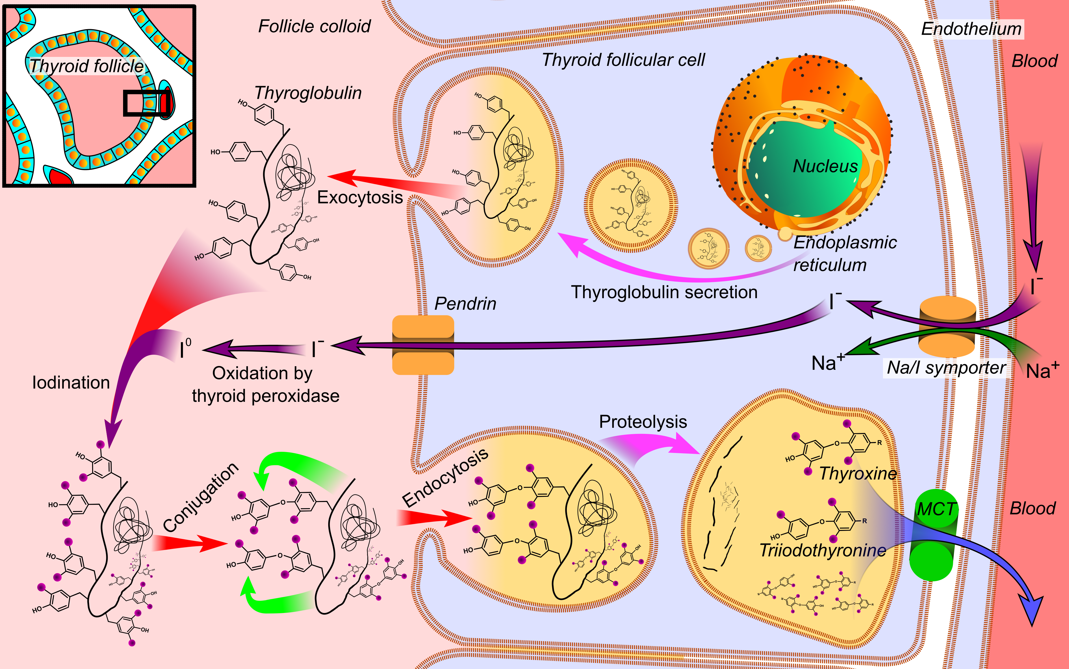
The sodium/iodide cotransporter, also known as the sodium/iodide symporter (NIS), is a protein that in humans is encoded by the ''SLC5A5'' gene. It is a transmembrane glycoprotein with a molecular weight of 87 kAtomic mass unit, Da and 13 transmembrane domains, which transports two sodium cations (Na+) for each iodide anion (I−) into the cell. NIS mediated uptake of iodide into Thyroid epithelial cell, follicular cells of the thyroid gland is the first step in the synthesis of thyroid hormone. Iodine uptake Iodine uptake mediated by thyroid Thyroid epithelial cell, follicular cells from the blood plasma is the first step for the synthesis of thyroid hormones. This ingested iodine is bound to serum proteins, especially to albumins. The rest of the iodine which remains unlinked and free in bloodstream, is removed from the body through urine (the kidney is essential in the removal of iodine from extracellular space). Iodine uptake is a result of an active transport mechanism media ... [...More Info...] [...Related Items...] OR: [Wikipedia] [Google] [Baidu] |


