|
Fractional Flow Reserve
Fractional flow reserve (FFR) is a diagnostic technique used in coronary catheterization. FFR measures pressure differences across a coronary artery stenosis (narrowing, usually due to atherosclerosis) to determine the likelihood that the stenosis impedes oxygen delivery to the heart muscle (myocardial ischemia). Fractional flow reserve is defined as the pressure after (distal to) a stenosis relative to the pressure before the stenosis. The result is an absolute number; an FFR of 0.80 means that a given stenosis causes a 20% drop in blood pressure. In other words, FFR expresses the maximal flow down a vessel in the presence of a stenosis compared to the maximal flow in the hypothetical absence of the stenosis. Procedure During coronary catheterization, a catheter is inserted into the femoral (groin) or radial arteries (wrist) using a sheath and guidewire. FFR uses a small sensor on the tip of the wire (commonly a transducer) to measure pressure, temperature and flow to determin ... [...More Info...] [...Related Items...] OR: [Wikipedia] [Google] [Baidu] |
Coronary Catheterization
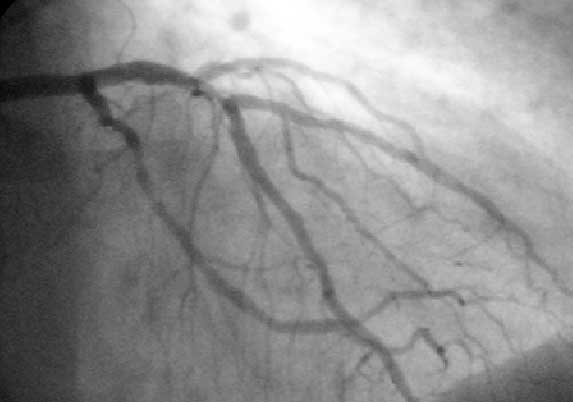
A coronary catheterization is a minimally invasive procedure to access the coronary circulation and blood filled chambers of the heart using a catheter. It is performed for both diagnostic and interventional (treatment) purposes. Coronary catheterization is one of the several cardiology diagnostic tests and procedures. Specifically, through the injection of a liquid radiocontrast agent and illumination with X-rays, angiocardiography allows the recognition of Vascular occlusion, occlusion, stenosis, restenosis, thrombosis or aneurysmal enlargement of the coronary artery lumen (anatomy), lumens; heart chamber size; heart muscle contraction performance; and some aspects of heart valve function. Important internal heart and lung blood pressures, not measurable from outside the body, can be accurately measured during the test. The relevant problems that the test deals with most commonly occur as a result of advanced atherosclerosis – atheroma activity within the wall of the coronary ... [...More Info...] [...Related Items...] OR: [Wikipedia] [Google] [Baidu] |
Papaverine
Papaverine (Latin '' papaver'', "poppy") is an opium alkaloid antispasmodic drug, used primarily in the treatment of visceral spasms and vasospasms (especially those involving the intestines, heart, or brain), occasionally in the treatment of erectile dysfunction and acute mesenteric ischemia. While it is found in the opium poppy, papaverine differs in both structure and pharmacological action from the analgesic morphine and its derivatives (such as codeine). In addition to opium, papaverine is purported to be present in high concentrations in star gooseberry. History Papaverine was discovered in 1848 by Georg Merck (1825–1873). Merck was a student of the German chemists Justus von Liebig and August Hofmann, and he was the son of Emanuel Merck (1794–1855), founder of the Merck corporation, a major German chemical and pharmaceutical company. Uses Papaverine is approved to treat spasms of the gastrointestinal tract, bile ducts and ureter and for use as a ce ... [...More Info...] [...Related Items...] OR: [Wikipedia] [Google] [Baidu] |
Revascularization
In medical and surgical therapy, revascularization is the restoration of perfusion to a body part or organ that has had ischemia. It is typically accomplished by surgical means. Vascular bypass and angioplasty are the two primary means of revascularization. The term derives from the prefix re-, in this case meaning "restoration" and vasculature, which refers to the circulatory structures of an organ. It is often combined with "urgent" to form urgent vascularization. Revascularization involves a thorough analysis and diagnosis and treatment of the existing diseased vasculature of the affected organ, and can be aided by the use of different imaging modalities such as magnetic resonance imaging, PET scan, CT scan, and X-ray fluoroscopy. Applications For coronary artery disease Coronary artery disease (CAD), also called coronary heart disease (CHD), or ischemic heart disease (IHD), is a type of cardiovascular disease, heart disease involving Ischemia, the reduction of ... [...More Info...] [...Related Items...] OR: [Wikipedia] [Google] [Baidu] |
Myocardial Infarction
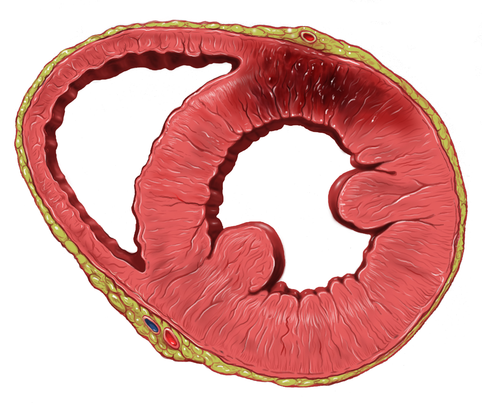
A myocardial infarction (MI), commonly known as a heart attack, occurs when Ischemia, blood flow decreases or stops in one of the coronary arteries of the heart, causing infarction (tissue death) to the heart muscle. The most common symptom is retrosternal Angina, chest pain or discomfort that classically radiates to the left shoulder, arm, or jaw. The pain may occasionally feel like heartburn. This is the dangerous type of acute coronary syndrome. Other symptoms may include shortness of breath, nausea, presyncope, feeling faint, a diaphoresis, cold sweat, Fatigue, feeling tired, and decreased level of consciousness. About 30% of people have atypical symptoms. Women more often present without chest pain and instead have neck pain, arm pain or feel tired. Among those over 75 years old, about 5% have had an MI with little or no history of symptoms. An MI may cause heart failure, an Cardiac arrhythmia, irregular heartbeat, cardiogenic shock or cardiac arrest. Most MIs occur d ... [...More Info...] [...Related Items...] OR: [Wikipedia] [Google] [Baidu] |
Drug Eluting Stent
A drug-eluting stent (DES) is a tube made of a mesh-like material used to treat narrowed arteries in medical procedures both mechanically (by providing a supporting scaffold inside the artery) and pharmacologically (by slowly releasing a pharmaceutical compound). A DES is inserted into a narrowed artery using a delivery catheter usually inserted through a larger artery in the groin or wrist. The stent assembly has the DES mechanism attached towards the front of the stent, and usually is composed of the collapsed stent over a collapsed polymeric balloon mechanism, the balloon mechanism is inflated and used to expand the meshed stent once in position. The stent expands, embedding into the occluded artery wall, keeping the artery open, thereby improving blood flow. The mesh design allows for stent expansion and also for new healthy vessel endothelial cells to grow through and around it, securing it in place. A DES is different from other types of stents in that it has a coating ... [...More Info...] [...Related Items...] OR: [Wikipedia] [Google] [Baidu] |
Nuclear Imaging
Nuclear medicine (nuclear radiology, nucleology), is a medical specialty involving the application of radioactive substances in the diagnosis and treatment of disease. Nuclear imaging is, in a sense, ''radiology done inside out'', because it records radiation emitted from within the body rather than radiation that is transmitted through the body from external sources like X-ray generators. In addition, nuclear medicine scans differ from radiology, as the emphasis is not on imaging anatomy, but on the function. For such reason, it is called a physiological imaging modality. Single photon emission computed tomography (SPECT) and positron emission tomography (PET) scans are the two most common imaging modalities in nuclear medicine. Diagnostic medical imaging Diagnostic In nuclear medicine imaging, radiopharmaceuticals are taken internally, for example, through inhalation, intravenously, or orally. Then, external detectors (gamma cameras) capture and form images from the radi ... [...More Info...] [...Related Items...] OR: [Wikipedia] [Google] [Baidu] |
Cardiac Stress Test
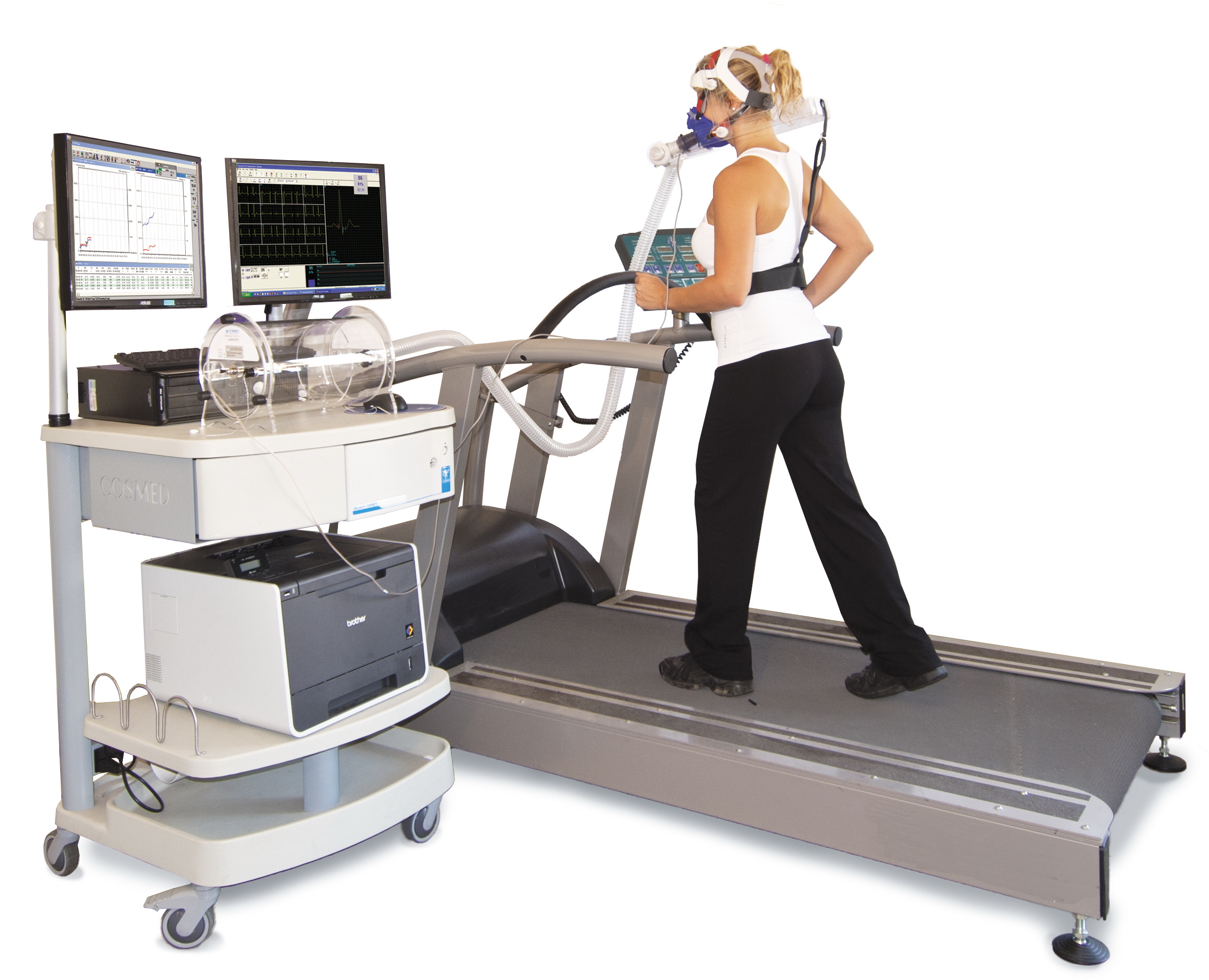
A cardiac stress test is a cardiological examination that evaluates the cardiovascular system's response to external stress within a controlled clinical setting. This stress response can be induced through physical exercise (usually a treadmill) or intravenous pharmacological stimulation of heart rate. As the heart works progressively harder (stressed) it is monitored using an electrocardiogram (ECG) monitor. This measures the heart's electrical rhythms and broader electrophysiology. Pulse rate, blood pressure and symptoms such as chest discomfort or fatigue are simultaneously monitored by attending clinical staff. Clinical staff will question the patient throughout the procedure asking questions that relate to pain and perceived discomfort. Abnormalities in blood pressure, heart rate, ECG or worsening physical symptoms could be indicative of coronary artery disease. Stress testing does not accurately diagnose all cases of coronary artery disease, and can often indicate that it e ... [...More Info...] [...Related Items...] OR: [Wikipedia] [Google] [Baidu] |
Invasive (medical)
Minimally invasive procedures (also known as minimally invasive surgeries) encompass Surgery, surgical techniques that limit the size of incisions needed, thereby reducing wound healing time, associated pain, and risk of infection. Surgery by definition is invasive, and many operations requiring surgical incision, incisions of some size are referred to as ''open surgery''. Incisions made during open surgery can sometimes leave large wounds that may be painful and take a long time to heal. Advancements in medical technologies have enabled the development and regular use of minimally invasive procedures. For example, endovascular aneurysm repair, a minimally invasive surgery, has become the most common method of repairing abdominal aortic aneurysms in the US as of 2003. The procedure involves much smaller incisions than the corresponding cardiac surgery#open surgery, open surgery procedure of open aortic surgery. interventional radiology, Interventional radiologists were the forer ... [...More Info...] [...Related Items...] OR: [Wikipedia] [Google] [Baidu] |
Vulnerable Plaque
A vulnerable plaque is a kind of atheromatous plaque – a collection of white blood cells (primarily macrophages) and lipids (including cholesterol) in the wall of an artery – that is particularly unstable and prone to produce sudden major problems such as a heart attack or stroke. The defining characteristics of a vulnerable plaque include but are not limited to: a thin fibrous cap, large lipid-rich necrotic core, increased plaque inflammation, positive vascular remodeling, increased vasa-vasorum neovascularization, and intra-plaque hemorrhage. These characteristics together with the usual hemodynamic pulsating expansion during systole and elastic recoil contraction during diastole contribute to a high mechanical stress zone on the fibrous cap of the atheroma, making it prone to rupture. Increased hemodynamic stress, e.g. increased blood pressure, especially pulse pressure (systolic blood pressure vs. diastolic blood pressure difference), correlates with increased rates of ... [...More Info...] [...Related Items...] OR: [Wikipedia] [Google] [Baidu] |
Computed Tomography Of The Heart
A computed tomography scan (CT scan), formerly called computed axial tomography scan (CAT scan), is a medical imaging technique used to obtain detailed internal images of the body. The personnel that perform CT scans are called radiographers or radiology technologists. CT scanners use a rotating X-ray tube and a row of detectors placed in a gantry to measure X-ray attenuations by different tissues inside the body. The multiple X-ray measurements taken from different angles are then processed on a computer using tomographic reconstruction algorithms to produce tomographic (cross-sectional) images (virtual "slices") of a body. CT scans can be used in patients with metallic implants or pacemakers, for whom magnetic resonance imaging (MRI) is contraindicated. Since its development in the 1970s, CT scanning has proven to be a versatile imaging technique. While CT is most prominently used in medical diagnosis, it can also be used to form images of non-living objects. The 1979 Nobel ... [...More Info...] [...Related Items...] OR: [Wikipedia] [Google] [Baidu] |
Intravascular Ultrasound
Intravascular ultrasound (IVUS) or intravascular echocardiography is a medical imaging methodology using a specially designed catheter with a miniaturized ultrasound probe attached to the distal end of the catheter. The proximal end of the catheter is attached to computerized ultrasound equipment. It allows the application of ultrasound technology, such as piezoelectric transducer or Capacitive micromachined ultrasonic transducers, CMUT, to see from inside blood vessels out through the surrounding blood column, visualizing the endothelium (inner wall) of blood vessels. The artery, arteries of the heart (the coronary circulation, coronary arteries) are the most frequent imaging target for IVUS. IVUS is used in the coronary arteries to determine the amount of atheroma, atheromatous plaque built up at any particular point in the epicardial coronary artery. Intravascular ultrasound provides a unique method to study the regression or progression of atherosclerotic lesions in vivo. T ... [...More Info...] [...Related Items...] OR: [Wikipedia] [Google] [Baidu] |


