|
Focal Neurological Deficit
Focal neurologic signs, also known as focal neurological deficits or focal CNS signs, are impairments of nerve, spinal cord, or brain function that affects a specific region of the body, e.g. weakness in the left arm, the right leg, paresis, or plegia. Focal neurological deficits may be caused by a variety of medical conditions such as head trauma, tumors or stroke; or by various diseases such as meningitis or encephalitis or as a side effect of certain medications such as those used in anesthesia. Neurological soft signs are a group of non-focal neurologic signs. Frontal lobe signs Frontal lobe signs usually involve the motor system and may include many special types of deficit, depending on which part of the frontal lobe is affected: * unsteady gait (unsteadiness in walking) * muscular rigidity, resistance to passive movements of the limbs (hypertonia) * paralysis of a limb (monoparesis) or a larger area on one side of the body (hemiparesis) * paralysis head and eye movements ... [...More Info...] [...Related Items...] OR: [Wikipedia] [Google] [Baidu] |
Central Nervous System
The central nervous system (CNS) is the part of the nervous system consisting primarily of the brain, spinal cord and retina. The CNS is so named because the brain integrates the received information and coordinates and influences the activity of all parts of the bodies of bilateria, bilaterally symmetric and triploblastic animals—that is, all multicellular animals except sponges and Coelenterata, diploblasts. It is a structure composed of nervous tissue positioned along the Anatomical_terms_of_location#Rostral,_cranial,_and_caudal, rostral (nose end) to caudal (tail end) axis of the body and may have an enlarged section at the rostral end which is a brain. Only arthropods, cephalopods and vertebrates have a true brain, though precursor structures exist in onychophorans, gastropods and lancelets. The rest of this article exclusively discusses the vertebrate central nervous system, which is radically distinct from all other animals. Overview In vertebrates, the brain and spinal ... [...More Info...] [...Related Items...] OR: [Wikipedia] [Google] [Baidu] |
Jacksonian Seizure
Focal seizures are seizures that originate within brain networks limited to one hemisphere of the brain. In most cases, each seizure type has a consistent site of onset and characteristic patterns of spread, although some individuals experience more than one type of focal seizure arising from distinct networks. Seizure activity may remain localized or propagate to the opposite hemisphere. Symptoms will vary according to where the seizure occurs. When seizures occur in the frontal lobe, the patient may experience a wave-like sensation in the head. When seizures occur in the temporal lobe, a feeling of déjà vu may be experienced. When seizures are localized to the parietal lobe, a numbness or tingling may occur. With seizures occurring in the occipital lobe, visual disturbances or hallucinations have been reported. , ... [...More Info...] [...Related Items...] OR: [Wikipedia] [Google] [Baidu] |
Agnosia
Agnosia is a neurological disorder characterized by an inability to process sensory information. Often there is a loss of ability to recognize objects, persons, sounds, shapes, or smells while the specific sense is neither defective nor is there any significant memory loss. It is usually associated with brain injury or neurological illness, particularly after damage to the occipitotemporal border, which is part of the ventral stream. Agnosia affects only a single modality, such as vision or hearing. More recently, a top-down interruption is considered to cause the disturbance of handling perceptual information. Types Visual agnosia Visual agnosia is a broad category that refers to a deficiency in the ability to recognize visual objects. Visual agnosia can be further subdivided into two different subtypes: apperceptive visual agnosia and associative visual agnosia. Individuals with apperceptive visual agnosia display the ability to see contours and outlines w ... [...More Info...] [...Related Items...] OR: [Wikipedia] [Google] [Baidu] |
Dyscalculia
Dyscalculia () is a learning disability resulting in difficulty learning or comprehending arithmetic, such as difficulty in understanding numbers, numeracy, learning how to manipulate numbers, performing mathematical calculations, and learning facts in mathematics. It is sometimes colloquially referred to as "math dyslexia", though this analogy can be misleading as they are distinct syndromes. Dyscalculia is associated with dysfunction in the region around the intraparietal sulcus and potentially also the frontal lobe. Dyscalculia does not reflect a general deficit in cognitive abilities or difficulties with time, measurement, and Spatial visualization ability, spatial reasoning. Estimates of the prevalence of dyscalculia range between 3 and 6% of the population. In 2015 it was established that 11% of children with dyscalculia also have attention deficit hyperactivity disorder (ADHD). Dyscalculia has also been associated with Turner syndrome and people who have spina bifida. Mat ... [...More Info...] [...Related Items...] OR: [Wikipedia] [Google] [Baidu] |
Dysgraphia
Dysgraphia is a neurological disorder and learning disability that concerns impairments in written expression, which affects the ability to write, primarily handwriting, but also coherence. It is a specific learning disability (SLD) as well as a transcription disability, meaning that it is a writing disorder associated with impaired handwriting, orthographic coding and finger sequencing (the movement of muscles required to write). It often overlaps with other learning disabilities and neurodevelopmental disorders such as speech impairment, attention deficit hyperactivity disorder (ADHD) or developmental coordination disorder (DCD). In the ''Diagnostic and Statistical Manual of Mental Disorders'' (''DSM-5''), dysgraphia is characterized as a neurodevelopmental disorder under the umbrella category of specific learning disorder. Dysgraphia is when one's writing skills are below those expected given a person's age measured through intelligence and age-appropriate education. The DSM ... [...More Info...] [...Related Items...] OR: [Wikipedia] [Google] [Baidu] |
Dyslexia
Dyslexia (), previously known as word blindness, is a learning disability that affects either reading or writing. Different people are affected to different degrees. Problems may include difficulties in spelling words, reading quickly, writing words, "sounding out" words in the head, pronouncing words when reading aloud and understanding what one reads. Often these difficulties are first noticed at school. The difficulties are involuntary, and people with this disorder have a normal desire to learn. People with dyslexia have higher rates of attention deficit hyperactivity disorder (ADHD), developmental language disorders, and difficulties with numbers. Dyslexia is believed to be caused by the interaction of genetic and environmental factors. Some cases run in families. Dyslexia that develops due to a traumatic brain injury, stroke, or dementia is sometimes called "acquired dyslexia" or alexia. The underlying mechanisms of dyslexia result from differences within the ... [...More Info...] [...Related Items...] OR: [Wikipedia] [Google] [Baidu] |
Hemispatial Neglect
Hemispatial neglect is a neuropsychological condition in which, after damage to one hemisphere of the brain (e.g. after a stroke), a deficit in attention and awareness towards the side of space opposite brain damage (contralesional space) is observed. It is defined by the inability of a person to process and perceive stimuli towards the contralesional side of the body or environment. Hemispatial neglect is very commonly contralateral to the damaged hemisphere, but instances of ipsilesional neglect (on the same side as the lesion) have been reported. Presentation Hemispatial neglect results most commonly from strokes and brain unilateral injury to the right cerebral hemisphere, with rates in the critical stage of up to 80% causing visual neglect of the left-hand side of space. Neglect is often produced by massive strokes in the middle cerebral artery region and is variegated, so that most sufferers do not exhibit all of the syndrome's traits. Right-sided spatial neglect is rare ... [...More Info...] [...Related Items...] OR: [Wikipedia] [Google] [Baidu] |
Proprioception
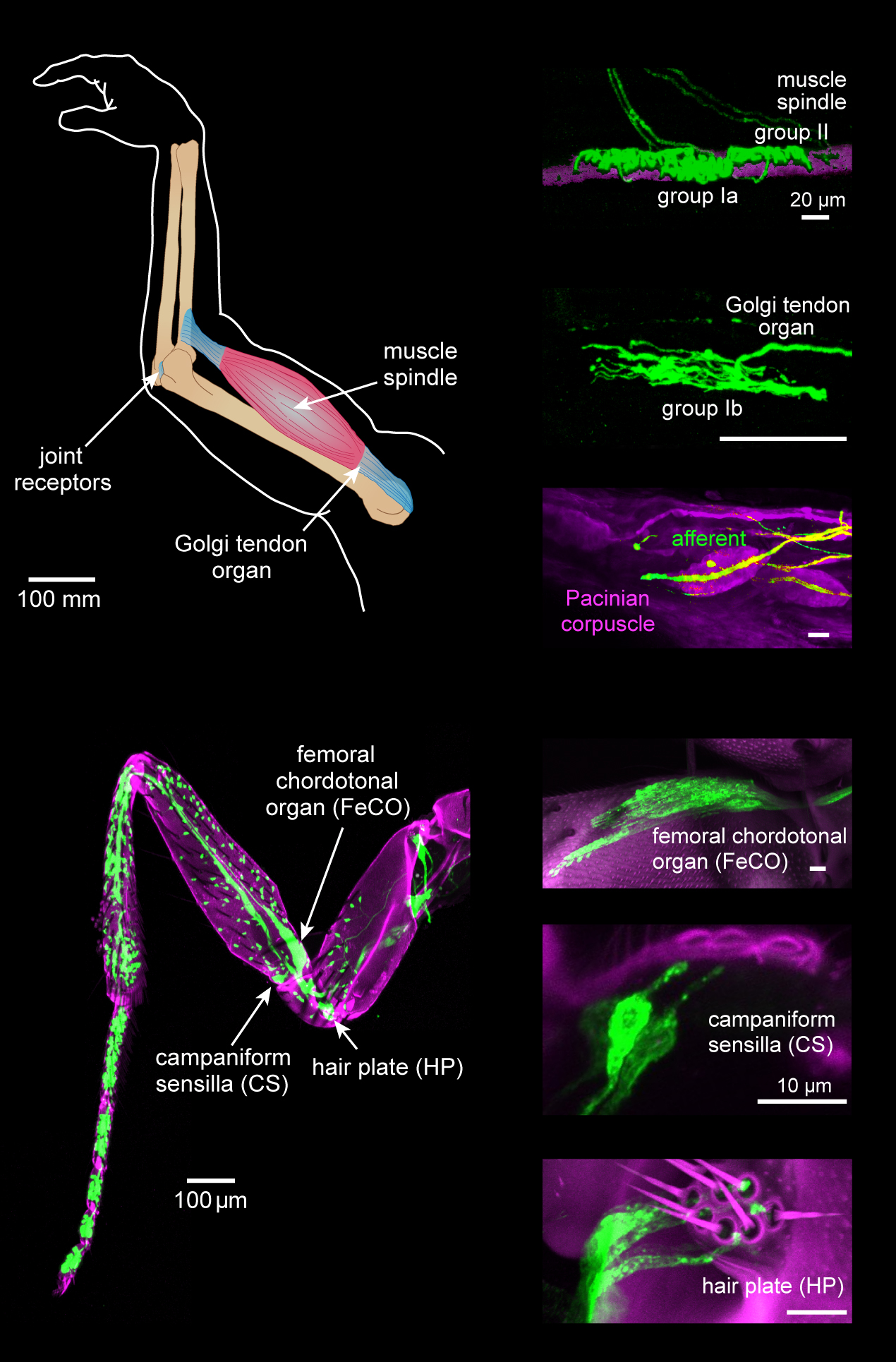
Proprioception ( ) is the sense of self-movement, force, and body position. Proprioception is mediated by proprioceptors, a type of sensory receptor, located within muscles, tendons, and joints. Most animals possess multiple subtypes of proprioceptors, which detect distinct kinesthetic parameters, such as joint position, movement, and load. Although all mobile animals possess proprioceptors, the structure of the sensory organs can vary across species. Proprioceptive signals are transmitted to the central nervous system, where they are integrated with information from other Sensory nervous system, sensory systems, such as Visual perception, the visual system and the vestibular system, to create an overall representation of body position, movement, and acceleration. In many animals, sensory feedback from proprioceptors is essential for stabilizing body posture and coordinating body movement. System overview In vertebrates, limb movement and velocity (muscle length and the rate ... [...More Info...] [...Related Items...] OR: [Wikipedia] [Google] [Baidu] |
Parietal Lobe
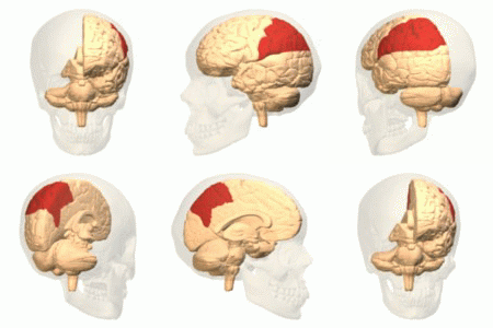
The parietal lobe is one of the four Lobes of the brain, major lobes of the cerebral cortex in the brain of mammals. The parietal lobe is positioned above the temporal lobe and behind the frontal lobe and central sulcus. The parietal lobe integrates sensory information among various sensory modality, modalities, including spatial sense and navigation (proprioception), the main sensory receptive area for the sense of touch in the somatosensory cortex which is just posterior to the central sulcus in the postcentral gyrus, and the two-streams hypothesis#Dorsal stream, dorsal stream of the visual system. The major sensory inputs from the skin (mechanoreceptor, touch, thermoreceptor, temperature, and nociceptor, pain receptors), relay through the thalamus to the parietal lobe. Several areas of the parietal lobe are important in language processing in the brain, language processing. The somatosensory cortex can be illustrated as a distorted figure – the cortical homunculus (Latin: "li ... [...More Info...] [...Related Items...] OR: [Wikipedia] [Google] [Baidu] |
Palmomental Reflex
The palmomental reflex (PMR) or Marinescu-Radovici Sign or Kinn reflex or Marinescu Reflex is a primitive reflex consisting of a twitch of the chin muscle elicited by stroking a specific part of the palm. It is present in infancy and disappears as the brain matures during childhood but may reappear due to processes that disrupt the normal cortical inhibitory pathways. Therefore, it is an example of a frontal release sign. Eliciting and observing response The thenar eminence is stroked briskly with a thin stick, from proximal (edge of wrist) to distal (base of thumb) using moderate pressure. A positive response is considered if there is a single visible twitch of the ipsilateral mentalis muscle (chin muscle on the same side as the hand tested). Clinical relevance In their seminal 1920 paper, Gheorghe Marinescu and Anghel Radovici hypothesized that both the afferent (receptive) and efferent (motor) arms of the reflex are on the same side (ipsilateral) to the hand stimulated; ... [...More Info...] [...Related Items...] OR: [Wikipedia] [Google] [Baidu] |
Palmar Grasp Reflex
The palmar grasp reflex (or grasp reflex) is a primitive and involuntary reflex found in infants of humans and most primates. When an object, such as an adult finger, is placed in an infant's palm, the infant's fingers reflexively grasp the object. Placement of the object triggers a spinal reflex, resulting from stimulation of tendons in the palm, that gets transmitted through motor neurons in the median and ulnar sensory nerves. The reverse motion can be induced by stroking the back or side of the hand. A fetus exhibits the reflex ''in utero'' by 28 weeks into gestation (sometimes, as early as 16 weeks), and persists until development of rudimentary fine motor skills between two and six months of age. Evolutionary significance Biologists have found that the reflex is significantly more frequent in infants of fur carrying primate species. It is theorized that the grasping reflex evolved as it is essential to survival in species, usually primates, where the young are carried in th ... [...More Info...] [...Related Items...] OR: [Wikipedia] [Google] [Baidu] |
Snout Reflex
The Snout reflex (also orbicularis oris reflex) or a "Pout" is a pouting or pursing of the lips that is elicited by light tapping of the closed lips near the midline. The contraction of the muscles causes the mouth to resemble a snout. This reflex is tested in a neurological exam and if present, is a sign of brain damage or dysfunction. Along with the "suck", palmomental reflexes and other reflexes, snout is considered a frontal release sign. These reflexes are normally inhibited by frontal lobe activity in the brain, but can be "released" from inhibition if the frontal lobes are damaged. They are normally present in infancy, however, and until about one year of age, leading to the hypothesis that they are primitive or archaic reflexes. Frontal release signs are seen in disorders that affect the frontal lobes, such as dementia Dementia is a syndrome associated with many neurodegenerative diseases, characterized by a general decline in cognitive abilities that affects a ... [...More Info...] [...Related Items...] OR: [Wikipedia] [Google] [Baidu] |


