|
Flexor Pollicis Brevis Muscle
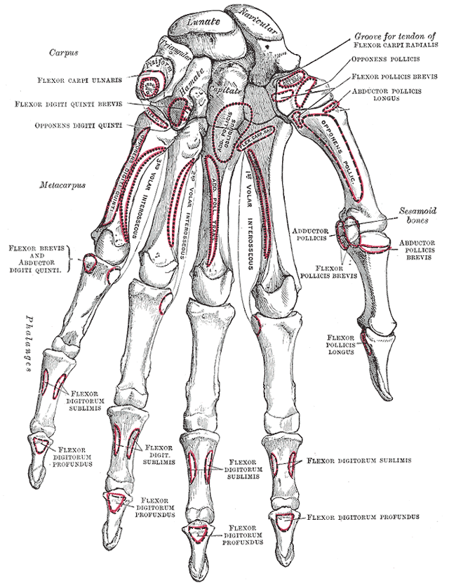
The flexor pollicis brevis is a muscle in the hand that flexes the thumb. It is one of three thenar muscles. It has both a superficial part and a deep part. Origin and insertion The muscle's superficial head arises from the distal edge of the flexor retinaculum and the tubercle of the trapezium, the most lateral bone in the distal row of carpal bones. It passes along the radial side of the tendon of the flexor pollicis longus. The deeper (and medial) head "varies in size and may be absent."Gray's 37th British Edition, p. 630" It arises from the trapezoid and capitate bones on the floor of the carpal tunnel, as well as the ligaments of the distal carpal row. Both heads become tendinous and insert together into the radial side of the base of the proximal phalanx of the thumb; at the junction between the tendinous heads there is a sesamoid bone.''Gray's Anatomy'' 1918, see infobox Innervation The superficial head is usually innervated by the lateral terminal branch of the med ... [...More Info...] [...Related Items...] OR: [Wikipedia] [Google] [Baidu] |
Trapezium Bone
The trapezium bone (greater multangular bone) is a carpal bone in the hand. It forms the radial border of the carpal tunnel. Structure The trapezium is distinguished by a deep groove on its anterior surface. It is situated at the radial side of the carpus, between the scaphoid and the first metacarpal bone (the metacarpal bone of the thumb). It is homologous with the first distal carpal of reptiles and amphibians. Surfaces The trapezium is an irregular-shaped carpal bone found within the hand. The trapezium is found within the distal row of carpal bones, and is directly adjacent to the metacarpal bone of the thumb. On its ulnar surface are found the trapezoid and scaphoid bones. The '' superior surface'' is directed upward and medialward; medially it is smooth, and articulates with the scaphoid; laterally it is rough and continuous with the lateral surface. The '' inferior surface'' is oval, concave from side to side, convex from before backward, so as to form a saddle-shaped ... [...More Info...] [...Related Items...] OR: [Wikipedia] [Google] [Baidu] |
Carpal Tunnel
In the human body, the carpal tunnel or carpal canal is a flattened body cavity on the flexor ( palmar/volar) side of the wrist, bounded by the carpal bones and flexor retinaculum. It forms the passageway that transmits the median nerve and the tendons of the extrinsic flexor muscles of the hand from the forearm to the hand. The median artery is an anatomical variant (increasingly found). When present it lies between the radial artery, and the ulnar artery and runs with the median nerve supplying the same structures innervated. When swelling or degeneration occurs in the tendons and sheaths of any of the nine flexor muscles ( flexor pollicis longus, four flexor digitorum profundus and four flexor digitorum superficialis) passing through the carpal tunnel, the canal can narrow and compress/entrap the median nerve, resulting in a compression neuropathy known as carpal tunnel syndrome (CTS). If untreated, neuropraxia, parasthesia and muscle atrophy (especially of the ... [...More Info...] [...Related Items...] OR: [Wikipedia] [Google] [Baidu] |
Anatomical Terms Of Motion
Motion, the process of movement, is described using specific anatomical terms. Motion includes movement of organs, joints, limbs, and specific sections of the body. The terminology used describes this motion according to its direction relative to the anatomical position of the body parts involved. Anatomists and others use a unified set of terms to describe most of the movements, although other, more specialized terms are necessary for describing unique movements such as those of the hands, feet, and eyes. In general, motion is classified according to the anatomical plane it occurs in. ''Flexion'' and ''extension'' are examples of ''angular'' motions, in which two axes of a joint are brought closer together or moved further apart. ''Rotational'' motion may occur at other joints, for example the shoulder, and are described as ''internal'' or ''external''. Other terms, such as ''elevation'' and ''depression'', describe movement above or below the horizontal plane. Many anatom ... [...More Info...] [...Related Items...] OR: [Wikipedia] [Google] [Baidu] |
Superficial Palmar Branch Of Radial Artery
The superficial palmar branch of the radial artery arises from the radial artery, just where this vessel is about to wind around the lateral side of the wrist. Running forward, it passes through, occasionally over, the thenar muscles, which it supplies, and sometimes anastomoses with the terminal portion of the ulnar artery, completing the superficial palmar arch The superficial palmar arch is formed predominantly by the ulnar artery, with a contribution from the superficial palmar branch of the radial artery. However, in some individuals the contribution from the radial artery might be absent, and instea .... This vessel varies considerably in size: usually it is very small, and ends in the muscles of the thumb; sometimes it is as large as the continuation of the radial artery itself. References Arteries of the upper limb {{circulatory-stub ... [...More Info...] [...Related Items...] OR: [Wikipedia] [Google] [Baidu] |
Spinal Nerve
A spinal nerve is a mixed nerve, which carries Motor neuron, motor, Sensory neuron, sensory, and Autonomic nervous system, autonomic signals between the spinal cord and the body. In the human body there are 31 pairs of spinal nerves, one on each side of the vertebral column. These are grouped into the corresponding cervical vertebrae, cervical, thoracic vertebrae, thoracic, lumbar vertebrae, lumbar, sacral vertebrae, sacral and coccygeal vertebrae, coccygeal regions of the spine. There are eight pairs of cervical nerves, twelve pairs of thoracic nerves, five pairs of lumbar nerves, five pairs of sacral nerves, and one pair of coccygeal nerves. The spinal nerves are part of the peripheral nervous system. Structure Each spinal nerve is a mixed nerve, formed from the combination of nerve root axon, fibers from its Dorsal root of spinal nerve, dorsal and Ventral root of spinal nerve, ventral roots. The dorsal root is the afferent nerve fiber, afferent sensory root and carries sen ... [...More Info...] [...Related Items...] OR: [Wikipedia] [Google] [Baidu] |
Cervical Spinal Nerve 8
The cervical spinal nerve 8 (C8) is a spinal nerve of the cervical segment. It originates from the spinal column from below the cervical vertebra 7 (C7). Innervation The C8 nerve forms part of the radial and ulnar nerves via the brachial plexus, and therefore has motor and sensory function in the upper limb. Sensory The C8 nerve receives sensory afferents from the C8 dermatome. This consists of all the skin on the little finger, and continuing up slightly past the wrist on the palmar and dorsal aspects of the hand and forearm.Drake et al. Gray's Anatomy for Students. Second Edition (2010). Clinically, a test of the pad of the little finger is often used to assess C8 integrity.Aland et al. University of Queensland School of Medicine Clinical Skills Handbook 2010 Motor The C8 nerve contributes to the motor innervation of many of the muscles in the trunk and upper limb. Its primary function is the flexion of the fingers, and this is used as the clinical test for C8 inte ... [...More Info...] [...Related Items...] OR: [Wikipedia] [Google] [Baidu] |
Ulnar Nerve
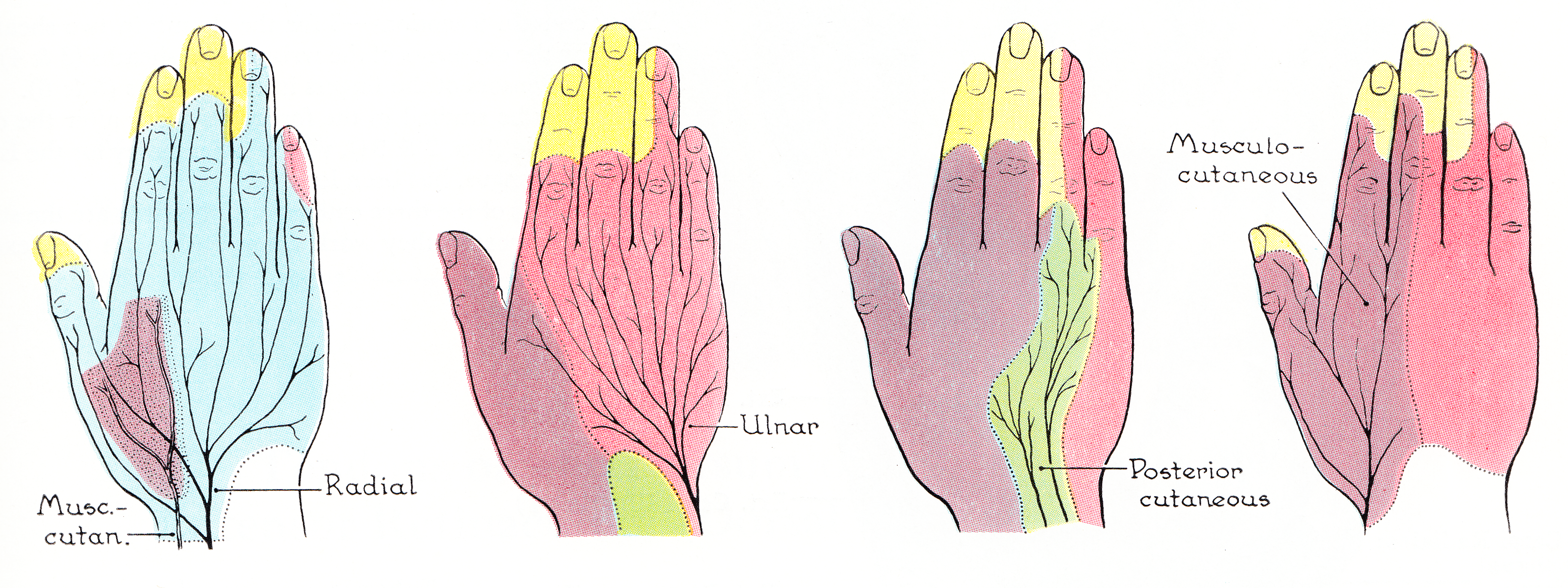
The ulnar nerve is a nerve that runs near the ulna, one of the two long bones in the forearm. The ulnar collateral ligament of elbow joint is in relation with the ulnar nerve. The nerve is the largest in the human body unprotected by muscle or bone, so injury is common. This nerve is directly connected to the little finger, and the adjacent half of the ring finger, innervating the palmar aspect of these fingers, including both front and back of the tips, perhaps as far back as the fingernail beds. This nerve can cause an electric shock-like sensation by striking the medial epicondyle of the humerus posteriorly, or inferiorly with the elbow flexed. The ulnar nerve is trapped between the bone and the overlying skin at this point. This is commonly referred to as bumping one's "funny bone". This name is thought to be a pun, based on the sound resemblance between the name of the bone of the upper arm, the humerus, and the word " humorous". Alternatively, according to the Oxfor ... [...More Info...] [...Related Items...] OR: [Wikipedia] [Google] [Baidu] |
Median Nerve
The median nerve is a nerve in humans and other animals in the upper limb. It is one of the five main nerves originating from the brachial plexus. The median nerve originates from the lateral and medial cords of the brachial plexus, and has contributions from ventral roots of C6-C7 (lateral cord) and C8 and T1 (medial cord). The median nerve is the only nerve that passes through the carpal tunnel. Carpal tunnel syndrome is the disability that results from the median nerve being pressed in the carpal tunnel. Structure The median nerve arises from the branches from lateral and medial cords of the brachial plexus, courses through the anterior part of arm, forearm, and hand, and terminates by supplying the muscles of the hand. Arm After receiving inputs from both the lateral and medial cords of the brachial plexus, the median nerve enters the arm from the axilla at the inferior margin of the teres major muscle. It then passes vertically down and courses lateral to the brac ... [...More Info...] [...Related Items...] OR: [Wikipedia] [Google] [Baidu] |
Gray's Anatomy
''Gray's Anatomy'' is a reference book of human anatomy written by Henry Gray, illustrated by Henry Vandyke Carter and first published in London in 1858. It has had multiple revised editions, and the current edition, the 42nd (October 2020), remains a standard reference, often considered "the doctors' bible". Earlier editions were called ''Anatomy: Descriptive and Surgical'', ''Anatomy of the Human Body'' and ''Gray's Anatomy: Descriptive and Applied'', but the book's name is commonly shortened to, and later editions are titled, ''Gray's Anatomy''. The book is widely regarded as an extremely influential work on the subject. Publication history Origins The English anatomist Henry Gray was born in 1827. He studied the development of the endocrine glands and spleen and in 1853 was appointed Lecturer on Anatomy at St George's Hospital Medical School in London. In 1855, he approached his colleague Henry Vandyke Carter with his idea to produce an inexpensive and access ... [...More Info...] [...Related Items...] OR: [Wikipedia] [Google] [Baidu] |
Sesamoid
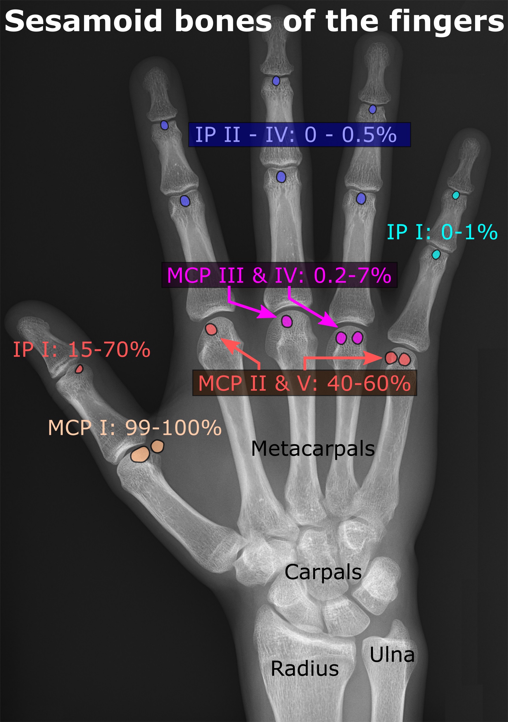
In anatomy, a sesamoid bone () is a bone embedded within a tendon or a muscle. Its name is derived from the Greek word for 'sesame seed', indicating the small size of most sesamoids. Often, these bones form in response to strain, or can be present as a normal variant. The patella is the largest sesamoid bone in the body. Sesamoids act like pulleys, providing a smooth surface for tendons to slide over, increasing the tendon's ability to transmit muscular forces. Structure Sesamoid bones can be found on joints throughout the human body, including: * In the knee—the patella (within the quadriceps tendon). This is the largest sesamoid bone. * In the hand—two sesamoid bones are commonly found in the distal portions of the first metacarpal bone (within the tendons of adductor pollicis and flexor pollicis brevis). There is also commonly a sesamoid bone in distal portions of the second metacarpal bone and fifth metacarpal bone. * In the wrist—The pisiform of the wrist is a ... [...More Info...] [...Related Items...] OR: [Wikipedia] [Google] [Baidu] |
Thumb
The thumb is the first digit of the hand, next to the index finger. When a person is standing in the medical anatomical position (where the palm is facing to the front), the thumb is the outermost digit. The Medical Latin English noun for thumb is ''pollex'' (compare ''hallux'' for big toe), and the corresponding adjective for thumb is ''pollical''. Definition Thumb and fingers The English word ''finger'' has two senses, even in the context of appendages of a single typical human hand: 1) Any of the five terminal members of the hand. 2) Any of the four terminal members of the hand, other than the thumb. Linguistically, it appears that the original sense was the first of these two: (also rendered as ) was, in the inferred Proto-Indo-European language, a suffixed form of (or ), which has given rise to many Indo-European-family words (tens of them defined in English dictionaries) that involve, or stem from, concepts of fiveness. The thumb shares the following with each of ... [...More Info...] [...Related Items...] OR: [Wikipedia] [Google] [Baidu] |
Phalanx Bones
The phalanges (: phalanx ) are digital bones in the hands and feet of most vertebrates. In primates, the thumbs and big toes have two phalanges while the other digits have three phalanges. The phalanges are classed as long bones. Structure The phalanges are the bones that make up the fingers of the hand and the toes of the foot. There are 56 phalanges in the human body, with fourteen on each hand and foot. Three phalanges are present on each finger and toe, with the exception of the thumb and big toe, which possess only two. The middle and far phalanges of the fifth toes are often fused together (symphalangism). The phalanges of the hand are commonly known as the finger bones. The phalanges of the foot differ from the hand in that they are often shorter and more compressed, especially in the proximal phalanges, those closest to the torso. A phalanx is named according to whether it is proximal, middle, or distal and its associated finger or toe. The proximal phalang ... [...More Info...] [...Related Items...] OR: [Wikipedia] [Google] [Baidu] |


