|
Fibrosarcoma
Fibrosarcoma (fibroblastic sarcoma) is a malignant mesenchymal tumour derived from fibrous connective tissue and characterized by the presence of immature proliferating fibroblasts or undifferentiated anaplastic spindle cells in a storiform pattern. Fibrosarcomas mainly arise in people between the ages of 25 and 79. It originates in fibrous tissues of the bone and invades long or flat bones such as the femur, tibia, and mandible. It also involves the periosteum and overlying muscle. Presentation Adult-type Individuals presenting with fibrosarcoma are usually adults thirty to fifty-five years old, often presenting with pain. Among adults, fibrosarcomas develop equally in men and women. Infantile-type In infants, fibrosarcoma (often termed congenital infantile fibrosarcoma) is usually congenital. Infants presenting with this fibrosarcoma usually do so in the first two years of their life. Cytogenetically, congenital infantile fibrosarcoma is characterized by the majority of ... [...More Info...] [...Related Items...] OR: [Wikipedia] [Google] [Baidu] |
Mesoblastic Nephroma
Congenital mesoblastic nephroma, while rare, is the most common kidney neoplasm diagnosed in the first three months of life and accounts for 3-5% of all childhood renal neoplasms. It is generally non-aggressive and amenable to surgical removal, though there is a subtype that is more aggressive and tends to metastasis, spread to other organs. Congenital mesoblastic nephroma was first named as such in 1967 but was recognized decades before this as fetal renal hamartoma or leiomyomatous renal hamartoma. It is embryologically derived from the metanephrogenic blastema, the same tissue that gives rise to nephroblastomatosis and Wilms tumor. Presentation Congenital mesoblastic nephroma typically (76% of cases) presents as an abdominal mass which is detected prenatally (16% of cases) by ultrasound or by clinical inspection (84% of cases) either at birth or by 3.8 years of age (median age ~1 month). The neoplasm shows a slight male preference. Concurrent findings include hypertension (19% ... [...More Info...] [...Related Items...] OR: [Wikipedia] [Google] [Baidu] |
Chromosomal Translocation
In genetics, chromosome translocation is a phenomenon that results in unusual rearrangement of chromosomes. This includes "balanced" and "unbalanced" translocation, with three main types: "reciprocal", "nonreciprocal" and "Robertsonian" translocation. Reciprocal translocation is a chromosome abnormality caused by exchange of parts between non-homologous chromosomes. Two detached fragments of two different chromosomes are switched. Robertsonian translocation occurs when two non-homologous chromosomes get attached, meaning that given two healthy pairs of chromosomes, one of each pair "sticks" and blends together homogeneously. Each type of chromosomal translocation can result in disorders for growth, function and the development of an individuals body, often resulting from a change in their genome. A gene fusion may be created when the translocation joins two otherwise-separated genes. It is detected on cytogenetics or a karyotype of affected cells. Translocations can be bala ... [...More Info...] [...Related Items...] OR: [Wikipedia] [Google] [Baidu] |
Synovial Sarcoma
A synovial sarcoma (also known as malignant synovioma) is a rare form of cancer which occurs primarily in the extremities of the arms or legs, often in proximity to joint capsules and tendon sheaths. It is a type of soft-tissue sarcoma. The name "synovial sarcoma" was coined early in the 20th century, as some researchers thought that the microscopic similarity of some tumors to synovium, and its propensity to arise adjacent to joints, indicated a synovial origin; however, the actual cells from which the tumor develops are unknown and not necessarily synovial. Primary synovial sarcomas are most common in the soft tissue near the large joints of the arm and leg but have been documented in most human tissues and organs, including the brain, prostate, and heart. Synovial sarcoma occurs in about 1–2 per 1,000,000 people a year. They occur most commonly in the third decade of life, with males being affected more often than females (ratio around 1.2:1). Signs and symptoms Synovial ... [...More Info...] [...Related Items...] OR: [Wikipedia] [Google] [Baidu] |
Leiomyosarcoma
A leiomyosarcoma (LMS) is a rare malignant (cancerous) smooth muscle tumor. The word is . The stomach, bladder, uterus, blood vessels, and Gastrointestinal tract, intestines are examples of hollow organs made up of smooth muscles where LMS can be located; however, the uterus and abdomen are the most common sites. Although leiomyosarcomas are rare, they belong to the more common types of soft-tissue sarcoma, representing 10–20% of new cases. This type of cancer is more frequently diagnosed in adults as compared to children. When considering LMS specifically in the context of the uterus, it affects approximately 6 individuals per 1 million people in the United States each year. LMSs are resistant cancers, meaning they are generally not very responsive to chemotherapy or radiation. The best outcomes occur when the tumor tissue can be removed surgically at an early stage, while it is small and has not yet spread from the original site (it remains ''in situ''). Mechanism Smooth mus ... [...More Info...] [...Related Items...] OR: [Wikipedia] [Google] [Baidu] |
Micrograph
A micrograph is an image, captured photographically or digitally, taken through a microscope or similar device to show a magnify, magnified image of an object. This is opposed to a macrograph or photomacrograph, an image which is also taken on a microscope but is only slightly magnified, usually less than 10 times. Micrography is the practice or art of using microscopes to make photographs. A photographic micrograph is a photomicrograph, and one taken with an electron microscope is an electron micrograph. A micrograph contains extensive details of microstructure. A wealth of information can be obtained from a simple micrograph like behavior of the material under different conditions, the phases found in the system, failure analysis, grain size estimation, elemental analysis and so on. Micrographs are widely used in all fields of microscopy. Types Photomicrograph A light micrograph or photomicrograph is a micrograph prepared using an optical microscope, a process referred to ... [...More Info...] [...Related Items...] OR: [Wikipedia] [Google] [Baidu] |
Chemotherapy
Chemotherapy (often abbreviated chemo, sometimes CTX and CTx) is the type of cancer treatment that uses one or more anti-cancer drugs (list of chemotherapeutic agents, chemotherapeutic agents or alkylating agents) in a standard chemotherapy regimen, regimen. Chemotherapy may be given with a cure, curative intent (which almost always involves combinations of drugs), or it may aim only to prolong life or to Palliative care, reduce symptoms (Palliative care, palliative chemotherapy). Chemotherapy is one of the major categories of the medical discipline specifically devoted to pharmacotherapy for cancer, which is called ''oncology#Specialties, medical oncology''. The term ''chemotherapy'' now means the non-specific use of intracellular poisons to inhibit mitosis (cell division) or to induce DNA damage (naturally occurring), DNA damage (so that DNA repair can augment chemotherapy). This meaning excludes the more-selective agents that block extracellular signals (signal transduction) ... [...More Info...] [...Related Items...] OR: [Wikipedia] [Google] [Baidu] |
Radiation Therapy
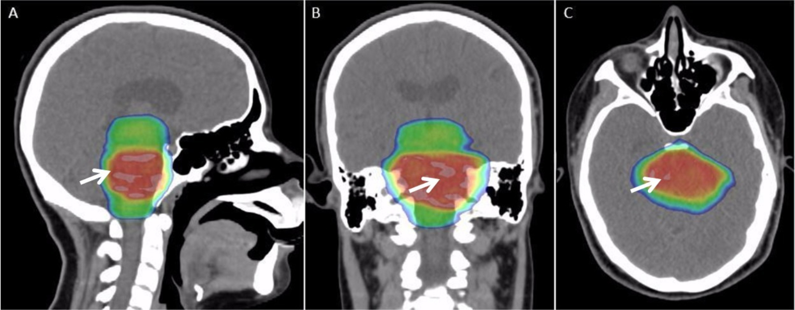
Radiation therapy or radiotherapy (RT, RTx, or XRT) is a therapy, treatment using ionizing radiation, generally provided as part of treatment of cancer, cancer therapy to either kill or control the growth of malignancy, malignant cell (biology), cells. It is normally delivered by a linear particle accelerator. Radiation therapy may be cure, curative in a number of types of cancer if they are localized to one area of the body, and have not metastasis, spread to other parts. It may also be used as part of adjuvant therapy, to prevent tumor recurrence after surgery to remove a primary malignant tumor (for example, early stages of breast cancer). Radiation therapy is synergistic with chemotherapy, and has been used before, during, and after chemotherapy in susceptible cancers. The subspecialty of oncology concerned with radiotherapy is called radiation oncology. A physician who practices in this subspecialty is a radiation oncologist. Radiation therapy is commonly applied to the canc ... [...More Info...] [...Related Items...] OR: [Wikipedia] [Google] [Baidu] |
Cat After Fibrosarcom Op
The cat (''Felis catus''), also referred to as the domestic cat or house cat, is a small Domestication, domesticated carnivorous mammal. It is the only domesticated species of the family Felidae. Advances in archaeology and genetics have shown that the domestication of the cat occurred in the Near East around 7500 BC. It is commonly kept as a pet and working cat, but also ranges freely as a feral cat avoiding human contact. It is valued by humans for companionship and its ability to kill vermin. Its retractable claws are adapted to killing small prey species such as mice and rats. It has a strong, flexible body, quick reflexes, and sharp teeth, and its night vision and sense of smell are well developed. It is a social species, but a solitary hunter and a crepuscular predator. Cat intelligence is evident in their ability to adapt, learn through observation, and solve problems. Research has shown they possess strong memories, exhibit neuroplasticity, and display cognitive skil ... [...More Info...] [...Related Items...] OR: [Wikipedia] [Google] [Baidu] |
Actin
Actin is a family of globular multi-functional proteins that form microfilaments in the cytoskeleton, and the thin filaments in muscle fibrils. It is found in essentially all eukaryotic cells, where it may be present at a concentration of over 100 μM; its mass is roughly 42 kDa, with a diameter of 4 to 7 nm. An actin protein is the monomeric subunit of two types of filaments in cells: microfilaments, one of the three major components of the cytoskeleton, and thin filaments, part of the contractile apparatus in muscle cells. It can be present as either a free monomer called G-actin (globular) or as part of a linear polymer microfilament called F-actin (filamentous), both of which are essential for such important cellular functions as the mobility and contraction of cells during cell division. Actin participates in many important cellular processes, including muscle contraction, cell motility, cell division and cytokinesis, vesicle and organelle mov ... [...More Info...] [...Related Items...] OR: [Wikipedia] [Google] [Baidu] |
S-100 Protein
The S100 proteins are a family of low molecular-weight proteins found in vertebrates characterized by two calcium-binding sites that have helix-loop-helix ("EF-hand-type") conformation. At least 21 different S100 proteins are known. They are encoded by a family of genes whose symbols use the ''S100'' prefix, for example, ''S100A1'', ''S100A2'', ''S100A3''. They are also considered as damage-associated molecular pattern molecules (DAMPs), and knockdown of aryl hydrocarbon receptor downregulates the expression of S100 proteins in THP-1 cells. Structure Most S100 proteins consist of two identical polypeptides (homodimeric), which are held together by noncovalent bonds. They are structurally similar to calmodulin. They differ from calmodulin, though, on the other features. For instance, their expression pattern is cell-specific, i.e. they are expressed in particular cell types. Their expression depends on environmental factors. In contrast, calmodulin is a ubiquitous and universa ... [...More Info...] [...Related Items...] OR: [Wikipedia] [Google] [Baidu] |
Cytokeratin
Cytokeratins are keratin proteins found in the intracytoplasmic cytoskeleton of epithelial tissue. They are an important component of intermediate filaments, which help cells resist mechanical stress. Expression of these cytokeratins within epithelial cells is largely specific to particular organs or tissues. Thus they are used clinically to identify the cell of origin of various human tumors. Naming The term ''cytokeratin'' began to be used in the late 1970s, when the protein subunits of keratin intermediate filaments inside cells were first being identified and characterized. In 2006 a new systematic nomenclature for mammalian keratins was created, and the proteins previously called ''cytokeratins'' are simply called ''keratins'' (human epithelial category). For example, cytokeratin-4 (CK-4) has been renamed keratin-4 (K4). However, they are still commonly referred to as cytokeratins in clinical practice. Types There are two categories of cytokeratins: the acidic type I ... [...More Info...] [...Related Items...] OR: [Wikipedia] [Google] [Baidu] |


