|
Fetal Pole
The fetal pole is a thickening on the margin of the yolk sac of a fetus during pregnancy. It is usually identified at six weeks with vaginal ultrasound and at six and a half weeks with abdominal ultrasound. However, it is not unheard of for the fetal pole to not be visible until about 9 weeks. The fetal pole may be seen at 2–4 mm crown-rump length Crown-rump length (CRL) is the measurement of the length of human embryos and fetuses from the top of the head (crown) to the bottom of the buttocks (rump). It is typically determined from ultrasound imagery and can be used to estimate gestatio ... (CRL). References Embryology {{Developmental-biology-stub ... [...More Info...] [...Related Items...] OR: [Wikipedia] [Google] [Baidu] |
Yolk Sac
The yolk sac is a membranous wikt:sac, sac attached to an embryo, formed by cells of the hypoblast layer of the bilaminar embryonic disc. This is alternatively called the umbilical vesicle by the Terminologia Embryologica (TE), though ''yolk sac'' is far more widely used. The yolk sac is one of the fetal membranes and is important in early embryonic blood supply. In humans much of it is incorporated into the primordial gut (anatomy), gut during the fourth week of embryonic development. In humans The yolk sac is the first element seen within the gestational sac during pregnancy, usually at three days gestation. The yolk sac is situated on the front (ventral) part of the embryo; it is lined by extra-embryonic endoderm, outside of which is a layer of extra-embryonic mesenchyme, derived from the epiblast. Blood is conveyed to the wall of the yolk sac by the primitive aorta and after circulating through a wide-meshed capillary plexus, is returned by the vitelline veins to the tubul ... [...More Info...] [...Related Items...] OR: [Wikipedia] [Google] [Baidu] |
Fetus
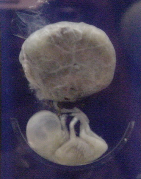
A fetus or foetus (; : fetuses, foetuses, rarely feti or foeti) is the unborn offspring of a viviparous animal that develops from an embryo. Following the embryonic development, embryonic stage, the fetal stage of development takes place. Prenatal development is a continuum, with no clear defining feature distinguishing an embryo from a fetus. However, in general a fetus is characterized by the presence of all the major body organs, though they will not yet be fully developed and functional, and some may not yet be situated in their final Anatomy, anatomical location. In human prenatal development, fetal development begins from the ninth week after Human fertilization, fertilization (which is the eleventh week of Gestational age (obstetrics), gestational age) and continues until the childbirth, birth of a newborn. Etymology The word ''wikt:fetus#English, fetus'' (plural ''wikt:fetuses#English, fetuses'' or rarely, the solecism ''wikt:feti#English, feti''''Oxford English Dict ... [...More Info...] [...Related Items...] OR: [Wikipedia] [Google] [Baidu] |
Pregnancy
Pregnancy is the time during which one or more offspring gestation, gestates inside a woman's uterus. A multiple birth, multiple pregnancy involves more than one offspring, such as with twins. Conception (biology), Conception usually occurs following sexual intercourse, vaginal intercourse, but can also occur through assisted reproductive technology procedures. A pregnancy may end in a Live birth (human), live birth, a miscarriage, an Abortion#Induced, induced abortion, or a stillbirth. Childbirth typically occurs around 40 weeks from the start of the Menstruation#Onset and frequency, last menstrual period (LMP), a span known as the Gestational age (obstetrics), ''gestational age''; this is just over nine months. Counting by Human fertilization#Fertilization age, ''fertilization age'', the length is about 38 weeks. Implantation (embryology), Implantation occurs on average 8–9 days after Human fertilization, fertilization. An ''embryo'' is the term for the deve ... [...More Info...] [...Related Items...] OR: [Wikipedia] [Google] [Baidu] |
Vaginal Ultrasonography
Vaginal ultrasonography is a medical ultrasonography that applies an ultrasound transducer (or "probe") in the vagina to visualize organs within the pelvic cavity. It is also called transvaginal ultrasonography because the ultrasound waves go ''across'' the vaginal wall to study tissues beyond it. Uses Vaginal ultrasonography is used both as a means of gynecologic ultrasonography and obstetric ultrasonography. It is preferred over abdominal ultrasonography in the diagnosis of ectopic pregnancy. It also can be used to evaluate patients with post-menopausal bleeding. The finding on transvaginal ultrasound of a thin endometrial lining gives the physician a 99% negative predictive value that the patient does not have endometrial cancer. If a patient had a prior endometrial sampling that was inconclusive, then a transvaginal ultrasound can be used to triage a woman with post-menopausal bleeding. See also * Gynecologic ultrasonography * Post-menopausal bleeding Vaginal bleeding ... [...More Info...] [...Related Items...] OR: [Wikipedia] [Google] [Baidu] |
Abdominal Ultrasonography
Abdominal ultrasonography (also called abdominal ultrasound imaging or abdominal sonography) is a form of medical ultrasonography (medical application of ultrasound technology) to visualise abdominal anatomical structures. It uses transmission and reflection of ultrasound waves to visualise internal organs through the abdominal wall (with the help of gel, which helps transmission of the sound waves). For this reason, the procedure is also called a transabdominal ultrasound, in contrast to endoscopic ultrasound, the latter combining ultrasound with endoscopy through visualize internal structures from within hollow organs. Abdominal ultrasound examinations are performed by gastroenterologists or other specialists in internal medicine, radiologists, or sonographers trained for this procedure. Medical uses Abdominal ultrasound can be used to diagnose abnormalities in various internal organs, such as the kidneys, liver, gallbladder, pancreas, spleen and abdominal aorta. If Doppler ... [...More Info...] [...Related Items...] OR: [Wikipedia] [Google] [Baidu] |
Crown-rump Length
Crown-rump length (CRL) is the measurement of the length of human embryos and fetuses from the top of the head (crown) to the bottom of the buttocks (rump). It is typically determined from ultrasound imagery and can be used to estimate gestational age. Introduction The embryo and fetus float in the amniotic fluid inside the uterus of the mother usually in a curved posture resembling the letter ''C''. The measurement can actually vary slightly if the fetus is temporarily stretching (straightening) its body. The measurement needs to be in the natural state with an unstretched body which is actually ''C'' shaped. The measurement of CRL is useful in determining the gestational age (menstrual age starting from the first day of the last menstrual period) and thus the expected date of delivery (EDD). Different human fetuses grow at different rates and thus the gestational age is an approximation. Recent evidence has indicated that CRL growth (and thus the approximation of gestational ... [...More Info...] [...Related Items...] OR: [Wikipedia] [Google] [Baidu] |


