|
Eyelids
An eyelid ( ) is a thin fold of skin that covers and protects an eye. The levator palpebrae superioris muscle retracts the eyelid, exposing the cornea to the outside, giving vision. This can be either voluntarily or involuntarily. "Palpebral" (and "blepharal") means relating to the eyelids. Its key function is to regularly spread the tears and other secretions on the eye surface to keep it moist, since the cornea must be continuously moist. They keep the eyes from drying out when asleep. Moreover, the blink reflex protects the eye from foreign bodies. A set of specialized hairs known as lashes grow from the upper and lower eyelid margins to further protect the eye from dust and debris. The appearance of the human upper eyelid often varies between different populations. The prevalence of an epicanthic fold covering the inner corner of the eye account for the majority of East Asian and Southeast Asian populations, and is also found in varying degrees among other populatio ... [...More Info...] [...Related Items...] OR: [Wikipedia] [Google] [Baidu] |
Orbicularis Oculi Muscle
The orbicularis oculi is a Sphincter, sphincter-like muscle in the face that closes the eyelids. It arises from the nasal part of the frontal bone, from the frontal process of the maxilla in front of the lacrimal groove, and from the anterior surface and borders of a short fibrous band, the medial palpebral ligament. From this origin, the fibers are directed laterally, forming a broad and thin layer, which occupies the eyelids or palpebræ, surrounds the circumference of the orbit, and spreads over the temple, and downward on the cheek. Structure There are at least 3 clearly defined sections of the orbicularis muscle. However, it is not clear whether the lacrimal section is a separate section, or whether it is just an extension of the preseptal and pretarsal sections. Orbital orbicularis The orbital portion is thicker and of a reddish color; its fibers form a complete ellipse without interruption at the lateral palpebral commissure; the upper fibers of this portion blend with the ... [...More Info...] [...Related Items...] OR: [Wikipedia] [Google] [Baidu] |
Eyelash
An eyelash (also called lash) (Neo-Latin: ''cilium'', plural ''cilia'') is one of the hairs that grows at the edges of the top and bottom eyelids, spanning outwards and away from the eyes. The lashes grow in up to six layers on each of the upper and lower eyelids. Eyelashes serve to protect the eye from debris, dust, and small particles, and are highly sensitive to touch, thus providing a warning that an object (such as an insect or lint) is near the eye, which then reflexively closes or flutters to rid the area of the object. The eyelid margin from which lashes grow is among the most sensitive parts of the human body, with many Nerve, nerve endings enveloping the roots of the lashes, giving it sensitivity to very light tactile input even at the tips of the lashes, enabling it to trigger the Corneal reflex, blink reflex when touched. Eyelashes are also an important component of physical attractiveness, with long prominent lashes giving the illusion of large, gazing eyes, and drawin ... [...More Info...] [...Related Items...] OR: [Wikipedia] [Google] [Baidu] |
Sebaceous Glands
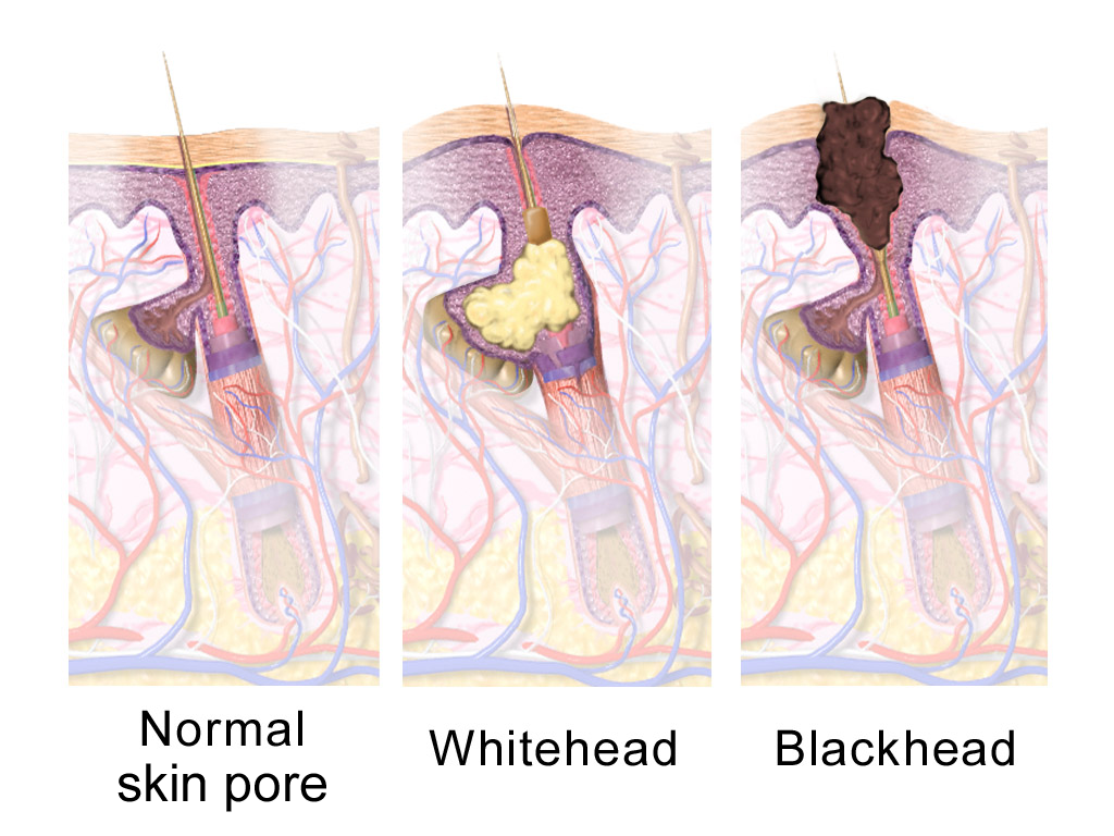
A sebaceous gland or oil gland is a microscopic exocrine gland in the skin that opens into a hair follicle to secrete an oily or waxy matter, called sebum, which lubricates the hair and skin of mammals. In humans, sebaceous glands occur in the greatest number on the face and scalp, but also on all parts of the skin except the palms of the hands and soles of the feet. In the eyelids, meibomian glands, also called tarsal glands, are a type of sebaceous gland that secrete a special type of sebum into tears. Surrounding the female nipples, areolar glands are specialized sebaceous glands for lubricating the nipples. Fordyce spots are benign, visible, sebaceous glands found usually on the lips, gums and inner cheeks, and genitals. Structure Location In humans, sebaceous glands are found throughout all areas of the skin, except the palms of the hands and soles of the feet. There are two types of sebaceous glands: those connected to hair follicles and those that exist indepen ... [...More Info...] [...Related Items...] OR: [Wikipedia] [Google] [Baidu] |
Medial Palpebral Arteries
The medial palpebral arteries (internal palpebral arteries) are arteries of the head that contribute arterial blood supply to the eyelids. They are derived from the ophthalmic artery; a single medial palpebral artery issues from the ophthalmic artery before splitting into a superior and an inferior medial palpebral artery, each supplying one eyelid. Anatomy Origin A single medial palpebral artery issues from the ophthalmic artery before bifurcating into a superior and an inferior medial palpebral artery. The origin occurs near the trochlea of the superior oblique muscle. Course The medial palpebral arteries leave the orbit to encircle the eyelids near their free margins, forming a superior and an inferior arch, which lie between the orbicularis oculi and the tarsi. Anastomoses The superior medial palpebral artery anastomoses (at lateral angle of the orbit) with the upper lateral palpebral artery, and the zygomaticoorbital branch of the temporal artery. The inferio ... [...More Info...] [...Related Items...] OR: [Wikipedia] [Google] [Baidu] |
Orbital Septum
In anatomy, the orbital septum (palpebral fascia) is a membranous sheet that acts as the anterior (frontal) boundary of the orbit. It extends from the orbital rims to the eyelids. It forms the fibrous portion of the eyelids. Structure In the upper eyelid, the orbital septum blends with the tendon of the levator palpebrae superioris, and in the lower eyelid with the tarsal plate. When the eyes are closed, the whole orbital opening is covered by the septum and tarsi. Medially it is thin, and, becoming separated from the medial palpebral ligament, attaches to the lacrimal bone at its posterior crest. The medial ligament and its much weaker lateral counterpart, attached to the septum and orbit, keep the lids stable as the eye moves. The septum is perforated by the vessels and nerves which pass from the orbital cavity to the face and scalp. Clinical significance Orbital septum supports the orbital contents located posterior to it, especially orbital fat. The septum can be w ... [...More Info...] [...Related Items...] OR: [Wikipedia] [Google] [Baidu] |
Epicanthic Fold
An epicanthic fold or epicanthus is a skin fold of the upper eyelid that covers the inner corner (medial canthus) of the Human eye, eye. However, variation occurs in the nature of this feature and the presence of "partial epicanthic folds" or "slight epicanthic folds" is noted in the relevant literature. Various factors influence whether epicanthic folds form, including ancestry, age, and certain medical conditions. The primary cause of the epicanthic fold is the hypertrophy of the preseptal portion of the orbicularis oculi muscle. Etymology ''Epicanthus'' means 'above the canthus', with epi-canthus being the Latinized form of the Ancient Greek : 'corner of the eye'. Classification Variation in the shape of the epicanthic fold has led to four types being recognised: * ''Epicanthus supraciliaris'' runs from the brow, curving downwards towards the lachrymal sac. * ''Epicanthus palpebralis'' begins above the upper Tarsus (eyelids), tarsus and extends to the inferior orbita ... [...More Info...] [...Related Items...] OR: [Wikipedia] [Google] [Baidu] |
Infratrochlear
The infratrochlear nerve is a branch of the nasociliary nerve (itself a branch of the ophthalmic nerve (CN V1)) in the orbit. It exits the orbit inferior to the trochlea of superior oblique. It provides sensory innervation to structures of the orbit and skin of adjacent structures. Structure The nasociliary nerve terminates by bifurcating into the infratrochlear and the anterior ethmoidal nerves. The infratrochlear nerve travels anteriorly in the orbit along the upper border of the medial rectus muscle and underneath the trochlea of the superior oblique muscle. It exits the orbit medially and divides into small sensory branches. Distribution The infratrochlear nerve provides sensory innervation to the skin of the eyelids, the conjunctiva, lacrimal sac, lacrimal caruncle, and the side of the nose superior to the medial canthus. Communications The infratrochlear nerve receives a descending communicating branch from the supratrochlear nerve The supratrochlear nerve is a ... [...More Info...] [...Related Items...] OR: [Wikipedia] [Google] [Baidu] |
Meibomian Gland
Meibomian glands (also called tarsal glands, palpebral glands, and tarsoconjunctival glands) are sebaceous glands along the rims of the eyelid inside the tarsal plate. They produce meibum, an oily substance that prevents evaporation of the eye, eye's tear film. Meibum prevents tears from spilling onto the cheek, traps them between the oiled edge and the eyeball, and makes the closed lids airtight. There are about 25 such glands on the upper eyelid, and 20 on the lower eyelid. Meibomian gland dysfunction, Dysfunctional meibomian glands is believed to be the most often cause of dry eyes. They are also the cause of blepharitis, posterior blepharitis. History The glands were mentioned by Galen in 200 AD and were described in more detail by Heinrich Meibom (doctor), Heinrich Meibom (1638–1700), a German physician, in his work ''De Vasis Palpebrarum Novis Epistola'' in 1666. This work included a drawing with the basic characteristics of the glands. Anatomy Although the upper lid ... [...More Info...] [...Related Items...] OR: [Wikipedia] [Google] [Baidu] |
Meibomian Glands
Meibomian glands (also called tarsal glands, palpebral glands, and tarsoconjunctival glands) are sebaceous glands along the rims of the eyelid inside the tarsal plate. They produce meibum, an oily substance that prevents evaporation of the eye's tear film. Meibum prevents tears from spilling onto the cheek, traps them between the oiled edge and the eyeball, and makes the closed lids airtight. There are about 25 such glands on the upper eyelid, and 20 on the lower eyelid. Dysfunctional meibomian glands is believed to be the most often cause of dry eyes. They are also the cause of posterior blepharitis. History The glands were mentioned by Galen in 200 AD and were described in more detail by Heinrich Meibom (1638–1700), a German physician, in his work ''De Vasis Palpebrarum Novis Epistola'' in 1666. This work included a drawing with the basic characteristics of the glands. Anatomy Although the upper lid has a greater number and volume of meibomian glands than the low ... [...More Info...] [...Related Items...] OR: [Wikipedia] [Google] [Baidu] |
Tears
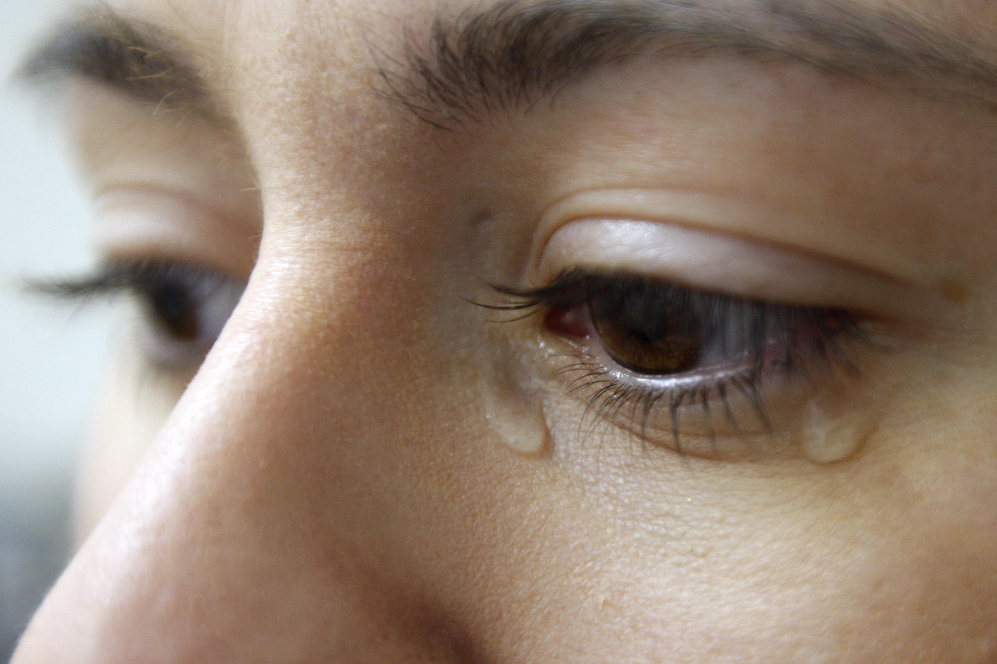
Tears are a clear liquid secreted by the lacrimal glands (tear gland) found in the eyes of all land mammals. Tears are made up of water, electrolytes, proteins, lipids, and mucins that form layers on the surface of eyes. The different types of tears—basal, reflex, and emotional—vary significantly in composition. The functions of tears include lubricating the eyes (basal tears), removing irritants (reflex tears), and also aiding the immune system. Tears also occur as a part of the body's natural pain response. Emotional secretion of tears may serve a biological function by excreting stress-inducing hormones built up through times of emotional distress. Tears have symbolic significance among humans. Physiology Chemical composition Tears are made up of three layers: lipid, aqueous, and mucous. Tears are composed of water, salts, antibodies, and lysozymes (antibacterial enzymes); though composition varies among different tear types. The composition of tears caused by an ... [...More Info...] [...Related Items...] OR: [Wikipedia] [Google] [Baidu] |
Lateral Palpebral Arteries
The lateral palpebral arteries are the two large branches of those terminal branches of the lacrimal gland The lacrimal glands are paired exocrine glands, one for each eye, found in most terrestrial vertebrates and some marine mammals, that secrete the aqueous layer of the tear film. In humans, they are situated in the upper lateral region of each o ... that supply the eyelid, with one lateral palpebral artery supplying one eyelid or the other. They pass medial-ward within the eyelid. They anastomose with medial palpebral arteries to form an arterial circle. See also * Medial palpebral arteries References Arteries of the head and neck {{circulatory-stub ... [...More Info...] [...Related Items...] OR: [Wikipedia] [Google] [Baidu] |


