|
Extensor Pollicis Brevis
In human anatomy, the extensor pollicis brevis (EPB) is a skeletal muscle on the dorsal side of the forearm. It lies on the medial side of, and is closely connected with, the abductor pollicis longus. The extensor pollicis brevis belongs to the deep group of the posterior fascial compartment of the forearm. It is a part of the lateral border of the anatomical snuffbox. Structure The extensor pollicis brevis arises from the ulna distal to the abductor pollicis longus, from the interosseous membrane, and from the dorsal surface of the radius. Its direction is similar to that of the abductor pollicis longus, its tendon passing the same groove on the lateral side of the lower end of the radius, to be inserted into the base of the first phalanx of the thumb. Variation Absence; fusion of tendon with that of the extensor pollicis longus or abductor pollicis longus muscle. Function In a close relationship to the abductor pollicis longus, the extensor pollicis brevis both e ... [...More Info...] [...Related Items...] OR: [Wikipedia] [Google] [Baidu] |
Radius (bone)
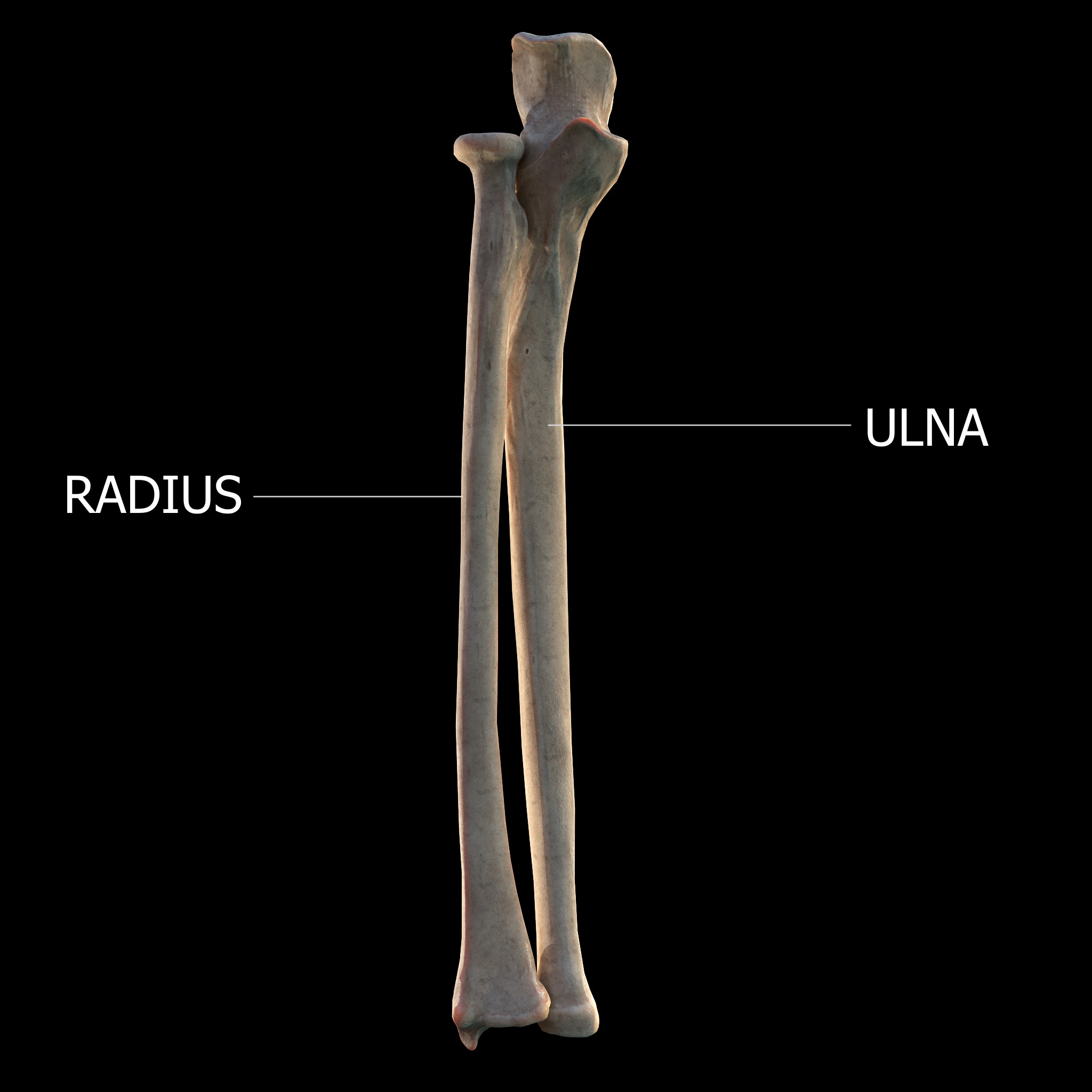
The radius or radial bone (: radii or radiuses) is one of the two large bones of the forearm, the other being the ulna. It extends from the Anatomical terms of location, lateral side of the Elbow-joint, elbow to the thumb side of the wrist and runs parallel to the ulna. The ulna is longer than the radius, but the radius is thicker. The radius is a long bone, Prism (geometry), prism-shaped and slightly curved longitudinally. The radius is part of two joint (anatomy), joints: the elbow and the wrist. At the elbow, it joins with the capitulum of the humerus, and in a separate region, with the ulna at the radial notch. At the wrist, the radius forms a joint with the ulna bone. The corresponding bone in the human leg, lower leg is the tibia. Structure The long narrow medullary cavity is enclosed in a strong wall of compact bone. It is thickest along the interosseous border and thinnest at the extremities, same over the cup-shaped articular surface (fovea) of the head. The tra ... [...More Info...] [...Related Items...] OR: [Wikipedia] [Google] [Baidu] |
Skeletal Muscle
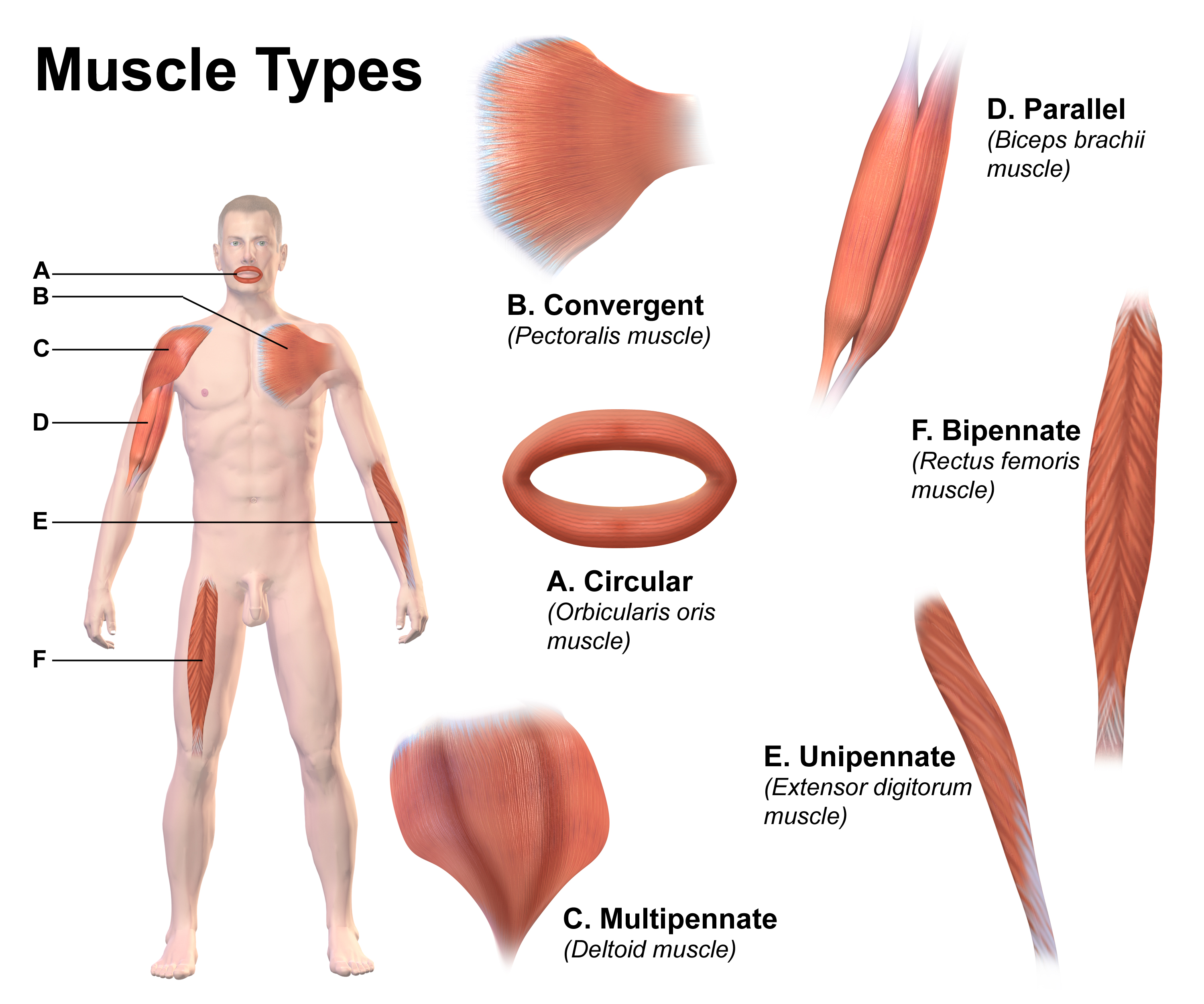
Skeletal muscle (commonly referred to as muscle) is one of the three types of vertebrate muscle tissue, the others being cardiac muscle and smooth muscle. They are part of the somatic nervous system, voluntary muscular system and typically are attached by tendons to bones of a skeleton. The skeletal muscle cells are much longer than in the other types of muscle tissue, and are also known as ''muscle fibers''. The tissue of a skeletal muscle is striated muscle tissue, striated – having a striped appearance due to the arrangement of the sarcomeres. A skeletal muscle contains multiple muscle fascicle, fascicles – bundles of muscle fibers. Each individual fiber and each muscle is surrounded by a type of connective tissue layer of fascia. Muscle fibers are formed from the cell fusion, fusion of developmental myoblasts in a process known as myogenesis resulting in long multinucleated cells. In these cells, the cell nucleus, nuclei, termed ''myonuclei'', are located along the inside ... [...More Info...] [...Related Items...] OR: [Wikipedia] [Google] [Baidu] |
Anatomical Snuff Box
The anatomical snuff box or snuffbox or foveola radialis is a triangular deepening on the radial, dorsal aspect of the hand—at the level of the carpal bones, specifically, the scaphoid and trapezium bones forming the floor. The name originates from the use of this surface for placing and then sniffing powdered tobacco, or " snuff." It is sometimes referred to by its French name ''tabatière''. Structure Boundaries * The medial border (ulnar side) of the snuffbox is the tendon of the extensor pollicis longus * The lateral border (radial side) is a pair of parallel and intimate tendons, of the extensor pollicis brevis and the abductor pollicis longus. (Accordingly, the anatomical snuffbox is most visible, having a more pronounced concavity, during thumb extension.) * The proximal border is formed by the styloid process of the radius * The distal border is formed by the approximate apex of the schematic snuffbox isosceles triangle. * The floor of the snuffbox varies dependi ... [...More Info...] [...Related Items...] OR: [Wikipedia] [Google] [Baidu] |
Carpometacarpal Joint
The carpometacarpal (CMC) joints are five joints in the wrist that articulate the distal row of carpal bones and the proximal bases of the five metacarpal bones. The CMC joint of the thumb or the first CMC joint, also known as the trapeziometacarpal (TMC) joint, differs significantly from the other four CMC joints and is therefore described separately. Thumb The carpometacarpal joint of the thumb (''pollex''), also known as the first carpometacarpal joint, or the trapeziometacarpal joint (TMC) because it connects the trapezium to the first metacarpal bone, plays an irreplaceable role in the normal functioning of the thumb. The most important joint connecting the wrist to the metacarpus, osteoarthritis of the TMC is a severely disabling condition; it is up to twenty times more common among elderly women than in the average. Pronation-supination of the first metacarpal is especially important for the action of opposition. The movements of the first CMC are limited by the sha ... [...More Info...] [...Related Items...] OR: [Wikipedia] [Google] [Baidu] |
Interosseous Membrane Of The Forearm
The interosseous membrane of the forearm (rarely middle or intermediate radioulnar joint) is a fibrous sheet that connects the interosseous margins of the radius and the ulna. It is the main part of the radio-ulnar syndesmosis, a fibrous joint In anatomy, fibrous joints are joints connected by Fibrous connective tissue, fibrous tissue, consisting mainly of collagen. These are fixed joints where bones are united by a layer of white fibrous tissue of varying thickness. In the skull, the ... between the two bones. Function The interosseous membrane divides the forearm into anterior and posterior compartments, serves as a site of attachment for muscles of the forearm, and transfers loads placed on the forearm. The interosseous membrane is designed to shift compressive loads (as in doing a hand-stand) from the distal radius to the proximal ulna. The fibers within the interosseous membrane are oriented obliquely so that when force is applied the fibers are drawn taut, shifting mor ... [...More Info...] [...Related Items...] OR: [Wikipedia] [Google] [Baidu] |
Abductor Pollicis Longus Muscle
In human anatomy, the abductor pollicis longus (APL) is one of the extrinsic muscles of the hand. Its major function is to abduct the thumb at the wrist. Its tendon forms the anterior border of the anatomical snuffbox. Structure The abductor pollicis longus lies immediately below the supinator and is sometimes united with it. It arises from the lateral part of the dorsal surface of the body of the ulna, below the insertion of the anconeus, from the interosseous membrane, and from the middle third of the dorsal surface of the body of the radius.''Gray's Anatomy'' (1918), see infobox Passing obliquely downward and lateralward, it ends in a tendon, which runs through a groove on the lateral side of the lower end of the radius, accompanied by the tendon of the extensor pollicis brevis. The insertion is divided into a distal, superficial part and a proximal, deep part. The superficial part is inserted with one or more tendons into the radial side of the base of the first ... [...More Info...] [...Related Items...] OR: [Wikipedia] [Google] [Baidu] |
Ulna
The ulna or ulnar bone (: ulnae or ulnas) is a long bone in the forearm stretching from the elbow to the wrist. It is on the same side of the forearm as the little finger, running parallel to the Radius (bone), radius, the forearm's other long bone. Longer and thinner than the radius, the ulna is considered to be the smaller long bone of the lower arm. The corresponding bone in the Human leg#Structure, lower leg is the fibula. Structure The ulna is a long bone found in the forearm that stretches from the elbow to the wrist, and when in standard anatomical position, is found on the Medial (anatomy), medial side of the forearm. It is broader close to the elbow, and narrows as it approaches the wrist. Close to the elbow, the ulna has a bony Process (anatomy), process, the olecranon process, a hook-like structure that fits into the olecranon fossa of the humerus. This prevents hyperextension and forms a hinge joint with the trochlea of the humerus. There is also a radial notch for ... [...More Info...] [...Related Items...] OR: [Wikipedia] [Google] [Baidu] |
Anatomical Snuffbox
The anatomical snuff box or snuffbox or foveola radialis is a triangular deepening on the radial, dorsal aspect of the hand—at the level of the carpal bones, specifically, the scaphoid and trapezium bones forming the floor. The name originates from the use of this surface for placing and then sniffing powdered tobacco, or " snuff." It is sometimes referred to by its French name ''tabatière''. Structure Boundaries * The medial border (ulnar side) of the snuffbox is the tendon of the extensor pollicis longus * The lateral border (radial side) is a pair of parallel and intimate tendons, of the extensor pollicis brevis and the abductor pollicis longus. (Accordingly, the anatomical snuffbox is most visible, having a more pronounced concavity, during thumb extension.) * The proximal border is formed by the styloid process of the radius * The distal border is formed by the approximate apex of the schematic snuffbox isosceles triangle. * The floor of the snuffbox varies dependi ... [...More Info...] [...Related Items...] OR: [Wikipedia] [Google] [Baidu] |
Abductor Pollicis Longus Muscle
In human anatomy, the abductor pollicis longus (APL) is one of the extrinsic muscles of the hand. Its major function is to abduct the thumb at the wrist. Its tendon forms the anterior border of the anatomical snuffbox. Structure The abductor pollicis longus lies immediately below the supinator and is sometimes united with it. It arises from the lateral part of the dorsal surface of the body of the ulna, below the insertion of the anconeus, from the interosseous membrane, and from the middle third of the dorsal surface of the body of the radius.''Gray's Anatomy'' (1918), see infobox Passing obliquely downward and lateralward, it ends in a tendon, which runs through a groove on the lateral side of the lower end of the radius, accompanied by the tendon of the extensor pollicis brevis. The insertion is divided into a distal, superficial part and a proximal, deep part. The superficial part is inserted with one or more tendons into the radial side of the base of the first ... [...More Info...] [...Related Items...] OR: [Wikipedia] [Google] [Baidu] |
Forearm
The forearm is the region of the upper limb between the elbow and the wrist. The term forearm is used in anatomy to distinguish it from the arm, a word which is used to describe the entire appendage of the upper limb, but which in anatomy, technically, means only the region of the upper arm, whereas the lower "arm" is called the forearm. It is homologous to the region of the leg that lies between the knee and the ankle joints, the crus. The forearm contains two long bones, the radius and the ulna, forming the two radioulnar joints. The interosseous membrane connects these bones. Ultimately, the forearm is covered by skin, the anterior surface usually being less hairy than the posterior surface. The forearm contains many muscles, including the flexors and extensors of the wrist, flexors and extensors of the digits, a flexor of the elbow ( brachioradialis), and pronators and supinators that turn the hand to face down or upwards, respectively. In cross-section, the forearm can ... [...More Info...] [...Related Items...] OR: [Wikipedia] [Google] [Baidu] |
Human Anatomy
Human anatomy (gr. ἀνατομία, "dissection", from ἀνά, "up", and τέμνειν, "cut") is primarily the scientific study of the morphology of the human body. Anatomy is subdivided into gross anatomy and microscopic anatomy. Gross anatomy (also called macroscopic anatomy, topographical anatomy, regional anatomy, or anthropotomy) is the study of anatomical structures that can be seen by the naked eye. Microscopic anatomy is the study of minute anatomical structures assisted with microscopes, which includes histology (the study of the organization of tissues), and cytology (the study of cells). Anatomy, human physiology (the study of function), and biochemistry (the study of the chemistry of living structures) are complementary basic medical sciences that are generally together (or in tandem) to students studying medical sciences. In some of its facets human anatomy is closely related to embryology, comparative anatomy and comparative embryology, through common ... [...More Info...] [...Related Items...] OR: [Wikipedia] [Google] [Baidu] |
Interosseous Membrane Of Forearm
The interosseous membrane of the forearm (rarely middle or intermediate radioulnar joint) is a fibrous sheet that connects the interosseous margins of the radius and the ulna. It is the main part of the radio-ulnar syndesmosis, a fibrous joint In anatomy, fibrous joints are joints connected by Fibrous connective tissue, fibrous tissue, consisting mainly of collagen. These are fixed joints where bones are united by a layer of white fibrous tissue of varying thickness. In the skull, the ... between the two bones. Function The interosseous membrane divides the forearm into anterior and posterior compartments, serves as a site of attachment for muscles of the forearm, and transfers loads placed on the forearm. The interosseous membrane is designed to shift compressive loads (as in doing a hand-stand) from the distal radius to the proximal ulna. The fibers within the interosseous membrane are oriented obliquely so that when force is applied the fibers are drawn taut, shifting mor ... [...More Info...] [...Related Items...] OR: [Wikipedia] [Google] [Baidu] |


