|
Epididymis
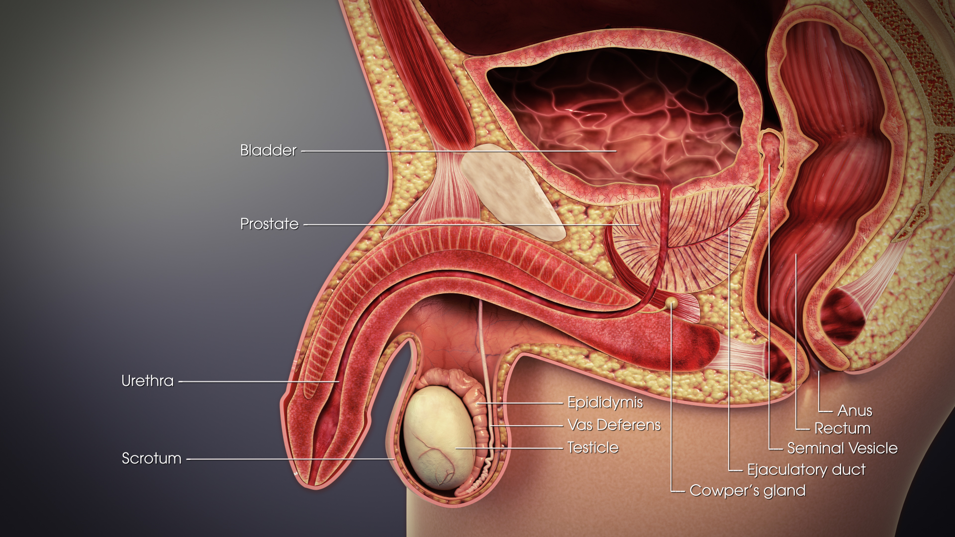
The epididymis (; : epididymides or ) is an elongated tubular genital organ attached to the posterior side of each one of the two male reproductive glands, the testicles. It is a single, narrow, tightly coiled tube in adult humans, in length; uncoiled the tube would be approximately 6 m (20 feet) long. It connects the testicle to the vas deferens in the male reproductive system. The epididymis serves as an interconnection between the multiple efferent ducts at the rear of a testicle (proximally), and the vas deferens (distally). Its primary function is the storage, maturation and transport of sperm cells. Structure The human epididymis is situated posterior and somewhat lateral to the testis. The epididymis is invested completely by the tunica vaginalis (which is continuous with the tunica vaginalis covering the testis). The epididymis can be divided into three main regions: * The head (). The head of the epididymis receives spermatozoa via the efferent ducts of the medi ... [...More Info...] [...Related Items...] OR: [Wikipedia] [Google] [Baidu] |
Testicle
A testicle or testis ( testes) is the gonad in all male bilaterians, including humans, and is Homology (biology), homologous to the ovary in females. Its primary functions are the production of sperm and the secretion of Androgen, androgens, primarily testosterone. The release of testosterone is regulated by luteinizing hormone (LH) from the anterior pituitary gland. Sperm production is controlled by follicle-stimulating hormone (FSH) from the anterior pituitary gland and by testosterone produced within the gonads. Structure Appearance Males have two testicles of similar size contained within the scrotum, which is an extension of the abdominal wall. Scrotal asymmetry, in which one testicle extends farther down into the scrotum than the other, is common. This is because of the differences in the vasculature's anatomy. For 85% of men, the right testis hangs lower than the left one. Measurement and volume The volume of the testicle can be estimated by palpating it and compari ... [...More Info...] [...Related Items...] OR: [Wikipedia] [Google] [Baidu] |
Mediastinum Testis
The mediastinum testis is a thick yet incomplete septum at the posterior part of the testis formed by the tunica albuginea of testis projecting into the testis at its posterior aspect where the testis is not lined by the serous membrane to allow for the attachment of the epididymis. It extends posteriorly between the testis' superior pole and inferior pole. It is wider superiorly than inferiorly. It supports the rete testis The rete testis ( ; : retia testes) is an anastomosis, anastomosing network of delicate tubules located in the hilum of the testicle (mediastinum testis) that carries spermatozoon, sperm from the seminiferous tubules to the efferent ducts. It is t ... and blood and lymphatic vessels of the testis in their passage into and out of the substance of the gland. The septa testis - extensions of the tunica albuginea into the substance of the testis that form fibrous partitions - converge towards the mediastinum testis. Additional images File:gray1145.png, Transve ... [...More Info...] [...Related Items...] OR: [Wikipedia] [Google] [Baidu] |
Efferent Ducts
The efferent ducts (also efferent ductules, ductuli efferentes, ductus efferentes, or vasa efferentia) connect the rete testis with the initial section of the epididymis.Hess 2018 There are two basic designs for efferent ductule structure: * a) multiple entries into the epididymis, as seen in most large mammals. In humans and other large mammals, there are approximately 15 to 20 efferent ducts, which also occupy nearly one-third of the head of the epididymis. * b) single entry, as seen in most small animals such as rodents, whereby the 3–6 ductules merge into a single small ductule before entering the epididymis. The ductuli are unilaminar and composed of columnar ciliated and non-ciliated (absorptive) cells. The ciliated cells serve to stir the luminal fluids, possibly to help ensure homogeneous absorption of water from the fluid produced by the testis, which increases the concentration of luminal sperm. The epithelium is surrounded by a band of smooth muscle that helps to p ... [...More Info...] [...Related Items...] OR: [Wikipedia] [Google] [Baidu] |
Ductus Deferens
The vas deferens (: vasa deferentia), ductus deferens (: ductūs deferentes), or sperm duct is part of the male reproductive system of many vertebrates. In mammals, spermatozoa are produced in the seminiferous tubules and flow into the epididymal duct. The end of the epididymis is connected to the vas deferens. The vas deferens ends with an opening into the ejaculatory duct at a point where the duct of the seminal vesicle also joins the ejaculatory duct. The vas deferens is a partially coiled tube which exits the abdominal cavity through the inguinal canal. Etymology ''Vas deferens'' is Latin, meaning "carrying-away vessel" while ''ductus deferens'', also Latin, means "carrying-away duct". Structure The human vas deferens measures 30–35 cm in length, and 2–3 mm in diameter. It is continuous proximally with the tail of the epididymis, and exhibits a tortuous, convoluted initial/proximal section (which measures 2–3 cm in length). Distally, it forms a dilated ... [...More Info...] [...Related Items...] OR: [Wikipedia] [Google] [Baidu] |
Mesonephric Duct
The mesonephric duct, also known as the Wolffian duct, archinephric duct, Leydig's duct or nephric duct, is a paired organ that develops in the early stages of embryonic development in humans and other mammals. It is an important structure that plays a critical role in the formation of male reproductive organs. The duct is named after Caspar Friedrich Wolff, a German physiologist and embryologist who first described it in 1759. During embryonic development, the mesonephric ducts form as a part of the urogenital system. Structure The mesonephric duct connects the primitive kidney, the ''mesonephros'', to the cloaca. It also serves as the primordium for male urogenital structures including the epididymides, vasa deferentia, and seminal vesicles. Development In both males and females, the mesonephric ducts develop into the trigone of urinary bladder, a part of the bladder wall, but the sexes differentiate in other ways during development of the urinary and reproductive organs ... [...More Info...] [...Related Items...] OR: [Wikipedia] [Google] [Baidu] |
Tunica Vaginalis
The tunica vaginalis is a pouch of serous membrane within the scrotum that lines the testis and epididymis (visceral layer of tunica vaginalis), and the inner surface of the scrotum (parietal layer of tunica vaginalis). It is the outermost of the three layers that constitute the capsule of the testis, with the tunica albuginea of testis situated beneath it. It is the remnant of a pouch of peritoneum which is pulled into the scrotum by the testis as it descends out of the abdominal cavity during foetal development. Anatomy Visceral layer The visceral layer of tunica vaginalis of testis (lamina visceralis tunicae vaginalis testis) is the portion of the tunica vaginalis that covers the testis and epididymis. It is the superficial-most of the three layers that constitute the capsule of the testis, with the tunica albuginea of testis situated deep to it. Posteriorly, the visceral layer does not line the surface of the testis - instead, it passes onto the epididymis where t ... [...More Info...] [...Related Items...] OR: [Wikipedia] [Google] [Baidu] |
Pampiniform Plexus
The pampiniform plexus () is a venous plexus – a network of many small veins found in the human male spermatic cord, and the suspensory ligament of the ovary. In the male, it is formed by the union of multiple testicular veins from the back of the testis and tributaries from the epididymis. In the male The veins of the plexus ascend along the spermatic cord in front of the vas deferens. Below the superficial inguinal ring they unite to form three or four veins, which pass along the inguinal canal, and, entering the abdomen through the deep inguinal ring, coalesce to form two veins. These again unite to form a single vein, the testicular vein, which opens on the right side into the inferior vena cava, at an acute angle, and on the left side into the left renal vein, at a right angle. The pampiniform plexus forms the chief mass of the cord. In addition to its function in venous return from the testes, the pampiniform plexus also plays a role in the temperature regulation of the ... [...More Info...] [...Related Items...] OR: [Wikipedia] [Google] [Baidu] |
Pseudostratified Epithelium
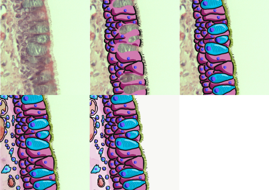
Pseudostratified columnar epithelium is a type of epithelium that, though comprising only a single layer of cells, has its cell nuclei positioned in a manner suggestive of stratified columnar epithelium. A stratified epithelium rarely occurs as squamous or cuboidal. The term ''pseudostratified'' is derived from the appearance of this epithelium in the section which conveys the erroneous (''pseudo'' means almost or approaching) impression that there is more than one layer of cells, when in fact this is a true simple epithelium since all the cells rest on the basement membrane. The nuclei of these cells, however, are disposed at different levels, thus creating the illusion of cellular stratification. All cells are not of equal size and not all cells extend to the luminal/apical surface; such cells are capable of cell division providing replacements for cells lost or damaged. Pseudostratified epithelia function in secretion or absorption. If a specimen looks stratified but has c ... [...More Info...] [...Related Items...] OR: [Wikipedia] [Google] [Baidu] |
Stereocilia
Stereocilia (or stereovilli or villi) are non-motile apical cell modifications. They are distinct from cilia and microvilli, but are closely related to microvilli. They form single "finger-like" projections that may be branched, with normal cell membrane characteristics. They contain actin. Stereocilia are found in the vas deferens, the epididymis, and the sensory cells of the inner ear. Structure Stereocilia are cylindrical and non-motile. They are much longer and thicker than microvilli, form single "finger-like" projections that may be branched, and have more of the characteristics of the cellular membrane proper. Like microvilli, they contain actin and lack an axoneme. This distinguishes them from cilia. They do not have a basal body at their base since they do not contain microtubules. They may or may not be covered by a glycocalyx coating. They have no fixed arrangement, different to the structure present in kinocilium. Organs containing stereocilia Stereocilia are ... [...More Info...] [...Related Items...] OR: [Wikipedia] [Google] [Baidu] |
Connective Tissue
Connective tissue is one of the four primary types of animal tissue, a group of cells that are similar in structure, along with epithelial tissue, muscle tissue, and nervous tissue. It develops mostly from the mesenchyme, derived from the mesoderm, the middle embryonic germ layer. Connective tissue is found in between other tissues everywhere in the body, including the nervous system. The three meninges, membranes that envelop the brain and spinal cord, are composed of connective tissue. Most types of connective tissue consists of three main components: elastic and collagen fibers, ground substance, and cells. Blood and lymph are classed as specialized fluid connective tissues that do not contain fiber. All are immersed in the body water. The cells of connective tissue include fibroblasts, adipocytes, macrophages, mast cells and leukocytes. The term "connective tissue" (in German, ) was introduced in 1830 by Johannes Peter Müller. The tissue was already recognized as ... [...More Info...] [...Related Items...] OR: [Wikipedia] [Google] [Baidu] |


