|
Elbow Pit
The cubital fossa, antecubital fossa, chelidon, inside of elbow, or, humorously, wagina, is the area on the anterior side of the upper part between the arm and forearm of a human or other hominid animals. It lies anteriorly to the elbow (antecubital) (Latin ) when in standard anatomical position. The cubital fossa is a triangular area having three borders. Boundaries * superior (proximal) boundary – an imaginary horizontal line connecting the medial epicondyle of the humerus to the lateral epicondyle of the humerus * medial (ulnar) boundary – lateral border of pronator teres muscle originating from the medial epicondyle of the humerus. * lateral (radial) boundary – medial border of brachioradialis muscle originating from the lateral supraepicondylar ridge of the humerus. * apex – it is directed inferiorly, and is formed by the meeting point of the lateral and medial boundaries * superficial boundary (roof) – skin, superficial fascia containing the median cubital vein, th ... [...More Info...] [...Related Items...] OR: [Wikipedia] [Google] [Baidu] |
Anterior
Standard anatomical terms of location are used to describe unambiguously the anatomy of humans and other animals. The terms, typically derived from Latin or Greek roots, describe something in its standard anatomical position. This position provides a definition of what is at the front ("anterior"), behind ("posterior") and so on. As part of defining and describing terms, the body is described through the use of anatomical planes and axes. The meaning of terms that are used can change depending on whether a vertebrate is a biped or a quadruped, due to the difference in the neuraxis, or if an invertebrate is a non-bilaterian. A non-bilaterian has no anterior or posterior surface for example but can still have a descriptor used such as proximal or distal in relation to a body part that is nearest to, or furthest from its middle. International organisations have determined vocabularies that are often used as standards for subdisciplines of anatomy. For example, '' Termin ... [...More Info...] [...Related Items...] OR: [Wikipedia] [Google] [Baidu] |
Tendon
A tendon or sinew is a tough band of fibrous connective tissue, dense fibrous connective tissue that connects skeletal muscle, muscle to bone. It sends the mechanical forces of muscle contraction to the skeletal system, while withstanding tension (physics), tension. Tendons, like ligaments, are made of collagen. The difference is that ligaments connect bone to bone, while tendons connect muscle to bone. There are about 4,000 tendons in the adult human body. Structure A tendon is made of dense regular connective tissue, whose main cellular components are special fibroblasts called tendon cells (tenocytes). Tendon cells synthesize the tendon's extracellular matrix, which abounds with densely-packed collagen fibers. The collagen fibers run parallel to each other and are grouped into fascicles. Each fascicle is bound by an endotendineum, which is a delicate loose connective tissue containing thin collagen fibrils and elastic fibers. A set of fascicles is bound by an epitenon, whi ... [...More Info...] [...Related Items...] OR: [Wikipedia] [Google] [Baidu] |
Lymph Node
A lymph node, or lymph gland, is a kidney-shaped organ of the lymphatic system and the adaptive immune system. A large number of lymph nodes are linked throughout the body by the lymphatic vessels. They are major sites of lymphocytes that include B and T cells. Lymph nodes are important for the proper functioning of the immune system, acting as filters for foreign particles including cancer cells, but have no detoxification function. In the lymphatic system, a lymph node is a secondary lymphoid organ. A lymph node is enclosed in a fibrous capsule and is made up of an outer cortex and an inner medulla. Lymph nodes become inflamed or enlarged in various diseases, which may range from trivial throat infections to life-threatening cancers. The condition of lymph nodes is very important in cancer staging, which decides the treatment to be used and determines the prognosis. Lymphadenopathy refers to glands that are enlarged or swollen. When inflamed or enlarged, lymph nodes can ... [...More Info...] [...Related Items...] OR: [Wikipedia] [Google] [Baidu] |
Neck
The neck is the part of the body in many vertebrates that connects the head to the torso. It supports the weight of the head and protects the nerves that transmit sensory and motor information between the brain and the rest of the body. Additionally, the neck is highly flexible, allowing the head to turn and move in all directions. Anatomically, the human neck is divided into four compartments: vertebral, visceral, and two vascular compartments. Within these compartments, the neck houses the cervical vertebrae, the cervical portion of the spinal cord, upper parts of the respiratory and digestive tracts, endocrine glands, nerves, arteries and veins. The muscles of the neck, which are separate from the compartments, form the boundaries of the neck triangles. In anatomy, the neck is also referred to as the or . However, when the term ''cervix'' is used alone, it often refers to the uterine cervix, the neck of the uterus. Therefore, the adjective ''cervical'' ... [...More Info...] [...Related Items...] OR: [Wikipedia] [Google] [Baidu] |
Armpit
The axilla (: axillae or axillas; also known as the armpit, underarm or oxter) is the area on the human body directly under the shoulder joint. It includes the axillary space, an anatomical space within the shoulder girdle between the arm and the thoracic cage, bounded superiorly by the imaginary plane between the superior borders of the first rib, clavicle and scapula (above which are considered part of the neck), medially by the serratus anterior muscle and thoracolumbar fascia, anteriorly by the pectoral muscles and posteriorly by the subscapularis, teres major and latissimus dorsi muscle. The soft skin covering the lateral axilla contains many hair and sweat glands. In humans, the formation of body odor happens mostly in the axilla. These odorant substances have been suggested by some to serve as pheromones, which play a role related to mate selection, although this is a controversial topic within the scientific community. The underarms seem more important than ... [...More Info...] [...Related Items...] OR: [Wikipedia] [Google] [Baidu] |
Popliteal Fossa
The popliteal fossa (also referred to as hough or kneepit in analogy to the cubital fossa) is a shallow depression located at the back of the knee joint. The bones of the popliteal fossa are the femur and the tibia. Like other flexion surfaces of large joints (groin, armpit, cubital fossa and essentially the anterior part of the neck), it is an area where blood vessels and nerves pass relatively superficially, and with an increased number of lymph nodes. Structure Boundaries The boundaries of the fossa are: Roof Moving from superficial to deep structures, the roof is formed by: # the skin. # the superficial fascia. This contains the small saphenous vein, the terminal branch of the posterior cutaneous nerve of the thigh, posterior division of the medial cutaneous nerve, lateral sural cutaneous nerve, and medial sural cutaneous nerve. # the popliteal fascia. Floor The floor is formed by: # the popliteal surface of the femur. # the capsule of the knee joint and the obli ... [...More Info...] [...Related Items...] OR: [Wikipedia] [Google] [Baidu] |
Groin
In human anatomy, the groin, also known as the inguinal region or iliac region, is the junctional area between the torso and the thigh. The groin is at the front of the body on either side of the pubic tubercle, where the lower part of the abdominal wall meets the thigh. A fold or crease is formed at this junction known as the inguinal groove, or crease. This is also the area of the medial compartment of thigh, medial compartment of the thigh that contains attachments of the adductor muscles of the hip or the groin muscles. The groin is the common site for a hernia. Gross anatomy Where the lower part of the anterior abdominal wall meets the thigh, a crease is formed known as the inguinal groove or crease. The junction is the area of the medial compartment of thigh, medial compartment of the thigh that contains the attachments of the adductor muscles of the hip, also known as the groin muscles. The adductor muscles that make up the groin consist of the adductor brevis, adductor l ... [...More Info...] [...Related Items...] OR: [Wikipedia] [Google] [Baidu] |
Basilic Vein
The basilic vein is a large superficial vein of the upper limb that helps drain parts of the hand and forearm. It originates on the medial ( ulnar) side of the dorsal venous network of the hand and travels up the base of the forearm, where its course is generally visible through the skin as it travels in the subcutaneous fat and fascia lying superficial to the muscles. The basilic vein terminates by uniting with the brachial veins to form the axillary vein. Anatomy Course As it ascends the medial side of the biceps in the arm proper (between the elbow and shoulder), the basilic vein normally perforates the brachial fascia ( deep fascia) in the middle of the medial bicipital groove, and run upwards medial to the brachial artery to the lower border of teres major, continuing as the axillary vein. Tributaries and anastomoses Near the region anterior to the cubital fossa (in the bend of the elbow joint), the basilic vein usually communicates with the cephalic vein (th ... [...More Info...] [...Related Items...] OR: [Wikipedia] [Google] [Baidu] |
Cephalic Vein
In human anatomy, the cephalic vein (also called the antecubital vein) is a superficial vein in the arm. It is the longest vein of the upper limb. It starts at the anatomical snuffbox from the radial end of the dorsal venous network of hand, and ascends along the radial (lateral) side of the arm before emptying into the axillary vein. At the elbow, it communicates with the basilic vein via the median cubital vein. Anatomy The cephalic vein is situated within the superficial fascia along the anterolateral surface of the biceps. Origin The cephalic vein forms at the roof of the anatomical snuffbox at the radial end of the dorsal venous network of hand. Course and relations From its origin, it ascends up the lateral aspect of the radius. Near the shoulder, the cephalic vein passes between the deltoid and pectoralis major muscles ( deltopectoral groove) through the clavipectoral triangle, where it empties into the axillary vein. Anastomoses It communicates wit ... [...More Info...] [...Related Items...] OR: [Wikipedia] [Google] [Baidu] |
Ulnar Nerve
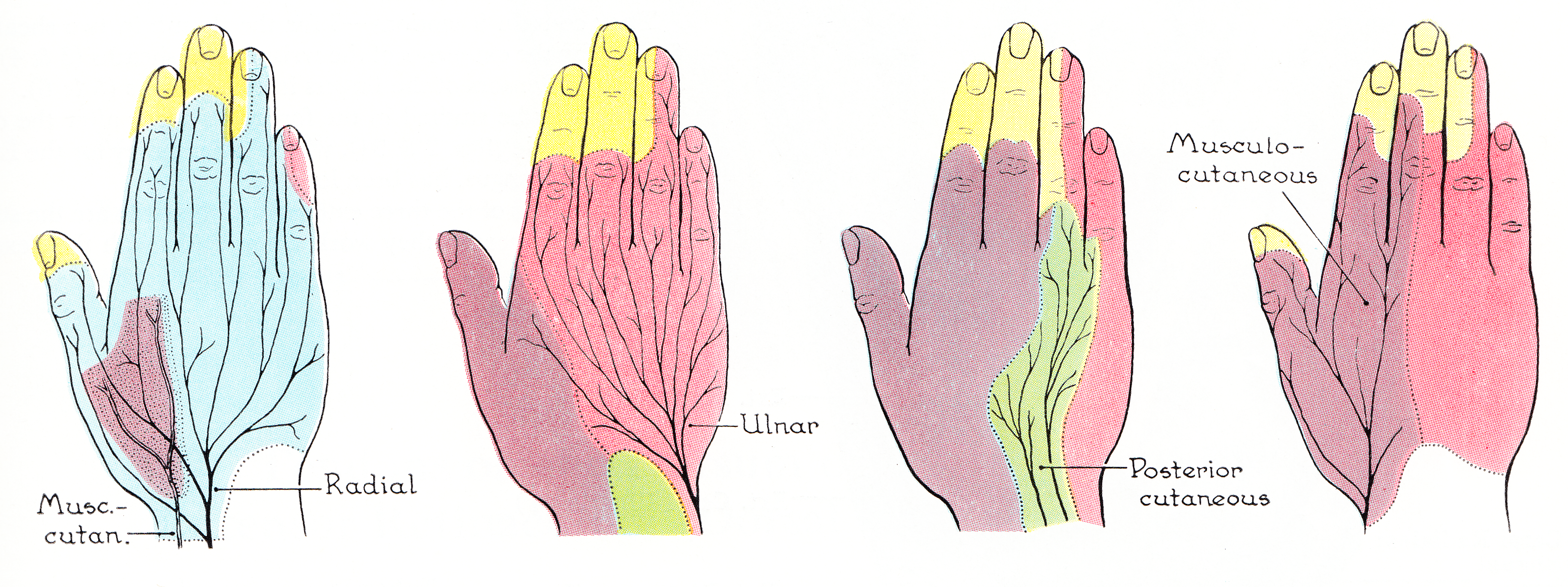
The ulnar nerve is a nerve that runs near the ulna, one of the two long bones in the forearm. The ulnar collateral ligament of elbow joint is in relation with the ulnar nerve. The nerve is the largest in the human body unprotected by muscle or bone, so injury is common. This nerve is directly connected to the little finger, and the adjacent half of the ring finger, innervating the palmar aspect of these fingers, including both front and back of the tips, perhaps as far back as the fingernail beds. This nerve can cause an electric shock-like sensation by striking the medial epicondyle of the humerus posteriorly, or inferiorly with the elbow flexed. The ulnar nerve is trapped between the bone and the overlying skin at this point. This is commonly referred to as bumping one's "funny bone". This name is thought to be a pun, based on the sound resemblance between the name of the bone of the upper arm, the humerus, and the word " humorous". Alternatively, according to the Oxfor ... [...More Info...] [...Related Items...] OR: [Wikipedia] [Google] [Baidu] |
Median Nerve
The median nerve is a nerve in humans and other animals in the upper limb. It is one of the five main nerves originating from the brachial plexus. The median nerve originates from the lateral and medial cords of the brachial plexus, and has contributions from ventral roots of C6-C7 (lateral cord) and C8 and T1 (medial cord). The median nerve is the only nerve that passes through the carpal tunnel. Carpal tunnel syndrome is the disability that results from the median nerve being pressed in the carpal tunnel. Structure The median nerve arises from the branches from lateral and medial cords of the brachial plexus, courses through the anterior part of arm, forearm, and hand, and terminates by supplying the muscles of the hand. Arm After receiving inputs from both the lateral and medial cords of the brachial plexus, the median nerve enters the arm from the axilla at the inferior margin of the teres major muscle. It then passes vertically down and courses lateral to the brac ... [...More Info...] [...Related Items...] OR: [Wikipedia] [Google] [Baidu] |


