|
Duodenojejunal Flexure
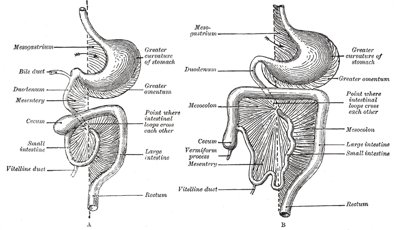
The duodenojejunal flexure or duodenojejunal junction, also known as the angle of Treitz, is the border between the duodenum and the jejunum. Structure The ascending portion of the duodenum ascends on the left side of the aorta, as far as the level of the upper border of the second lumbar vertebra. At this point, it turns abruptly forward to merge with the jejunum, forming the duodenojejunal flexure. This forms the beginning of the jejunum. The duodenojejunal flexure is surrounded by the suspensory muscle of the duodenum. It is retroperitoneal, so is less mobile than the jejunum that comes after it, helping to stabilise the jejunum. The duodenojejunal flexure lies in front of the left psoas major muscle, the left renal artery, and the left renal vein. It is covered in front, and partly at the sides, by peritoneum continuous with the left portion of the mesentery. Clinical significance The ligament of Treitz, a peritoneal fold, from the right crus of diaphragm, is an identi ... [...More Info...] [...Related Items...] OR: [Wikipedia] [Google] [Baidu] |
Duodenum
The duodenum is the first section of the small intestine in most vertebrates, including mammals, reptiles, and birds. In mammals, it may be the principal site for iron absorption. The duodenum precedes the jejunum and ileum and is the shortest part of the small intestine. In humans, the duodenum is a hollow jointed tube about long connecting the stomach to the jejunum, the middle part of the small intestine. It begins with the duodenal bulb, and ends at the duodenojejunal flexure marked by the suspensory muscle of duodenum. The duodenum can be divided into four parts: the first (superior), the second (descending), the third (transverse) and the fourth (ascending) parts. Overview The duodenum is the first section of the small intestine in most higher vertebrates, including mammals, reptiles, and birds. In fish, the divisions of the small intestine are not as clear, and the terms ''anterior intestine'' or ''proximal intestine'' may be used instead of duodenum. In mammals the d ... [...More Info...] [...Related Items...] OR: [Wikipedia] [Google] [Baidu] |
Mesentery
In human anatomy, the mesentery is an Organ (anatomy), organ that attaches the intestines to the posterior abdominal wall, consisting of a double fold of the peritoneum. It helps (among other functions) in storing Adipose tissue, fat and allowing blood vessels, lymphatics, and nerves to supply the intestines. The (the part of the mesentery that attaches the colon to the abdominal wall) was formerly thought to be a fragmented structure, with all named parts—the ascending, transverse, descending, and sigmoid mesocolons, the mesoappendix, and the mesorectum—separately terminating their insertion into the posterior abdominal wall. However, in 2012, new microscopy, microscopic and electron microscope, electron microscopic histology, examinations showed the mesocolon to be a single structure derived from the duodenojejunal flexure and extending to the distal mesorectal layer. Thus the mesentery is an internal organ. Structure The mesentery of the small intestine arises from th ... [...More Info...] [...Related Items...] OR: [Wikipedia] [Google] [Baidu] |
Duodenum
The duodenum is the first section of the small intestine in most vertebrates, including mammals, reptiles, and birds. In mammals, it may be the principal site for iron absorption. The duodenum precedes the jejunum and ileum and is the shortest part of the small intestine. In humans, the duodenum is a hollow jointed tube about long connecting the stomach to the jejunum, the middle part of the small intestine. It begins with the duodenal bulb, and ends at the duodenojejunal flexure marked by the suspensory muscle of duodenum. The duodenum can be divided into four parts: the first (superior), the second (descending), the third (transverse) and the fourth (ascending) parts. Overview The duodenum is the first section of the small intestine in most higher vertebrates, including mammals, reptiles, and birds. In fish, the divisions of the small intestine are not as clear, and the terms ''anterior intestine'' or ''proximal intestine'' may be used instead of duodenum. In mammals the d ... [...More Info...] [...Related Items...] OR: [Wikipedia] [Google] [Baidu] |
Kidneys
In humans, the kidneys are two reddish-brown bean-shaped blood-filtering organs that are a multilobar, multipapillary form of mammalian kidneys, usually without signs of external lobulation. They are located on the left and right in the retroperitoneal space, and in adult humans are about in length. They receive blood from the paired renal arteries; blood exits into the paired renal veins. Each kidney is attached to a ureter, a tube that carries excreted urine to the bladder. The kidney participates in the control of the volume of various body fluids, fluid osmolality, acid-base balance, various electrolyte concentrations, and removal of toxins. Filtration occurs in the glomerulus: one-fifth of the blood volume that enters the kidneys is filtered. Examples of substances reabsorbed are solute-free water, sodium, bicarbonate, glucose, and amino acids. Examples of substances secreted are hydrogen, ammonium, potassium and uric acid. The nephron is the structural and functi ... [...More Info...] [...Related Items...] OR: [Wikipedia] [Google] [Baidu] |
Pancreas
The pancreas (plural pancreases, or pancreata) is an Organ (anatomy), organ of the Digestion, digestive system and endocrine system of vertebrates. In humans, it is located in the abdominal cavity, abdomen behind the stomach and functions as a gland. The pancreas is a mixed or heterocrine gland, i.e., it has both an endocrine and a digestive exocrine function. Ninety-nine percent of the pancreas is exocrine and 1% is endocrine. As an endocrine gland, it functions mostly to regulate blood sugar levels, secreting the hormones insulin, glucagon, somatostatin and pancreatic polypeptide. As a part of the digestive system, it functions as an exocrine gland secreting pancreatic juice into the duodenum through the pancreatic duct. This juice contains bicarbonate, which neutralizes acid entering the duodenum from the stomach; and digestive enzymes, which break down carbohydrates, proteins and lipids, fats in food entering the duodenum from the stomach. Inflammation of the pancreas is kno ... [...More Info...] [...Related Items...] OR: [Wikipedia] [Google] [Baidu] |
Abdominal Surgery
The term abdominal surgery broadly covers surgical procedures that involve opening the abdomen (laparotomy). Surgery of each abdominal organ is dealt with separately in connection with the description of that organ (see stomach, kidney, liver, etc.) Diseases affecting the abdominal cavity are dealt with generally under their own names. Types The most common abdominal surgeries are described below. *Appendectomy: surgical opening of the abdominal cavity and removal of the appendix. Typically performed as definitive treatment for appendicitis, although sometimes the appendix is prophylactically removed incidental to another abdominal procedure. *Caesarean section (also known as C-section): a surgical procedure in which one or more incisions are made through a mother's abdomen (laparotomy) and uterus ( hysterotomy) to deliver one or more babies, or, rarely, to remove a dead fetus. * Inguinal hernia surgery: the repair of an inguinal hernia. *Exploratory laparotomy: the openin ... [...More Info...] [...Related Items...] OR: [Wikipedia] [Google] [Baidu] |
Crus Of Diaphragm
The crus of diaphragm (: crura), refers to one of two tendinous structures that extends below the diaphragm to the vertebral column. There is a right crus and a left crus, which together form a tether for muscular contraction. They take their name from their leg-shaped appearance – '' crus'' meaning ''leg'' in Latin. Structure The crura originate from the front of the bodies and intervertebral fibrocartilage of the lumbar vertebrae. They are tendinous and blend with the anterior longitudinal ligament of the vertebral column. * The ''right crus'', larger and longer than the left, arises from the front of the bodies and intervertebral fibrocartilages of the upper three lumbar vertebrae. * The ''left crus'' arises from the corresponding parts of the upper two lumbar vertebrae only. The medial tendinous margins of the crura pass anteriorly and medialward, and meet in the middle line to form an arch across the front of the aorta known as the median arcuate ligament; this arch is ... [...More Info...] [...Related Items...] OR: [Wikipedia] [Google] [Baidu] |
Ligament Of Treitz
The suspensory muscle of duodenum (also known as suspensory ligament of duodenum, Treitz's muscle or ligament of Treitz) is a thin muscle connecting the junction between the duodenum and jejunum (the small intestine's first and second parts, respectively), as well as the duodenojejunal flexure to connective tissue surrounding the superior mesenteric and coeliac arteries. The suspensory muscle most often connects to both the third and fourth parts of the duodenum, as well as the duodenojejunal flexure, although the attachment is quite variable. The suspensory muscle marks the formal division between the duodenum and the jejunum. This division is used to mark the difference between the upper and lower gastrointestinal tracts, which is relevant in clinical medicine as it may determine the source of gastrointestinal bleeding. The suspensory muscle is derived from mesoderm and plays a role in the embryological rotation of the gut, by offering a point of fixation for the rotating g ... [...More Info...] [...Related Items...] OR: [Wikipedia] [Google] [Baidu] |
Peritoneum
The peritoneum is the serous membrane forming the lining of the abdominal cavity or coelom in amniotes and some invertebrates, such as annelids. It covers most of the intra-abdominal (or coelomic) organs, and is composed of a layer of mesothelium supported by a thin layer of connective tissue. This peritoneal lining of the cavity supports many of the abdomen#Contents, abdominal organs and serves as a conduit for their blood vessels, lymphatic vessels, and nerves. The abdominal cavity (the space bounded by the vertebrae, abdominal muscles, Thoracic diaphragm, diaphragm, and pelvic floor) is different from the intraperitoneal space (located within the abdominal cavity but wrapped in peritoneum). The structures within the intraperitoneal space are called "intraperitoneal" (e.g., the stomach and intestines), the structures in the abdominal cavity that are located behind the intraperitoneal space are called "retroperitoneal" (e.g., the kidneys), and those structures below the intrape ... [...More Info...] [...Related Items...] OR: [Wikipedia] [Google] [Baidu] |
Jejunum
The jejunum is the second part of the small intestine in humans and most higher vertebrates, including mammals, reptiles, and birds. Its lining is specialized for the absorption by enterocytes of small nutrient molecules which have been previously digested by enzymes in the duodenum. The jejunum lies between the duodenum and the ileum and is considered to start at the suspensory muscle of the duodenum, a location called the duodenojejunal flexure. The division between the jejunum and ileum is not anatomically distinct. In adult humans, the small intestine is usually long (post mortem), about two-fifths of which (about ) is the jejunum. Structure The interior surface of the jejunum—which is exposed to ingested food—is covered in finger–like projections of mucosa, called villi, which increase the surface area of tissue available to absorb nutrients from ingested foodstuffs. The epithelial cells which line these villi have microvilli. The transport of nutrien ... [...More Info...] [...Related Items...] OR: [Wikipedia] [Google] [Baidu] |
Renal Vein
The renal veins in the renal circulation, are large-calibre veins that drain blood filtered by the kidneys into the inferior vena cava. There is one renal vein draining each kidney. Each renal vein is formed by the convergence of the interlobar veins of one kidney. Because the inferior vena cava is on the right half of the body, the left renal vein is longer than the right one. Structure One renal vein drains each kidney. A renal vein is situated anterior to its corresponding accompanying renal artery. The renal veins empty into the inferior vena cava, entering it at nearly a 90° angle. Due to the right-ward displacement of the inferior vena cava from the midline, the left renal vein is some 3 times longer than the right one (~7.5 cm and ~2.5 cm, respectively). The renal vein divides into 4 divisions upon entering the kidney: * the anterior branch which receives blood from the anterior portion of the kidney and, * the posterior branch which receives blood from the posterior ... [...More Info...] [...Related Items...] OR: [Wikipedia] [Google] [Baidu] |
Renal Artery
The renal arteries are paired arteries that supply the kidneys with blood. Each is directed across the crus of the diaphragm, so as to form nearly a right angle. The renal arteries carry a large portion of total blood flow to the kidneys. Up to a third of total cardiac output can pass through the renal arteries to be filtered by the kidneys. Structure The renal arteries normally arise at a 90° angle off of the left interior side of the abdominal aorta, immediately below the superior mesenteric artery. They have a radius of approximately 0.25 cm, 0.26 cm at the root. The measured mean diameter can differ depending on the imaging method used. For example, the diameter was found to be 5.04 ± 0.74 mm using ultrasound but 5.68 ± 1.19 mm using angiography. Due to the anatomical position of the aorta, the inferior vena cava, and the kidneys, the right renal artery is normally longer than the left renal artery. * The right passes behind the inferior vena cava, ... [...More Info...] [...Related Items...] OR: [Wikipedia] [Google] [Baidu] |


