|
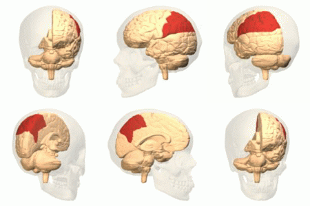
Dorsal Posterior Cingulate Area 31
Brodmann area 31, also known as dorsal posterior cingulate area 31, is a subdivision of the cytoarchitecturally defined cingulate region of the cerebral cortex. In the human, it occupies portions of the posterior cingulate gyrus and medial aspect of the parietal lobe. Approximate boundaries are the cingulate sulcus dorsally and the parieto-occipital sulcus caudally. It partially surrounds the subparietal sulcus, the ventral continuation of the cingulate sulcus in the parietal lobe. Cytoarchitecturally it is bounded rostrally by the ventral anterior cingulate area 24, ventrally by the ventral posterior cingulate area 23, dorsally by the gigantopyramidal area 4 and preparietal area 5 and caudally by the superior parietal area 7 (H) (Brodmann-1909). See also * Brodmann area A Brodmann area is a region of the cerebral cortex, in the human or other primate brain, defined by its cytoarchitecture, or histological structure and organization of cells. The concept was first introduc ... [...More Info...] [...Related Items...] OR: [Wikipedia] [Google] [Baidu] |
Cytoarchitecture
Cytoarchitecture (from Greek κύτος 'cell' and ἀρχιτεκτονική 'architecture'), also known as cytoarchitectonics, is the study of the cellular composition of the central nervous system's tissues under the microscope. Cytoarchitectonics is one of the ways to parse the brain, by obtaining sections of the brain using a microtome and staining them with chemical agents which reveal where different neurons are located. The study of the parcellation of ''nerve fibers'' (primarily axons) into layers forms the subject of myeloarchitectonics (from Greek μυελός 'marrow' and ἀρχιτεκτονική 'architecture'), an approach complementary to cytoarchitectonics. History of the cerebral cytoarchitecture Defining cerebral cytoarchitecture began with the advent of histology—the science of slicing and staining brain slices for examination. It is credited to the Viennese psychiatrist Theodor Meynert (1833–1892), who in 1867 noticed regional variations in the ... [...More Info...] [...Related Items...] OR: [Wikipedia] [Google] [Baidu] |
Cerebral Cortex
The cerebral cortex, also known as the cerebral mantle, is the outer layer of neural tissue of the cerebrum of the brain in humans and other mammals. It is the largest site of Neuron, neural integration in the central nervous system, and plays a key role in attention, perception, awareness, thought, memory, language, and consciousness. The six-layered neocortex makes up approximately 90% of the Cortex (anatomy), cortex, with the allocortex making up the remainder. The cortex is divided into left and right parts by the longitudinal fissure, which separates the two cerebral hemispheres that are joined beneath the cortex by the corpus callosum and other commissural fibers. In most mammals, apart from small mammals that have small brains, the cerebral cortex is folded, providing a greater surface area in the confined volume of the neurocranium, cranium. Apart from minimising brain and cranial volume, gyrification, cortical folding is crucial for the Neural circuit, brain circuitry ... [...More Info...] [...Related Items...] OR: [Wikipedia] [Google] [Baidu] |
Posterior Cingulate Gyrus
The posterior cingulate cortex (PCC) is the caudal part of the cingulate cortex, located posterior to the anterior cingulate cortex. This is the upper part of the "limbic lobe". The cingulate cortex is made up of an area around the midline of the brain. Surrounding areas include the retrosplenial cortex and the precuneus. Cytoarchitectonically the posterior cingulate cortex is associated with Brodmann areas 23 and 31. The PCC forms a central node in the default mode network of the brain. It has been shown to communicate with various brain networks simultaneously and is involved in diverse functions. Along with the precuneus, the PCC has been implicated as a neural substrate for human awareness in numerous studies of both the anesthetized and vegetative (coma) states. Imaging studies indicate a prominent role for the PCC in pain and episodic memory retrieval. Increased size of the ventral PCC is related to a decline in working memory performance. The PCC has also been strongly ... [...More Info...] [...Related Items...] OR: [Wikipedia] [Google] [Baidu] |
Parietal Lobe
The parietal lobe is one of the four Lobes of the brain, major lobes of the cerebral cortex in the brain of mammals. The parietal lobe is positioned above the temporal lobe and behind the frontal lobe and central sulcus. The parietal lobe integrates sensory information among various sensory modality, modalities, including spatial sense and navigation (proprioception), the main sensory receptive area for the sense of touch in the somatosensory cortex which is just posterior to the central sulcus in the postcentral gyrus, and the two-streams hypothesis#Dorsal stream, dorsal stream of the visual system. The major sensory inputs from the skin (mechanoreceptor, touch, thermoreceptor, temperature, and nociceptor, pain receptors), relay through the thalamus to the parietal lobe. Several areas of the parietal lobe are important in language processing in the brain, language processing. The somatosensory cortex can be illustrated as a distorted figure – the cortical homunculus (Latin: "li ... [...More Info...] [...Related Items...] OR: [Wikipedia] [Google] [Baidu] |
Cingulate Sulcus
The cingulate cortex is a part of the brain situated in the medial aspect of the cerebral cortex. The cingulate cortex includes the entire cingulate gyrus, which lies immediately above the corpus callosum, and the continuation of this in the cingulate sulcus. The cingulate cortex is usually considered part of the limbic lobe. It receives inputs from the thalamus and the neocortex, and projects to the entorhinal cortex via the cingulum. It is an integral part of the limbic system, which is involved with emotion formation and processing, learning, and memory. The combination of these three functions makes the cingulate gyrus highly influential in linking motivational outcomes to behavior (e.g. a certain action induced a positive emotional response, which results in learning). This role makes the cingulate cortex highly important in disorders such as depression and schizophrenia. It also plays a role in executive function and respiratory control. Structure Based on cerebral cy ... [...More Info...] [...Related Items...] OR: [Wikipedia] [Google] [Baidu] |
Parieto-occipital Sulcus
In neuroanatomy, the parieto-occipital sulcus (also called the parieto-occipital fissure) is a deep sulcus in the cerebral cortex that marks the boundary between the cuneus and precuneus, and also between the parietal and occipital lobes. Only a small part can be seen on the lateral surface of the hemisphere, its chief part being on the medial surface. The lateral part of the parieto-occipital sulcus (Fig. 726) is situated about 5 cm in front of the occipital pole of the hemisphere, and measures about 1.25 cm. in length. The medial part of the parieto-occipital sulcus (Fig. 727) runs downward and forward as a deep cleft on the medial surface of the hemisphere, and joins the calcarine fissure below and behind the posterior end of the corpus callosum. In most cases, it contains a submerged gyrus. Function The parieto-occipital lobe has been found in various neuroimaging studies, including PET (positron-emission-tomography) studies, and SPECT (single-photon emission comput ... [...More Info...] [...Related Items...] OR: [Wikipedia] [Google] [Baidu] |
Subparietal Sulcus
In neuroanatomy, the subparietal sulcus () or suprasplenial sulcus is a sulcus, or crevice, on the medial surface of each cerebral hemisphere, above the splenium of the corpus callosum. It separates the precuneus from the posterior part of the cingulate gyrus. It is the posterior continuation of the cingulate sulcus. The cingulate sulcus actually "terminates" as the marginal sulcus of the cingulate sulcus (margin of cingulate gyrus). It extends posteriorly toward the calcarine sulcus. The precuneus is bordered anteriorly by the marginal branch of the cingulate sulcus (margin of cingulate sulcus), posteriorly by the parieto-occipital sulcus In neuroanatomy, the parieto-occipital sulcus (also called the parieto-occipital fissure) is a deep sulcus in the cerebral cortex that marks the boundary between the cuneus and precuneus, and also between the parietal and occipital lobes. Only ..., and inferiorly by the subparietal sulcus. Additional images References * Michio ... [...More Info...] [...Related Items...] OR: [Wikipedia] [Google] [Baidu] |
Ventral Anterior Cingulate Area 24
Brodmann area 24 is part of the anterior cingulate in the human brain. Human In the human this area is known as ventral anterior cingulate area 24, and it refers to a subdivision of the cytoarchitecturally defined cingulate cortex region of cerebral cortex (area cingularis anterior ventralis). It occupies most of the anterior cingulate gyrus in an arc around the genu of the corpus callosum. Its outer border corresponds approximately to the cingulate sulcus. Cytoarchitecturally it is bounded internally by the pregenual area 33, externally by the dorsal anterior cingulate area 32, and caudally by the ventral posterior cingulate area 23 and the dorsal posterior cingulate area 31. Guenon In the guenon this area is referred to as area 24 of Brodmann-1905. It includes portions of the cingulate gyrus and the frontal lobe. The cortex is thin; it lacks the internal granular layer (IV) so that the densely distributed, plump pyramidal cells of sublayer 3b of the external pyramidal l ... [...More Info...] [...Related Items...] OR: [Wikipedia] [Google] [Baidu] |
Ventral Posterior Cingulate Area 23
Brodmann area 23 (BA23) is a region in the brain that lies inside the posterior cingulate cortex. It lies between Brodmann area 30 and Brodmann area 31 and is located on the medial wall of the cingulate gyrus between the callosal sulcus and the cingulate sulcus. Human This area is also known as ventral posterior cingulate area 23. It is a subdivision of the cytoarchitecturally defined cingulate region of cerebral cortex. In the human it occupies most of the posterior cingulate gyrus adjacent to the corpus callosum. At the caudal extreme it is bounded approximately by the parieto-occipital sulcus. Cytoarchitecturally it is bounded dorsally by the dorsal posterior cingulate area 31, rostrally by the ventral anterior cingulate area 24, and ventrorostrally in its caudal half by the retrosplenial region (Brodmann-1909). Guenon Brodmann area 23 is a subdivision of the cerebral cortex of the guenon defined on the basis of cytoarchitecture. Brodmann regarded it as topographically and ... [...More Info...] [...Related Items...] OR: [Wikipedia] [Google] [Baidu] |
Gigantopyramidal Area 4
Brodmann area 4 refers to the primary motor cortex of the human brain. It is located in the posterior portion of the frontal lobe. Brodmann area 4 is part of the precentral gyrus. The borders of this area are: the precentral sulcus in front (anteriorly), the medial longitudinal fissure at the top ( medially), the central sulcus in back (posteriorly), and the lateral sulcus along the bottom (laterally). This area of cortex, as shown by Wilder Penfield and others, has the pattern of a homunculus. That is, the legs and trunk fold over the midline; the arms and hands are along the middle of the area shown here; and the face is near the bottom of the figure. Because Brodmann area 4 is in the same general location as primary motor cortex, the homunculus here is called the motor homunculus. The term area 4 of Brodmann-1909 refers to a cytoarchitecturally defined portion of the frontal lobe of the guenon. It is located predominantly in the precentral gyrus. Brodmann-1909 regarde ... [...More Info...] [...Related Items...] OR: [Wikipedia] [Google] [Baidu] |
Superior Parietal Area 7
Brodmann area 7 is one of Brodmann's cytologically defined regions of the brain corresponding to precuneus and superior parietal lobule (SPL). It is involved in locating objects in space. It serves as a point of convergence between vision and proprioception to determine where objects are in relation to parts of the body. In humans Brodmann area 7 is part of the parietal cortex in the human brain. Situated posterior to the primary somatosensory cortex ( Brodmann areas 3, 1 and 2), and superior to the occipital lobe, this region is believed to play a role in visuo-motor coordination (e.g., in reaching to grasp an object). In addition, area 7 along with area 5 has been linked to a wide variety of high-level processing tasks, including activation in association with language use. This function in language has been theorized to stem from how these two regions play a vital role in generating conscious constructs of objects in the world. Brodmann area 7 spans both the medial and lat ... [...More Info...] [...Related Items...] OR: [Wikipedia] [Google] [Baidu] |


