|
Diffuse Hemispheric Glioma
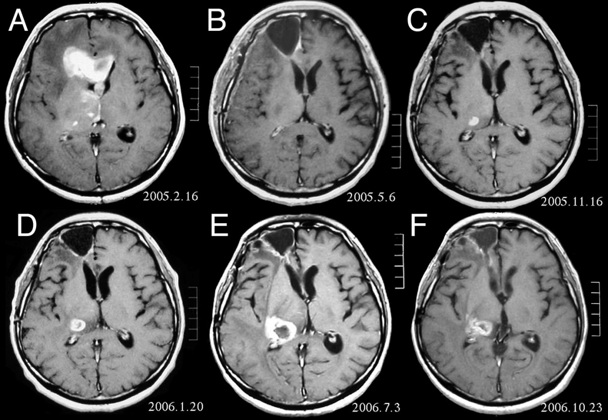
Diffuse hemispheric glioma, H3G34 mutant (DHG) is a rare, high-grade, infiltrative WHO grade 4 brain tumor most often found in adolescents and young adults. The majority are found in the frontal, parietal, and temporal lobes. Diagnosis Prior to 2021, most DHG's fell into the classification of Glioblastoma (GBM). However, modern sequencing techniques have revealed that the molecular alterations driving the cancer are entirely distinct to GBM, leading to changes in the World Health Organization classification system. They are further distinguished from GBM by their radiological presentations, including no or faint contrast enhancement A contrast agent (or contrast medium) is a substance used to increase the contrast of structures or fluids within the body in medical imaging. Contrast agents absorb or alter external electromagnetism or ultrasound, which is different from radioph .... A diagnosis can only be definitively made with genetic testing of the tumor. Treatment Avail ... [...More Info...] [...Related Items...] OR: [Wikipedia] [Google] [Baidu] |
WHO Classification Of Tumours Of The Central Nervous System
The WHO classification of tumours of the central nervous system is a WHO Blue Books, World Health Organization Blue Book that defines, describes and classifies Neoplasm, tumours of the central nervous system (CNS). Currently, as of 2023, clinicians are using the 5th edition, which incorporates recent advances in molecular pathology. The books lists ICD-O codes, Grading of the tumors of the central nervous system#WHO grading, CNS WHO grades and describes Epidemiology, epidemiological, clinical, Macroscopic scale, macroscopic and Histopathology, histopathological features, among others. The following is a simplified (deprecated) version of the fifth edition. Types 1. Gliomas, glioneuronal tumors, and neuronal tumours :1.1 Adult-type diffuse gliomas ::1.1.1 Astrocytoma, IDH-mutant ::1.1.2 Oligodendroglioma, IDH-mutant, and 1p/19q-codeleted ::1.1.3 Glioblastoma, IDH-wildtype :1.2 Pediatric-type diffuse low-grade gliomas ::1.2.1 Diffuse astrocytoma, MYB- or MYBL1-altered ::1. ... [...More Info...] [...Related Items...] OR: [Wikipedia] [Google] [Baidu] |
Frontal Lobe
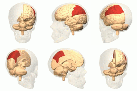
The frontal lobe is the largest of the four major lobes of the brain in mammals, and is located at the front of each cerebral hemisphere (in front of the parietal lobe and the temporal lobe). It is parted from the parietal lobe by a Sulcus (neuroanatomy), groove between tissues called the central sulcus and from the temporal lobe by a deeper groove called the lateral sulcus (Sylvian fissure). The most anterior rounded part of the frontal lobe (though not well-defined) is known as the frontal pole, one of the three Cerebral hemisphere#Poles, poles of the cerebrum. The frontal lobe is covered by the frontal cortex. The frontal cortex includes the premotor cortex and the primary motor cortex – parts of the motor cortex. The front part of the frontal cortex is covered by the prefrontal cortex. The nonprimary motor cortex is a functionally defined portion of the frontal lobe. There are four principal Gyrus, gyri in the frontal lobe. The precentral gyrus is directly anterior to the ... [...More Info...] [...Related Items...] OR: [Wikipedia] [Google] [Baidu] |
Parietal Lobe
The parietal lobe is one of the four Lobes of the brain, major lobes of the cerebral cortex in the brain of mammals. The parietal lobe is positioned above the temporal lobe and behind the frontal lobe and central sulcus. The parietal lobe integrates sensory information among various sensory modality, modalities, including spatial sense and navigation (proprioception), the main sensory receptive area for the sense of touch in the somatosensory cortex which is just posterior to the central sulcus in the postcentral gyrus, and the two-streams hypothesis#Dorsal stream, dorsal stream of the visual system. The major sensory inputs from the skin (mechanoreceptor, touch, thermoreceptor, temperature, and nociceptor, pain receptors), relay through the thalamus to the parietal lobe. Several areas of the parietal lobe are important in language processing in the brain, language processing. The somatosensory cortex can be illustrated as a distorted figure – the cortical homunculus (Latin: "li ... [...More Info...] [...Related Items...] OR: [Wikipedia] [Google] [Baidu] |
Temporal Lobe
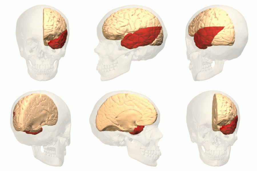
The temporal lobe is one of the four major lobes of the cerebral cortex in the brain of mammals. The temporal lobe is located beneath the lateral fissure on both cerebral hemispheres of the mammalian brain. The temporal lobe is involved in processing sensory input into derived meanings for the appropriate retention of visual memory, language comprehension, and emotion association. ''Temporal'' refers to the head's temples. Structure The temporal lobe consists of structures that are vital for declarative or long-term memory. Declarative (denotative) or explicit memory is conscious memory divided into semantic memory (facts) and episodic memory (events). The medial temporal lobe structures are critical for long-term memory, and include the hippocampal formation, perirhinal cortex, parahippocampal, and entorhinal neocortical regions. The hippocampus is critical for memory formation, and the surrounding medial temporal cortex is currently theorized to be critical f ... [...More Info...] [...Related Items...] OR: [Wikipedia] [Google] [Baidu] |
Contrast Agent
A contrast agent (or contrast medium) is a substance used to increase the contrast of structures or fluids within the body in medical imaging. Contrast agents absorb or alter external electromagnetism or ultrasound, which is different from radiopharmaceuticals, which emit radiation themselves. In X-ray imaging, contrast agents enhance the radiodensity in a target tissue or structure. In magnetic resonance imaging (MRI), contrast agents shorten (or in some instances increase) the relaxation times of nuclei within body tissues in order to alter the contrast in the image. Contrast agents are commonly used to improve the visibility of blood vessels and the gastrointestinal tract. The types of contrast agent are classified according to their intended imaging modalities. Radiocontrast media For radiography, which is based on X-rays, iodine and barium are the most common types of contrast agent. Various sorts of iodinated contrast agents exist, with variations occurring between the ... [...More Info...] [...Related Items...] OR: [Wikipedia] [Google] [Baidu] |
H3F3A
Histone H3.3 is a protein that in humans is encoded by the ''H3F3A'' and ''H3F3B'' genes. It plays an essential role in maintaining genome integrity during mammalian development. Histones are basic nuclear proteins that are responsible for the nucleosome structure of the chromosomal fiber in eukaryotes. Two molecules of each of the four core histones (H2A, H2B, H3, and H4) form an octamer, around which approximately 146 bp of DNA is wrapped in repeating units, called nucleosomes. The linker histone, H1, interacts with linker DNA between nucleosomes and functions in the compaction of chromatin into higher order structures. This gene contains introns and its mRNA is polyadenylated, unlike most histone genes. The protein encoded is a replication-independent member of the histone H3 family. Mutation of H3F3A are also linked to certain cancers. p.Lys27Met were discovered in Diffuse Intrinsic Pontine Glioma (DIPG), where they are present 65-75% of tumors and confer a worse prognosis. p. ... [...More Info...] [...Related Items...] OR: [Wikipedia] [Google] [Baidu] |


