|
Dibamids
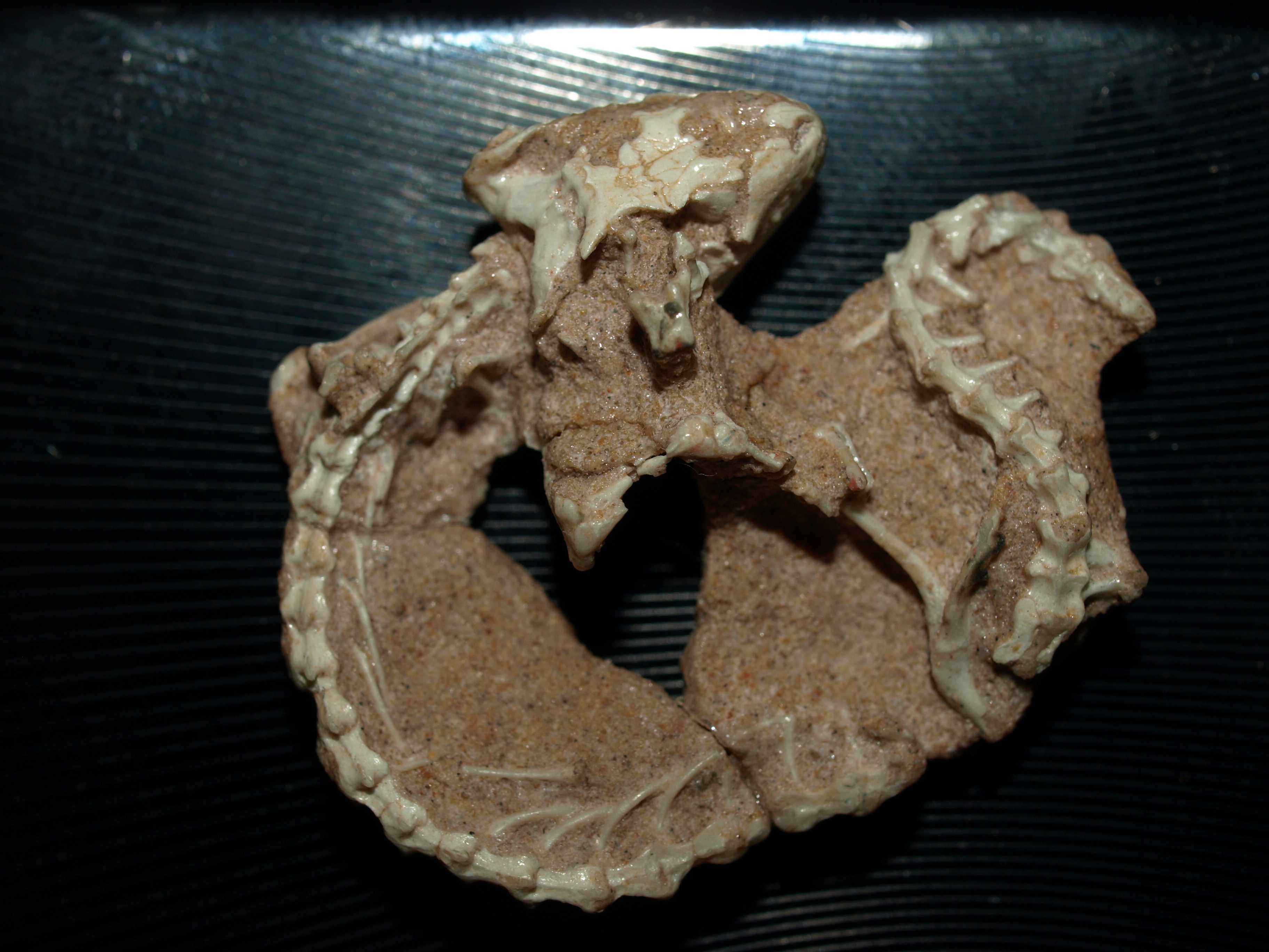
Dibamidae or blind skinks is a family of lizards characterized by their elongated cylindrical body and an apparent lack of limbs. Female dibamids are entirely limbless and the males retain small flap-like hind limbs, which they use to grip their partner during mating. They have a rigidly fused skull, lack pterygoid teeth and external ears. Their eyes are greatly reduced, and covered with a scale. They are small insectivorous lizards, with long, slender bodies, adapted for burrowing into the soil. They usually lay one egg with a hard, calcified shell, rather than the leathery shells typical of many other reptile groups. The family Dibamidae has two genera, ''Dibamus'' with 23 species native to Southeast Asia, Indonesia, the Philippines, and western New Guinea and the monotypic '' Anelytropsis'' native to Mexico. Recent phylogenetic analyses place the dibamids as the sister clade to all the other lizards and snakes or classify them as sharing a common ancestor with the infraorder ... [...More Info...] [...Related Items...] OR: [Wikipedia] [Google] [Baidu] |
Squamata
Squamata (, Latin ''squamatus'', 'scaly, having scales') is the largest order of reptiles, comprising lizards, snakes, and amphisbaenians (worm lizards), which are collectively known as squamates or scaled reptiles. With over 10,900 species, it is also the second-largest order of extant (living) vertebrates, after the perciform fish. Members of the order are distinguished by their skins, which bear horny scales or shields, and must periodically engage in molting. They also possess movable quadrate bones, making possible movement of the upper jaw relative to the neurocranium. This is particularly visible in snakes, which are able to open their mouths very wide to accommodate comparatively large prey. Squamata is the most variably sized order of reptiles, ranging from the dwarf gecko (''Sphaerodactylus ariasae'') to the Reticulated python (''Malayopython reticulatus'') and the now- extinct mosasaurs, which reached lengths over . Among other reptiles, squamates are most ... [...More Info...] [...Related Items...] OR: [Wikipedia] [Google] [Baidu] |
Hoeckosaurus
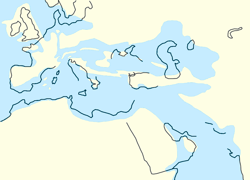
''Hoeckosaurus'' is an extinct genus of lizard from the Oligocene of Mongolia. It contains a single species, ''H. mongoliensis''. The genus name commemorates Austrian paleontologist Gudrun Höck, who collected the type material. ''Hoeckosaurus'' was a member of the Dibamidae (a family of primitive legless squamates), and represents the only known fossil record of the Dibamidae, despite the presumed ancient origins of the group. Despite the close resemblance of the fossils to extant dibamids in the genera '' Dibamus'' and '' Anelytropsis'', some distinguishing features such as an open Meckelian groove suggests that ''Hoeckosaurus'' is basal to both. The remains of ''Hoeckosaurus'' consist of some jawbones, and were collected in 1995-1997 from the Valley of the Lakes in central Mongolia. The sediments they were collected from are thought to be Early Oligocene in age. Previous studies postulated the jaws as belonging to an arretosaurid (an extinct family of Asian iguanians ... [...More Info...] [...Related Items...] OR: [Wikipedia] [Google] [Baidu] |
Oligocene
The Oligocene ( ) is a geologic epoch of the Paleogene Period and extends from about 33.9 million to 23 million years before the present ( to ). As with other older geologic periods, the rock beds that define the epoch are well identified but the exact dates of the start and end of the epoch are slightly uncertain. The name Oligocene was coined in 1854 by the German paleontologist Heinrich Ernst Beyrich from his studies of marine beds in Belgium and Germany. The name comes from the Ancient Greek (''olígos'', "few") and (''kainós'', "new"), and refers to the sparsity of extant forms of molluscs. The Oligocene is preceded by the Eocene Epoch and is followed by the Miocene The Miocene ( ) is the first geological epoch of the Neogene Period and extends from about (Ma). The Miocene was named by Scottish geologist Charles Lyell; the name comes from the Greek words (', "less") and (', "new") and means "less recent" ... Epoch. The Oligocene is the third and final epoch of ... [...More Info...] [...Related Items...] OR: [Wikipedia] [Google] [Baidu] |
Maxilla
The maxilla (plural: ''maxillae'' ) in vertebrates is the upper fixed (not fixed in Neopterygii) bone of the jaw formed from the fusion of two maxillary bones. In humans, the upper jaw includes the hard palate in the front of the mouth. The two maxillary bones are fused at the intermaxillary suture, forming the anterior nasal spine. This is similar to the mandible (lower jaw), which is also a fusion of two mandibular bones at the mandibular symphysis. The mandible is the movable part of the jaw. Structure In humans, the maxilla consists of: * The body of the maxilla * Four processes ** the zygomatic process ** the frontal process of maxilla ** the alveolar process ** the palatine process * three surfaces – anterior, posterior, medial * the Infraorbital foramen * the maxillary sinus * the incisive foramen Articulations Each maxilla articulates with nine bones: * two of the cranium: the frontal and ethmoid * seven of the face: the nasal, zygomatic, lacrimal, ... [...More Info...] [...Related Items...] OR: [Wikipedia] [Google] [Baidu] |
Pineal Foramen
A parietal eye, also known as a third eye or pineal eye, is a part of the epithalamus present in some vertebrates. The eye is located at the top of the head, is photoreceptive and is associated with the pineal gland, regulating circadian rhythmicity and hormone production for thermoregulation. The hole in the head which contains the eye is known as a pineal foramen or parietal foramen, since it is often enclosed by the parietal bones. Presence in various animals The parietal eye is found in the tuatara, most lizards, frogs, salamanders, certain bony fish, sharks, and lampreys. It is absent in mammals, but was present in their closest extinct relatives, the therapsids, suggesting it was lost during the course of the mammalian evolution due to it being useless in endothermic animals. It is also absent in the ancestrally endothermic ("warm-blooded") archosaurs such as birds. The parietal eye is also lost in ectothermic ("cold-blooded") archosaurs like crocodilians, and in turtle ... [...More Info...] [...Related Items...] OR: [Wikipedia] [Google] [Baidu] |
Quadrate Bone
The quadrate bone is a skull bone in most tetrapods, including amphibians, sauropsids (reptiles, birds), and early synapsids. In most tetrapods, the quadrate bone connects to the quadratojugal and squamosal bones in the skull, and forms upper part of the jaw joint. The lower jaw articulates at the articular bone, located at the rear end of the lower jaw. The quadrate bone forms the lower jaw articulation in all classes except mammals. Evolutionarily, it is derived from the hindmost part of the primitive cartilaginous upper jaw. Function in reptiles In certain extinct reptiles, the variation and stability of the morphology of the quadrate bone has helped paleontologists in the species-level taxonomy and identification of mosasaur squamates and spinosaurine dinosaurs. In some lizards and dinosaurs, the quadrate is articulated at both ends and movable. In snakes, the quadrate bone has become elongated and very mobile, and contributes greatly to their ability to swallow ve ... [...More Info...] [...Related Items...] OR: [Wikipedia] [Google] [Baidu] |
Neurocranium
In human anatomy, the neurocranium, also known as the braincase, brainpan, or brain-pan is the upper and back part of the skull, which forms a protective case around the brain. In the human skull, the neurocranium includes the calvaria or skullcap. The remainder of the skull is the facial skeleton. In comparative anatomy, neurocranium is sometimes used synonymously with endocranium or chondrocranium. Structure The neurocranium is divided into two portions: * the membranous part, consisting of flat bones, which surround the brain; and * the cartilaginous part, or chondrocranium, which forms bones of the base of the skull. In humans, the neurocranium is usually considered to include the following eight bones: * 1 ethmoid bone * 1 frontal bone * 1 occipital bone * 2 parietal bones * 1 sphenoid bone * 2 temporal bones The ossicles (three on each side) are usually not included as bones of the neurocranium. There may variably also be extra sutural bones present. Below the ... [...More Info...] [...Related Items...] OR: [Wikipedia] [Google] [Baidu] |
Post-temporal Fenestra
The skull is a bone protective cavity for the brain. The skull is composed of four types of bone i.e., cranial bones, facial bones, ear ossicles and hyoid bone. However two parts are more prominent: the cranium and the mandible. In humans, these two parts are the neurocranium and the viscerocranium (facial skeleton) that includes the mandible as its largest bone. The skull forms the anterior-most portion of the skeleton and is a product of cephalisation—housing the brain, and several sensory structures such as the eyes, ears, nose, and mouth. In humans these sensory structures are part of the facial skeleton. Functions of the skull include protection of the brain, fixing the distance between the eyes to allow stereoscopic vision, and fixing the position of the ears to enable sound localisation of the direction and distance of sounds. In some animals, such as horned ungulates (mammals with hooves), the skull also has a defensive function by providing the mount (on the frontal ... [...More Info...] [...Related Items...] OR: [Wikipedia] [Google] [Baidu] |
Fossorial
A fossorial () animal is one adapted to digging which lives primarily, but not solely, underground. Some examples are badgers, naked mole-rats, clams, meerkats, and mole salamanders, as well as many beetles, wasps, and bees. Prehistoric evidence The physical adaptation of fossoriality is widely accepted as being widespread among many prehistoric phyla and taxa, such as bacteria and early eukaryotes. Furthermore, fossoriality has evolved independently multiple times, even within a single family. Fossorial animals appeared simultaneously with the colonization of land by arthropods in the late Ordovician period (over 440 million years ago). Other notable early burrowers include '' Eocaecilia'' and possibly ''Dinilysia''. The oldest example of burrowing in synapsids, the lineage which includes modern mammals and their ancestors, is a cynodont, '' Thrinaxodon liorhinus'', found in the Karoo of South Africa, estimated to be 251 million years old. Evidence shows t ... [...More Info...] [...Related Items...] OR: [Wikipedia] [Google] [Baidu] |
Fibula
The fibula or calf bone is a human leg, leg bone on the Lateral (anatomy), lateral side of the tibia, to which it is connected above and below. It is the smaller of the two bones and, in proportion to its length, the most slender of all the long bones. Its upper extremity is small, placed toward the back of the Upper extremity of tibia, head of the tibia, below the knee, knee joint and excluded from the formation of this joint. Its lower extremity inclines a little forward, so as to be on a plane anterior to that of the upper end; it projects below the tibia and forms the lateral part of the ankle, ankle joint. Structure The bone has the following components: * Lateral malleolus * Interosseous membrane connecting the fibula to the tibia, forming a syndesmosis joint * The superior tibiofibular articulation is an arthrodial joint between the lateral condyle of tibia, lateral condyle of the tibia and the head of the fibula. * The inferior tibiofibular articulation (tibiofibular synd ... [...More Info...] [...Related Items...] OR: [Wikipedia] [Google] [Baidu] |
Tibia
The tibia (; ), also known as the shinbone or shankbone, is the larger, stronger, and anterior (frontal) of the two bones in the leg below the knee in vertebrates (the other being the fibula, behind and to the outside of the tibia); it connects the knee with the ankle. The tibia is found on the medial side of the leg next to the fibula and closer to the median plane. The tibia is connected to the fibula by the interosseous membrane of leg, forming a type of fibrous joint called a syndesmosis with very little movement. The tibia is named for the flute '' tibia''. It is the second largest bone in the human body, after the femur. The leg bones are the strongest long bones as they support the rest of the body. Structure In human anatomy, the tibia is the second largest bone next to the femur. As in other vertebrates the tibia is one of two bones in the lower leg, the other being the fibula, and is a component of the knee and ankle joints. The ossification or formation of the bo ... [...More Info...] [...Related Items...] OR: [Wikipedia] [Google] [Baidu] |


